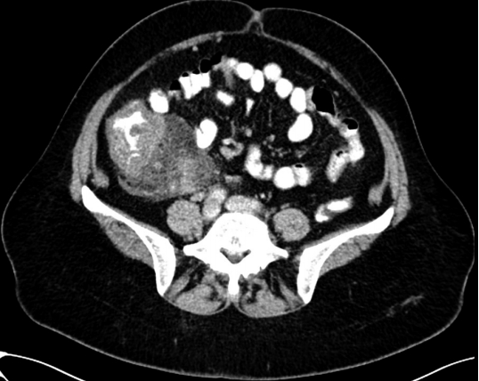
A 57-year-old woman with a history of arterial hypertension and appendectomy in childhood presented to the outpatient clinic, complaining of atypical abdominal pain and diarrhea that had persisted for 1 month. An abdominal computed tomography scan showed circular wall thickening in the ascending colon, as well as enlarged lymph nodes in the terminal ileum (Fig. 1).
Laboratory investigation results of hematological and biochemical parameters, as well as immunoglobulin levels, were all normal. Samples tested negative for the presence of hepatitis A, B, and C viruses, as well as for human immunodeficiency virus. Fecal sample also tested negative for bacteria and parasites. Upper gastrointestinal endoscopy was normal, and Helicobacter pylori infection was not identified.
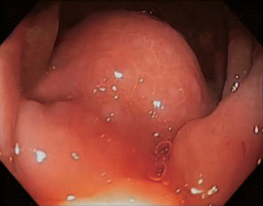
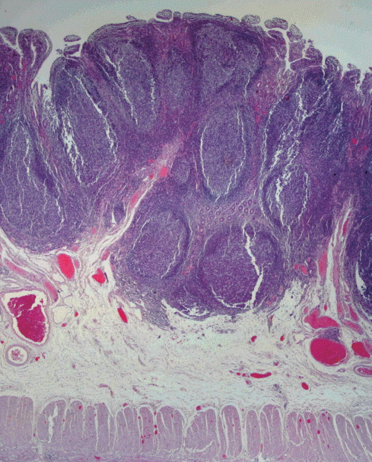
Colonoscopy showed a protruding mass in the hepatic flexure (Supplementary Video 1, Fig. 2). The colonoscope could not be advanced beyond the ascending colon; as a result, complete colonoscopy could not be performed. Histological examination of multiple biopsies of the mass revealed that it had features similar to those of lymphoid lesions. The patientтs clinical symptoms deteriorated and the intestine was almost completely obstructed; for this reason, a laparoscopic right hemicolectomy was performed. A macroscopic assessment of the surgical specimen revealed significant wall thickening in the ascending colon, cecum, and terminal ileum, as well as multiple mucosal nodules (1 to 7 mm) in the same area; the nodules were partially fused together and occupied 2-cm long area of the colon. A histology-based diagnosis of nodular lymphoid hyperplasia (NLH) of the terminal ileum and the colon was confirmed (Fig. 3). Furthermore, moderate chronic inflammation was detected, along with liponecrosis in the serosa. The patient did well post-operatively and since then has been free of symptoms.
Gastrointestinal NLH is characterized by the presence of multiple small nodules of 2 to 10 mm in diameter[1-3]. It primarily affects children, and it is usually asymptomatic; however, it may present with abdominal pain, chronic diarrhea, bleeding, or intestinal obstruction[1,4]. The presentation of NLH in the right colon with an aggressive endoscopic, radiological, and clinical appearance without congenital or acquired immunodeficiency in an adult woman is a rare case.