INTRODUCTION
Intragastric balloons (IGBs) are gaining popularity as a substitute for bariatric surgery [1]. Gastric perforation is a rare complication of IGB insertion and requires operative management. However, most serious complications present with obvious signs. We present a case of gastric perforation, 7 weeks after balloon deployment, with almost no complaint of abdominal pain despite food impaction at the perforation site. We also describe the endoscopic findings in gastric perforation caused by an IGB.
CASE REPORT
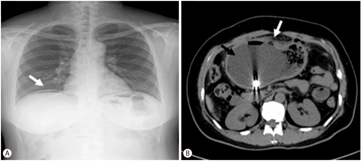
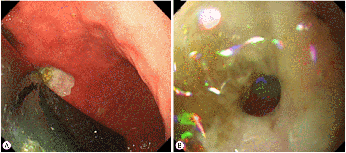
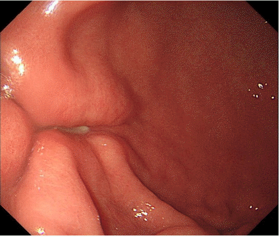
A 47-year-old woman with a body mass index (BMI) of 30 kg/m2 and a weight of 77 kg was admitted to the hospital because of mild epigastric pain. She had undergone IGB (Endball) insertion for weight loss 7 weeks prior, but had not been administered a proton-pump inhibitor (PPI), which is necessary after balloon insertion for at least one month. She had no history of other medication intake, such as non-steroidal anti-inflammatory medications, and she had not undergone any previous abdominal surgeries. She had no other known disease. Before presenting to our hospital, she visited another clinic where she had undergone chest radiography, during which a small amount of free air was seen (Fig. 1A). Physical examination showed mild tenderness, and her laboratory test results were unremarkable. Abdominal computed tomography showed the balloon within the stomach, with intraperitoneal free air (Fig. 1B). The provisional diagnosis was gastric perforation. Upper gastrointestinal endoscopy (GIF-Q260J; Olympus, Tokyo, Japan) was performed using carbon dioxide (CO2) gas to decompress the balloon and to attempt endoscopic closure of the defect. A 0.8-cm gastric ulcer covered with food particles was noted on the anterior wall of the lower gastric body (Fig. 2A). After food particles were removed with a clip device (HX-110LR; Olympus, Tokyo, Japan), a definite perforation was observed at the ulcer base (Fig. 2B, Supplementary Video 1). However, endoscopic closure could not be performed because the ulcer margin showed fibrosis and inflammation. The balloon was punctured and 500 mL of clear fluid was drained. It was difficult to remove the balloon; therefore, we decided to leave it in the stomach. The patient was transferred to the emergency surgery team. Laparoscopic primary repair was performed, and an omental patch was applied to the perforation. Irrigation and washing of the abdominal and pelvic cavities was done and a hemovac was inserted. Endoscopy was performed 7 days after surgery. The perforation was repaired (Fig. 3). Biopsy for histologic detection of Helicobacter pylori was positive. The eradication of H. pylori was then performed. The decompressed IGB was retrieved endoscopically. She was discharged in a stable condition 12 days later and was prescribed PPI.
DISCUSSION
Recently, IGB insertion has been used as an effective minimally invasive endoscopic treatment for morbid obesity. IGB insertion is popular in Western countries due to the simplicity of the procedure and its applicability to various degrees of obesity. Currently, there is a growing need for IGB deployment to treat obesity in Korea. Common adverse events after IGB placement are pain (33.7%) and nausea (29%). Serious adverse events, such as small bowel obstruction, perforation, and death, occur with an incidence of 0.3%, 0.1%, and 0.08%, respectively [2].
Bowel perforation after IGB insertion occurs in 0.1% of cases. The mechanism of perforation is not well understood. Constant pressure from continuous contact of the balloon with the gastric wall may result in a gastric ulcer and induce perforation [3]. Complications of balloon insertion constitute a diagnostic challenge because patients sometimes present with non-specific symptoms [4]. In order to decrease the risk of complications, balloons should not stay in place for more than 6 months [5] and patients should use PPIs [6]. Prior gastric surgery resulting in an anatomical abnormality of the gastrointestinal tract is a contraindication for intragastric implantation [7]. In addition, we recommend H. pylori testing and treatment for the prevention of a peptic ulcer. IGB placement is effective and feasible for the management of obesity. However, physicians should be aware of potential complications. As in our case, perforation can present with mild symptoms.
This is a case of gastric perforation secondary to poor compliance with PPIs, and it was evaluated through endoscopy. To our knowledge, this is the first such report [3]. Although the risk of perforation is low, PPI and H. pylori eradication are important for ulcer prevention following IGB deployment.