Clin Endosc > Volume 51(1); 2018 > Article
Gremida and Rustagi: White Bile in Malignant Biliary Obstruction: A Poor Prognostic Marker
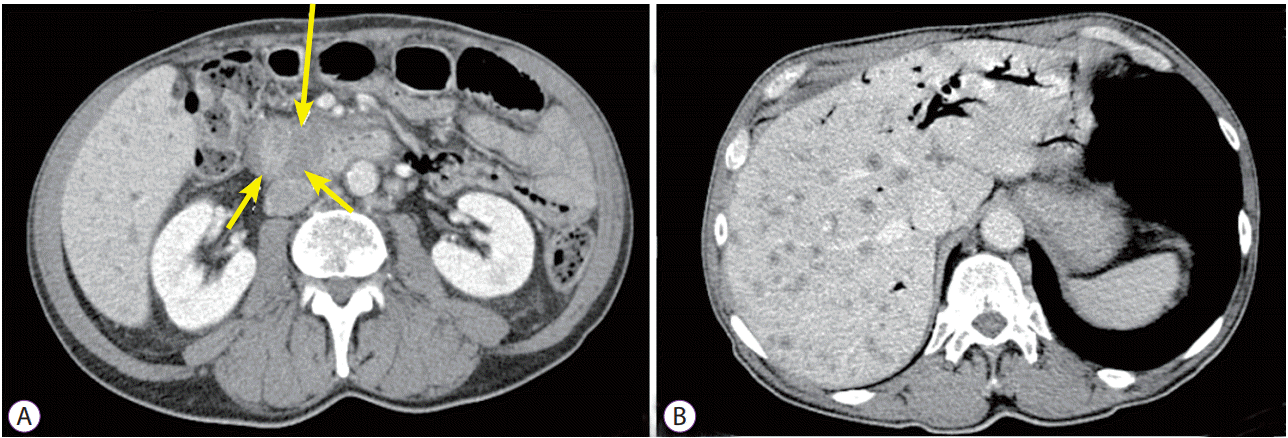
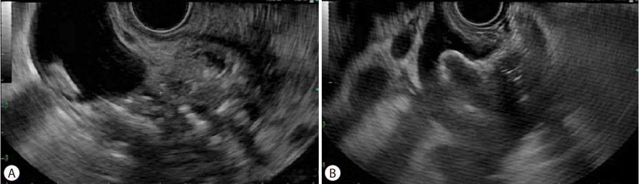
A 71-year-old man presented with progressively worsening painless jaundice. Laboratory findings were significant for severe cholestasis (serum total bilirubin, 24 mg/dL; direct bilirubin, 22 mg/dL). Computed tomography of the abdomen revealed a 4-cm mass in the head of the pancreas with marked extra- and intra-hepatic biliary dilatation (Fig. 1A). Diffuse hepatic metastases and extensive metastatic retroperitoneal and left supraclavicular adenopathy were also noted (Fig. 1B). Endoscopic ultrasound (EUS) revealed a 32×28 mm hypoechoic heterogeneous solid mass in the head of the pancreas, causing complete obstruction of the common bile duct, with marked upstream ductal dilation (Fig. 2A). EUS-guided fine needle aspiration cytology revealed a poorly differentiated pancreatic adenocarcinoma. Endoscopic retrograde cholangiopancreatography for transpapillary biliary stenting was unsuccessful. Subsequently, EUS-guided choledochoduodenostomy using a 15–10 mm, cautery-tipped, biflanged, fully covered, lumen-apposing metal stent (LAMS, Hot AXIOS; Boston Scientific, Marlborough, MA, USA), was performed resulting in the drainage of the colorless “white” bile (Figs. 2B, 3, Video 1).
White bile is a colorless fluid devoid of pigments, bile salts, and cholesterol found in an occluded biliary system. Decolorization of the bile in an obstructed biliary system occurs because the biliary epithelium continues to secrete mucus in stagnant bile. As a result, the bile becomes increasingly lighter in color, and eventually, colorless. This can only occur when there is a loss of communication with the gallbladder, as in cholecystectomy, in an obstructed cystic duct, obstruction above the level of the cystic duct, or non-functioning gallbladder (gallbladder hydrops). On the contrary, if the obstructed biliary duct is in communication with a functioning gallbladder, this will result in a more concentrated bile of darker color, known as “black bile” [1]. Patients with white bile have a predisposition to infection due to ineffective drainage and reduced antimicrobial activity. Furthermore, the finding of white bile in malignant jaundice has been associated with shorter survival than that in patients with green bile [2,3].

NOTES

Conflicts of Interest:The authors have no financial conflicts of interest.

Supplementary Material

Supplementary Video 1

Video 1. Endoscopy showing drainage of the colorless “white” bile through an endoscopic ultrasound-guided choledochoduodenostomy metal stent (https://doi.org/10.5946/ce.2017.111.v001).

Fig. 1.
Computed tomography scan of the abdomen indicating an infiltrating mass in the head of the pancreas (arrows) (A) and numerous hepatic metastatic lesions (B).
ce-2017-111f1.gif
Fig. 2.
Endoscopic ultrasound indicating a solid mass in the head of the pancreas, causing complete obstruction and marked dilation of the upstream bile duct (A). A transduodenal AXIOS (Boston Scientific, Marlborough, MA, USA) stent placed into the dilated bile duct, creating an endoscopic choledochoduodenostomy (B).
ce-2017-111f2.gif
Fig. 3.
Endoscopic appearance of the colorless “white” bile in the duodenum.
ce-2017-111f3.gif

REFERENCES

1. Hashmonai M, Kam I, Schramek A. The etiology of “white bile” in the biliary tree. J Surg Res 1984;37:479–486.
crossref pmid
2. Ahuja V, Garg PK, Kumar D, Goindi G, Tandon RK. Presence of white bile associated with lower survival in malignant biliary obstruction. Gastrointest Endosc 2002;55:186–191.
crossref pmid
3. Geraci G, Sciumè C, Pisello F, Li Volsi F, Facella T, Modica G. Presence of white bile in malignant biliary obstruction is associated with poor prognosis: personal preliminary observations. Langenbecks Arch Surg 2007;392:61–65.
crossref pmid
TOOLS
PDF Links  PDF Links
PubReader  PubReader
ePub Link  ePub Link
XML Download  XML Download
Full text via DOI  Full text via DOI
Download Citation  Download Citation
Supplement  Supplement
  Print
Share:      
METRICS
7
Web of Science
7
Crossref
7
Scopus
5,749
View
140
Download
Related articles
Endoscopic Stenting for Malignant Biliary Obstruction: Results of a Nationwide Experience  2021 September;54(5)
Diagnosis of Malignant Biliary Stricture: More is Better  2018 March;51(2)
Bilateral Metallic Stenting in Malignant Hilar Obstruction  2014 September;47(5)
Endoscopic Stent Placement in the Palliation of Malignant Biliary Obstruction  2011 December;44(2)
EUS-guided Stenting for Malignant Biliary Obstruction  2010 March;42(74)
Editorial Office
Korean Society of Gastrointestinal Endoscopy
#817, 156 Yanghwa-ro (LG Palace, Donggyo-dong), Mapo-gu, Seoul, 04050, Korea
TEL: +82-2-335-1552   FAX: +82-2-335-2690    E-mail: CE@gie.or.kr
Copyright © Korean Society of Gastrointestinal Endoscopy.                 Developed in M2PI
Close layer