Clin Endosc > Volume 54(4); 2021 > Article
Joo and Kim: Gastric Oxyntic Mucosa Pseudopolyps
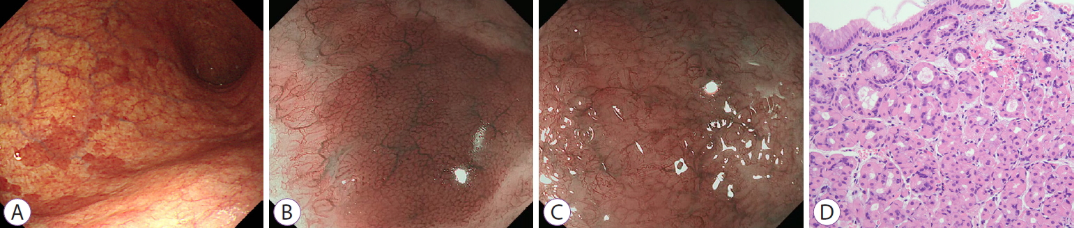
A 76-year-old woman presented to our department for the evaluation of multiple polyps in the stomach, incidentally detected during screening endoscopy. Endoscopy revealed multiple, reddish, nodular lesions with variable sizes in the background of atrophic gastritis in the gastric body and fundus (Fig. 1A). Magnifying endoscopy with narrow-band imaging (MENBI) of the reddish nodular lesions revealed small, round pits surrounded by honeycomb-type subepithelial capillary networks (SECNs) with a regular arrangement of collecting venules (Fig. 1B, Supplementary Video 1). However, ME-NBI of the surrounding atrophic mucosa revealed loss of the normal SECNs and round pits, with an irregular arrangement of the collecting venules (Fig. 1C). Rapid urease test and histological examination for Helicobacter pylori were negative. Endoscopic biopsy of the reddish nodular lesions revealed well-preserved oxyntic glands without evidence of atrophy or intestinal metaplasia, and pseudohypertrophy of parietal cells with protrusion into the gland lumen (Fig. 1D). On the contrary, endoscopic biopsy for surrounding flat mucosa revealed severe atrophic changes with hardly preserved oxyntic glands.
Oxyntic mucosa pseudopolyps are benign lesions observed infrequently during endoscopy and are characterized by relatively preserved non-atrophic oxyntic mucosa, which has polypoidal appearance in the background of atrophic mucosa. Since the biopsy results show normal oxyntic mucosa, these lesions are easily underdiagnosed due to the endoscopists’ lack of knowledge of this disease entity. Some reports suggest the association of oxyntic mucosa pseudopolyps with proton pump inhibitor usage in patients with atrophic gastritis [1]. Although the data regarding the natural course of this lesion are limited, oxyntic mucosa pseudopolyps are not progressive lesions [2]. In summary, when the atrophic mucosal change is present in a large area of the stomach, remnant areas of non-atrophic oxyntic mucosa occasionally appear like pseudopolyps, as in the present case.

NOTES

Conflicts of Interest: The authors have no potential conflicts of interest.
Funding
None.

Supplementary Material

Video 1. Magnifying endoscopy with narrow-band imaging of the reddish nodular lesions and surrounding mucosa (https://doi.org/10.5946/ce.2020.157.v001).

Fig. 1.
(A) On conventional endoscopy, multiple, reddish, nodular lesions with variable sizes are seen in the background of atrophic gastritis in the gastric body and fundus. (B) Magnifying endoscopy with narrow-band imaging (ME-NBI) of the reddish nodular lesions reveals small, round pits surrounded by honeycomb-type subepithelial capillary networks (SECNs) with a regular arrangement of collecting venules. (C) ME-NBI of the surrounding atrophic mucosa reveals loss of the normal SECNs and round pits, with an irregular arrangement of the collecting venules. (D) Endoscopic biopsy of the reddish nodular lesions reveals intact oxyntic mucosa without evidence of atrophy or intestinal metaplasia, and pseudohypertrophy of parietal cells with protrusion into the gland lumen (hematoxylin and eosin stain, ×200).
ce-2020-157f1.jpg

REFERENCES

1. Celikbilek M, Deniz K, Torun E. Image of the month. Gastric oxyntic mucosa pseudopolyposis. Clin Gastroenterol Hepatol 2010;8:e90.
crossref pmid
2. Krasinskas AM, Abraham SC, Metz DC, Furth EE. Oxyntic mucosa pseudopolyps: a presentation of atrophic autoimmune gastritis. Am J Surg Pathol 2003;27:236–241.
crossref pmid
TOOLS
PDF Links  PDF Links
PubReader  PubReader
ePub Link  ePub Link
XML Download  XML Download
Full text via DOI  Full text via DOI
Download Citation  Download Citation
Supplement  Supplement
  Print
Share:      
METRICS
0
Crossref
0
Scopus
4,007
View
146
Download
Related article
Editorial Office
Korean Society of Gastrointestinal Endoscopy
#817, 156 Yanghwa-ro (LG Palace, Donggyo-dong), Mapo-gu, Seoul, 04050, Korea
TEL: +82-2-335-1552   FAX: +82-2-335-2690    E-mail: CE@gie.or.kr
Copyright © Korean Society of Gastrointestinal Endoscopy.                 Developed in M2PI
Close layer