INTRODUCTION
Endoscopic retrograde cholangiopancreatography (ERCP) with endoscopic sphincterotomy (EST) and stone extraction with mechanical lithotripsy are well-established therapeutic procedures for managing a common bile duct (CBD) stone. However, the management of large impacted stones in the CBD is challenging [1,2]. Percutaneous cholangioscopy with electrohydraulic or laser lithotripsy may be effective in these cases.
In patients with severe kyphosis, percutaneous transhepatic bile duct drainage (PTBD) is difficult to perform because the liver is located posterior to the lung. The role of direct peroral cholangioscopy (POC) in the treatment of large impacted CBD stones has not yet been reported in patients with kyphosis. We report the case of a patient with severe kyphosis who had a large stone in the CBD that was fragmented and removed through direct POC by using an ultraslim endoscope with laser lithotripsy.
CASE REPORT
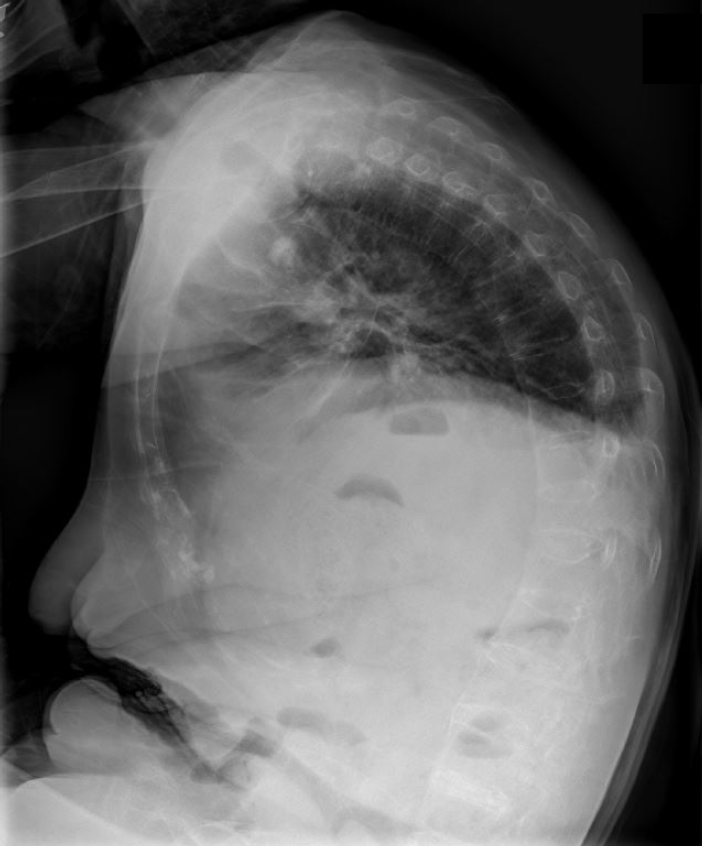
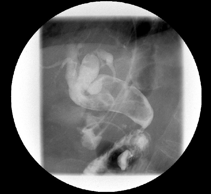
A 75-year-old woman with severe kyphosis was admitted because of acute cholangitis associated with CBD stones. Her vital signs included low blood pressure (90/60 mm Hg) and fever (body temperature, 38.5┬░C). The laboratory findings were as follows: white blood cells, 23,490/mm3; hemoglobin, 8.8 g/dL; platelets, 212,000/mm3; total/direct bilirubin, 10.9/8.31 mg/dL; aspartate aminotransferase, 98 IU/L; alanine aminotransferase, 55 IU/L; and alkaline phosphatase, 2,760 IU/L. Chest radiography revealed severe kyphosis (Fig. 1). Abdominal computed tomography revealed a large CBD stone with distal CBD angulation. Although we initially performed ERCP with EST, the stone could not be captured by using a basket, and a 5-Fr endoscopic nasobiliary drainage (ENBD) catheter was inserted in the right intrahepatic duct for infected bile drainage. Follow-up cholangiography through an ENBD catheter revealed a large filling defect (7├Ś3 cm) impacted in the CBD suggestive of stone (Fig. 2).
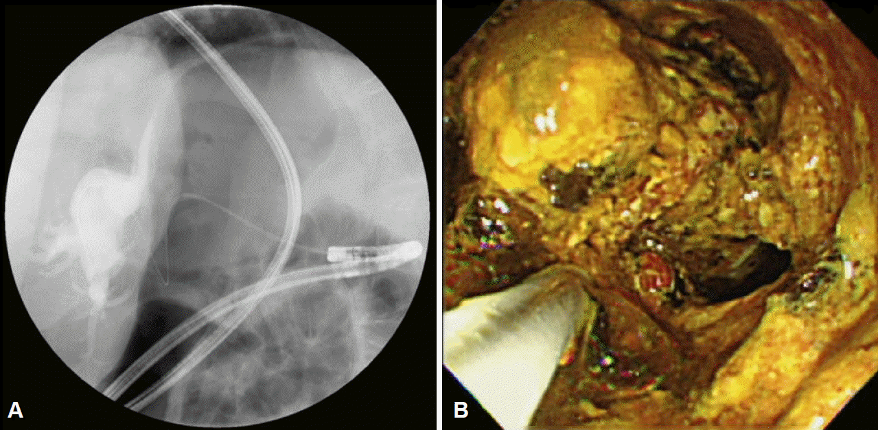
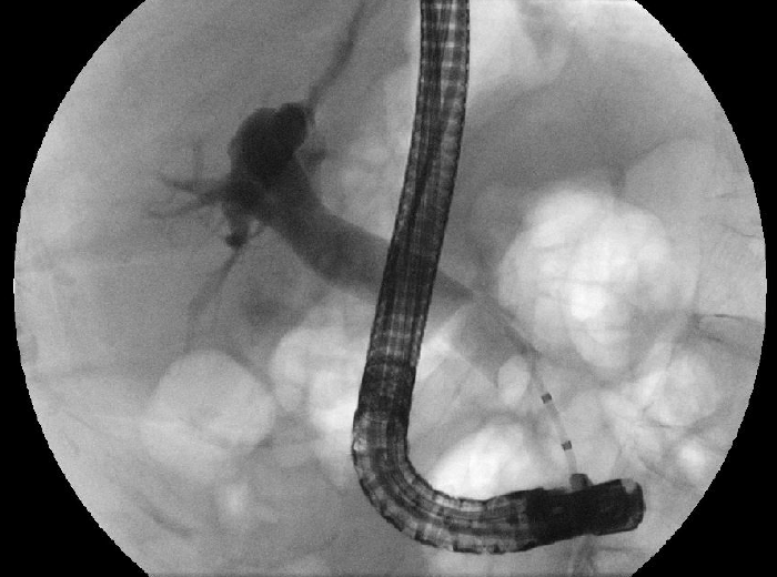
Three days later, her clinical findings had improved. As an alternative approach for the removal of the large stone, cholangioscopy after PTBD was considered. However, altered anatomy due to severe degenerative kyphosis made the percutaneous approach impossible. Therefore, we decided to attempt direct POC by using an ultraslim upper endoscope to break the stone with holmium laser (Lumenis, Chicago, IL, USA). After dilating the ampullary orifice with a 15-mm balloon (CRE wire-guided balloon dilator 15 to 18 mm; Boston Scientific, Natick, MA, USA), an ultraslim endoscope (GIF-XP180N; Olympus Co., Tokyo, Japan) with a 2-mm working channel was advanced into the CBD through the opened ampullary orifice over the guidewire under fluoroscopic and endoscopic control (Fig. 3A). The holmium laser fiber was placed inside a balloon catheter with a 5-Fr channel (MTW Endoskopie; MTW, Wesel, Germany). Then, this balloon catheter with the laser fiber was inserted in the working channel. The large stones impacted in the CBD and left intrahepatic duct were successfully fragmented by using the holmium laser under endoscopic visualization (Fig. 3B), captured by using a Dormia basket (Cook Medical, Bloomington, IN, USA), and then removed from the bile duct without complications. Two days later, we performed ERCP with a duodenoscope to remove any remaining stone fragments. We confirmed no remaining stone in the CBD under direct endoscopic visualization and cholangiography (Fig. 4). The patient was discharged 3 days later.
DISCUSSION
The incidence of bile duct stones increases with age and is associated with drug use (ceftriaxone, clofibrate, oral contraceptives, estrogen replacement, etc.), sex, obesity, and rapid weight loss. The Pima Indians of Arizona have the highest prevalence of bile duct stones worldwide. Whether kyphosis is linked with large bile duct stones is unknown [3].
Generally, most biliary stones can be removed with an extraction balloon, extraction basket, and mechanical lithotripsy after EST. Endoscopic papillary large balloon dilation with or without EST has been shown to be effective for the management of difficult or large bile duct stones in selected patients. However, removal of large CBD stones using the above-mentioned methods may be challenging. Although percutaneous cholangioscopy can be used [4], it is time-consuming, as it comes with a number of disadvantages such as perforation, bleeding, and infection. In the present patient with severe kyphosis, we safely removed a large stone with POC by using an ultraslim endoscope and laser lithotripsy.
Recently, direct POC using an ultraslim upper endoscope has been shown to provide optimal visualization of the biliary system, and the feasibility and efficacy of laser lithotripsy for the management of difficult stone have been demonstrated [5,6]. A therapeutic approach using a direct POC system can be widened by using an ultraslim endoscope with a larger working channel. The peroral approach with an ultraslim endoscope may be more comfortable and less stressful than the conventional ERCP approach because it is associated with fewer adverse effects on cardiopulmonary function and is better tolerated by the patients. However, approaching the bile duct using this endoscope can be difficult. This is due to stomach and duodenum loops that cause the endoscope to slip out of the bile duct [7]. Therefore, insertion of these endoscopes in the bile duct, over a previously placed guidewire and extraction balloon, has been suggested.
In patients with severe kyphosis, PTBD is difficult to perform because the liver is located posterior to the lung. Patients with severe kyphosis represent a challenging group to anesthesiologists and surgeons because the disease is associated with difficult intubation, restrictive ventilatory defects, and cardiac problems [8]. In the present case, we successfully inserted an ultraslim endoscope in the CBD without difficulty while the patient was in the lateral position. We thought that the patientŌĆÖs diaphragm or lung might prevent the looping of the endoscope, and this helped us to easily insert the endoscope in the CBD.
The laser probe can be easily inserted through the large working channel of the ultraslim endoscope, and the stone can be easily crushed under endoscopic vision. The holmium and frequency-doubled double pulsed Nd:YAG lasers have been used for managing urinary and bile duct stones [6,9-11]. Holmium laser lithotripsy may have high fragmentation rates for biliary stones, but its thermal effects on soft tissue may be associated with higher complication rates, including hemobilia [12]. Control of shock waves depends on visual guidance only. The ultraslim endoscope is equipped with an optimal working channel for inserting instruments required for stone fragmentation and permits easy targeting of the bile duct stones with good visualization. Effective visualization of the bile duct stone can decrease damage to the hepatobiliary system [13]. Therefore, we expect that holmium laser lithotripsy can be useful in treating patients with bile duct stones.
In conclusion, PTBD procedures with percutaneous transhepatic cholangioscopy are difficult to implement in patients with severe kyphosis. Therefore, holmium laser lithotripsy under direct POC using an ultraslim endoscope may be a feasible and effective procedure for managing large bile duct stones in patients with severe kyphosis.