Clin Endosc > Volume 50(5); 2017 > Article
Kwon, Kim, and Hong: Immediate Endoscopic Management of an Intramural Hematoma Developed during Colonoscopy
An intramural hematoma of the gastrointestinal tract could be precipitated by an endoscopic examination, especially in patients being administered anticoagulation therapy or those with concomitant hematological diseases [1]. Although an intramural hematoma can be primarily associated with a biopsy or therapeutic procedure, occurrence of a spontaneous intramural hematoma during a diagnostic colonoscopy has been reported in a few cases [2-4]. Intramural hematomas are known to cause partial or complete bowel obstruction [5], and surgical treatment should be considered in patients with evidence of intestinal necrosis, peritonitis, or a deteriorating general condition despite conservative management [6,7]. Early detection and treatment of a hematoma, can prevent bowel obstruction and obviate the need for surgery. Our report describes early endoscopic management of an intramural hematoma, which occurred during a diagnostic colonoscopy.
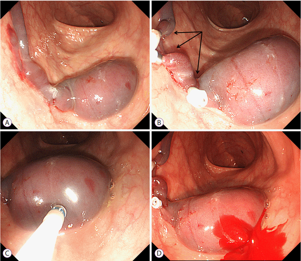
A 78-year-old man underwent colonoscopy during scheduled follow-up for the management of ulcerative colitis. He had not received anticoagulation therapy, and laboratory tests revealed a normal platelet count and coagulation profile. During colonoscopic examination, a large elongated intramural hematoma with a normal mucosa was seen to develop in the sigmoid colon (Fig. 1A). We concluded the intramural hematoma might have occurred as a consequence of a continuous suctioning process employed during the procedure due to poor bowel preparation. Even after 10 minutes of observation, we found that the hematoma continued to grow in size due to active submucosal bleeding. We placed three endoscopic clips (HX-610-090L; Olympus, Tokyo, Japan) at the suspected sites of active bleeding (Fig. 1B, arrows), which led to cessation of growth of the hematoma. To determine reliably, a Dual knife (Olympus, Tokyo, Japan) was applied to excise the hematoma at its center and thereby evacuate it (Fig. 1C). An electrosurgical unit (VIO 300D; ERBE Elektromedizin, Tübingen, Germany) with an Endocut I setting (effect 3, duration 3, interval 3) was used as a power source. After excision, we noted immediate active bleeding (Fig. 1D) followed by flattening of the hematoma without secondary bleeding (Fig. 2A). Follow-up examination after 3 hours of observation showed no re-growth of the hematoma, and no further bleeding was documented (Fig. 2B). The patient was discharged without admission or need for further treatment.

NOTES

Conflicts of Interest: The authors have no financial conflicts of interest

Fig. 1.
Colonoscopic images showing an intramural hematoma. (A) An intramural hematoma observed in the sigmoid colon. (B) Rapid increase in the size of the intramural hematoma, and placement of three endoscopic clips at the suspected sites of active bleeding (arrows). (C) Application of Dual knife (Olympus, Tokyo, Japan) to excise and evacuate the hematoma. (D) Active bleeding noted from the hematoma.
ce-2017-037f1.gif
Fig. 2.
Follow-up colonoscopic images after endoscopic treatment. (A) Flattened hematoma without secondary bleeding. (B) No evidence of further bleeding after 3 hours of observation.
ce-2017-037f2.gif

REFERENCES

1. Liu Y, Yang S, Tong Q. Spontaneous intramural hematoma of colon. Clin Gastroenterol Hepatol 2012;10:e38.
crossref
2. Gallo D, Tebrock C, Rivera D. Intramural cecal hematoma: an unusual complication of colonoscopy. Gastrointest Endosc 2003;57:254–257.
crossref pmid
3. Jongwutiwes U, Shaukat A, Pocha C. Image of the month. Intramural cecal hematoma: a rare complication after colonoscopy. Clin Gastroenterol Hepatol 2009;7:A32.
crossref
4. Katsurahara M, Horiki N, Kitade T, et al. Acute colonic intramural hematoma: a rare complication of colonoscopy. Endoscopy 2014;46 Suppl 1 UCTN:E180–E181.
crossref pmid pdf
5. Hughes CE 3rd, Conn J Jr, Sherman JO. Intramural hematoma of the gastrointestinal tract. Am J Surg 1977;133:276–279.
crossref pmid
6. Kwon K, Cheung DY, Seo Y, et al. Supportive management resolved a colonic intramural hematoma in an anticoagulant user. Intern Med 2014;53:1505–1509.
crossref pmid
7. Lobo L, Koudki R, Prasad Hl K, Shetty B. Colon obstruction due to an anticoagulant induced intramural haematoma; a rare case report. J Clin Diagn Res 2013;7:739–741.
crossref pmid pmc
TOOLS
PDF Links  PDF Links
PubReader  PubReader
ePub Link  ePub Link
XML Download  XML Download
Full text via DOI  Full text via DOI
Download Citation  Download Citation
  Print
Share:      
METRICS
4
Web of Science
4
Crossref
3
Scopus
5,666
View
101
Download
Related articles
Clinical Practice Guidelines for the Endoscopic Management of Peripancreatic Fluid Collections  2021 July;54(4)
Endoscopic Management of Post-Polypectomy Bleeding  2020 May;53(3)
Endoscopic Management of Iatrogenic Colon Perforation  2020 January;53(1)
Endoscopic Management of Gastroesophageal Reflux Disease: Revisited  2016 September;49(5)
External Validation of the Endoscopic Features of Sessile Serrated Adenomas in Expert and Trainee Colonoscopists  2017 May;50(3)
Editorial Office
Korean Society of Gastrointestinal Endoscopy
#817, 156 Yanghwa-ro (LG Palace, Donggyo-dong), Mapo-gu, Seoul, 04050, Korea
TEL: +82-2-335-1552   FAX: +82-2-335-2690    E-mail: CE@gie.or.kr
Copyright © Korean Society of Gastrointestinal Endoscopy.                 Developed in M2PI
Close layer