INTRODUCTION
Mirizzi syndrome (MS) is a rare complication of gallbladder (GB) disease caused by an impacted stone in the GB infundibulum or cystic duct, which causes partial or complete obstruction of the bile duct [1]. Open surgery is considered the gold standard treatment for MS, although it is invasive, has a high rate of complications, requires long postoperative hospital stay, and has high long-term morbidity [2]. Minimally invasive approaches that combine endoscopic treatment with laparoscopic cholecystectomy (LC) for the management of MS have been proposed in selected cases [3,4]. However, endoscopic treatment often fails because of the inability to access or to capture the impacted cystic duct stone. Digital single-operator cholangioscopy (DSOC)-guided electrohydraulic lithotripsy (EHL) is an effective modality for the management of difficult biliary stones [5]. Herein, we present a case of MS that was successfully managed by DSOC-guided EHL with endoscopic nasogallbladder drainage (ENGBD) and interval LC.
CASE REPORT
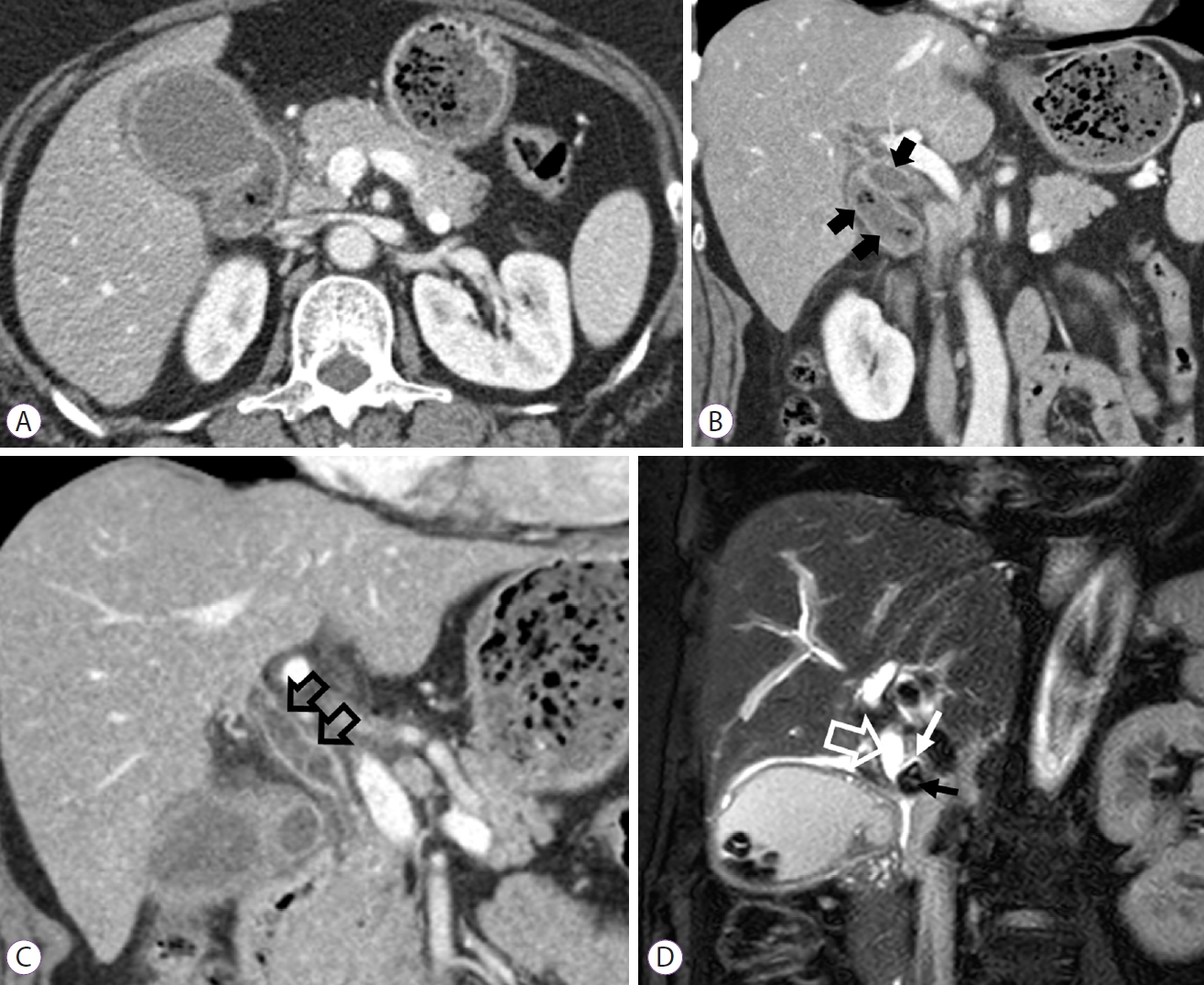
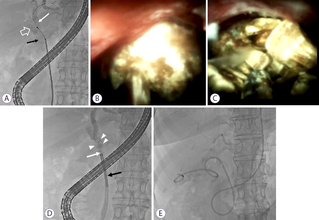
A 59-year-old woman was admitted to our hospital with upper abdominal pain and fever, with a temperature of 38.5┬░C. She had mild jaundice with scleral icterus and MurphyŌĆÖs sign. Laboratory findings included a white cell count of 14,000/┬ĄL, total bilirubin of 5.06 mg/dL, direct bilirubin of 3.51 mg/dL, aspartate aminotransferase of 918 U/L, alanine aminotransferase of 403 U/L, alkaline phosphatase of 290 U/L, and C-reactive protein of 12.25 mg/L. An abdominal computed tomography scan showed rim-like enhancement with inflammatory fat stranding around the GB (Fig. 1A). A dilated common hepatic duct and multiple gallstones along the dilated cystic duct and GB were observed, although the common bile duct (CBD) was not dilated (Fig. 1B, C). The T2-weight magnetic resonance cholangiopancreatography image demonstrated thickening of the GB wall with multiple stones. A stone measuring approximately 1.4 cm was observed at the junction of the cystic duct and common hepatic duct, with a cholecysto-cholecochal fistula. These findings were compatible with MS type III [6] and acute calculus cholecystitis (ACC) (Fig. 1D). First, we attempted to remove the stone using conventional endoscopic retrograde cholangiopancreatography (ERCP), although our efforts were unsuccessful (Fig. 2A). Thus, we decided to perform DSOD-guided EHL using the SpyglassTM DS Direct Visualization system (SpyDS) (Boston Scientific, Natrick, MA, USA). SpyDS was successfully inserted into the confluence of the cystic duct and common hepatic duct. Upon visualizing the impacted stone, SpyDS-guided EHL was conducted and fragmented stones were observed during cholangiography (Fig. 2B-D). Subsequently, the stones were completely removed using a basket and ENGBD was performed as a bridging therapy for ACC (Fig. 2E). No procedure-related complications occurred. Interval LC was performed 7 days after the procedure. According to the surgical findings, the adhesion between the GB and common hepatic duct remained, although LC was safely performed using the antegrade fundus-down technique and direct ligation of the cystic duct. The patient was discharged 5 days later.
DISCUSSION
To date, there are no consensus guidelines for management of MS. Treatment strategies for MS may be individualized depending on the grade of MS, adhesion, local anatomy, and/or surgeonŌĆÖs preference; however, most cases require surgery. Laparotomy is regarded as the gold standard treatment for MS. Minimally invasive approaches may also be considered but remain controversial because of severe adhesion in CalotŌĆÖs triangle, increased risk of bile duct injury, and a high open conversion rate [2]. However, several recent studies reported that various laparoscopic approaches following endoscopic management are feasible and safe, with the advantages of decreased invasiveness, fewer postoperative complications, less operative blood loss, and shorter hospital stay than laparotomy [7].
ERCP is not only the gold standard for diagnosing MS, but also a bridging therapeutic tool before surgery [8]. Placement of an endoscopic nasobiliary drainage tube or biliary stent during ERCP is important for biliary decompression and guiding the shape of the CBD during surgery, thus facilitating a minimally invasive laparoscopic approach for MS. In particular, removal of stones at the confluence of the cystic duct and common hepatic duct during ERCP makes it unnecessary to incise the bile duct during surgery, thus reducing bile duct injury. In our patient, removal of the impacted cystic duct gallstone was not successful by conventional ERCP because we could no entrap the stone using a basket. Therefore, we used SpyDS-guided EHL and achieved complete stone removal after fragmentation without any complications. When biliary stones are difficult to remove using conventional ERCP, EHL or laser lithotripsy using the SpyDS system may be a viable alternative with a high procedural success rate and lower radiation exposure [9]. Several studies have also shown a high success rate for management of MS (Table 1) [10-13]. The incidence of SpyDS-related complications such as acute cholangitis, acute pancreatitis, and bleeding is low, ranging from approximately 2.5% to 16.7%, and tends to mimic complications of conventional ERCP [14].
Early LC is the mainstay of the management of ACC. However, percutaneous cholecystostomy (PC) is an alternative for patients who are unfit for early LC due to severe comorbidities, liver function abnormalities, or advanced age. PC has high technical and clinical success rates with a low incidence of adverse events. However, post-procedural pain, inadvertent catheter removal, and/or low quality of life after the procedure are limitations of PC that should be considered. ENGBD is a useful alternative to PC. In a systematic review, the technical and clinical success rates of ENGBD were 81% and 75%, respectively. The procedural adverse event rate was 3.6% [15]. Recent studies have shown that SpyDS-assisted cystic duct cannulation increases the technical success rate of endoscopic transpapillary GB drainage when the cystic duct is invisible or is difficult to cannulate. Procedure-related complications are mainly associated with biliary cannulation, sphinterotomy, or inadvertent removal of the drainage tube or catheter [16,17]. In our case, early LC could not be performed because of elevated liver function test results; therefore, PC was required for GB decompression. However, ENGBD was successfully performed during the same session as ERCP after biliary stone removal. An advantage of ENGBD is that it is a more physiologic approach, and PC is not required. In addition, ENGBD may help the surgeon to visualize the cystic duct anatomy during LC, contributing to a safer procedure.
Treatment of MS is mainly surgical and comprises partial or complete cholecystectomy with or without CBD exploration. Conventionally, complex cases require choledochoplasty with a GB flap and T-tube placement, although extensive erosions of the bile duct are best managed with a bilioenteric anastomosis, such as a Roux-en-Y hepaticojejunostomy [18]. However, in the era of minimally invasive treatment, laparoscopic treatment of low-grade MS is continually being attempted. Csendes et al. [6] reported significantly higher morbidity for patients with MS types III and IV treated with an open procedure than for patients from other groups. In our case, preoperative intervention using DSOC-guided EHL downgraded the MS, resulting in a safe laparoscopic surgical approach. LC could be performed safely using an antegrade fundus-down technique even though there was inflammation in CalotŌĆÖs triangle and adhesion between the neck and common hepatic duct.
Based on our experience, DSOC-guided EHL with ENGBD is a feasible minimally invasive approach for the management of high-grade MS.