Clin Endosc > Volume 56(3); 2023 > Article
Nose, Ishida, Makino, Takahashi, Kurokawa, Eguchi, Doki, and Nakajima: Is simply covering the patient's mouth with a surgical mask during transnasal endoscopy sufficient as an anti-COVID-19 measure?
The transmission risk from infected patients to healthcare workers and/or other patients is a concern during the coronavirus disease 2019 (COVID-19) pandemic.1 Especially when performing digestive endoscopy, it seems inevitable that healthcare workers will be exposed to either respiratory or gastrointestinal fluids from patients.2 Therefore, appropriate infection control during digestive endoscopy based on experience is recommended by the World Endoscopy Organization.3 We read with interest the papers reported by Higashimori et al.4 and Nadatani et al.5 In these studies, nasal insertion of a thin endoscope and covering of the patient's mouth with a surgical mask prevented the spread of aerosol droplets due to coughing during the procedure. We support the studies’ findings and suggest that this simple infection control measure be implemented for all screening endoscopy cases. Conversely, several reports on gas leakage or release of fluid droplets from biopsy valves of full-sized endoscopes during insertion and withdrawal of endoscopic instruments have been published.6,7 Gas leakage may be observed even in thin endoscopes. If so, transnasal endoscopy with a surgical mask may be inadequate as an infection control measure, especially when performing procedures requiring instrument traffic, such as biopsies. Therefore, we aimed to visualize the gas leakage from the biopsy valve of the thin endoscope and to prove that simply covering the mouth with a mask is insufficient to prevent COVID-19 infection during endoscopy.
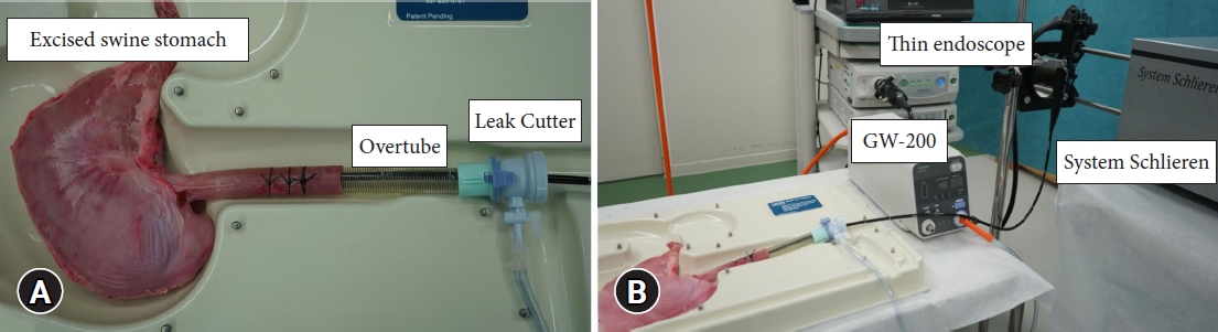
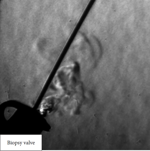
We used System Schlieren (SS100; Kato Koken Co., Ltd.) to visualize the gas leakage in this study. More information on this system can be found on the following website: https://kk-co.jp/products/schlierens.php. System Schlieren is a device that visualizes minute changes in airflow that are invisible to the naked eye by identifying differences in the refractive index of materials. This method is extremely useful for the visualization and evaluation of high-speed phenomena, mainly in the engineering field.8,9 The following experiments were conducted with the intra-gastric pressure of an excised swine stomach maintained at 8 mmHg using a carbon dioxide insufflation system (GW-200; Fujifilm). A thin endoscope (EG-6400N; Fujifilm) was advanced into the stomach, and gas leakage from the biopsy valve during insertion and withdrawal of the biopsy forceps was visualized (Fig. 1). In this experiment, the biopsy forceps for the thin endoscope with an outer diameter of 1.8 mm (Radial Jaw 4P; Boston Scientific) was inserted and withdrawn 3 cm every 3 seconds. This procedure was repeated five times.
Gas leakage from the biopsy valve of the thin endoscope was successfully visualized during forceps insertion and withdrawal (Fig. 2, Supplementary Video 1) in all five procedures, indicating that endoscopists and assistants may be exposed to potentially virus-infected aerosols. Even if the patient’s mouth is covered with a surgical mask, gas leakage cannot be prevented if the biopsy valve is airtight. Therefore, endoscopists and assistants must consider this and take further precautions against COVID-19. To optimize precaution, the development of a reinforced, improved, and leak-proof type biopsy valve is a pressing task.
The entire experimental protocol of this study was approved by the institutional animal care and ethical review board (IVTeC Co., Ltd., Animal Welfare Committee). This study was performed according to the Guide for the Care and Use of Laboratory Animals.

Supplementary Material

Supplementary Video 1. Gas leakage visualization from the biopsy valve of the thin endoscope during endoscopic procedures (https://doi.org/10.5946/ce.2022.199.v001).
Supplementary materials related to this article can be found online at https://doi.org/10.5946/ce.2022.199.

NOTES

Conflicts of Interest
The authors have no potential conflicts of interest.
Funding
None.
Acknowledgments
The authors would like to thank all the workers who participated in the study.
Author Contributions
Conceptualization: KN; Data curation: YN, TI, KN; Formal analysis: YN, TI, YD, KN; Investigation: YN, TI, KN; Methodology: YN, TI, KN; Project administration: YN, TI, KN; Resources: YN, TI, KN; Supervision: YN, TI, TM, TT, YK, HE, YD, KN; Validation: YN, TI, KN; Visualization: YN, TI, KN; Writing–original draft: YN; Writing–review & editing: all authors.

Fig. 1.
The experimental model. (A) A Leak Cutter (TOP) was connected to a standard endoscopic overtube (#16400; TOP). A thin endoscope was advanced into an excised swine stomach through the overtube. A carbon dioxide insufflation system was connected to the side channel of the Leak Cutter. (B) The gas leakage from the biopsy valve of the thin endoscope during endoscopic procedures was visualized using System Schlieren (SS100; Kato Koken Co., Ltd.), while the intra-gastric pressure of the excised swine stomach was maintained at 8 mmHg.
ce-2022-199f1.jpg
Fig. 2.
Gas leakage visualization from the biopsy valve of the thin endoscope during endoscopic procedures.
ce-2022-199f2.jpg

REFERENCES

1. Gómez-Ochoa SA, Franco OH, Rojas LZ, et al. COVID-19 in health-care workers: a living systematic review and meta-analysis of prevalence, risk factors, clinical characteristics, and outcomes. Am J Epidemiol 2021;190:161–175.
crossref pmid pmc pdf
2. Soetikno R, Teoh AY, Kaltenbach T, et al. Considerations in performing endoscopy during the COVID-19 pandemic. Gastrointest Endosc 2020;92:176–183.
crossref pmid pmc
3. Guda NM, Emura F, Reddy DN, et al. Recommendations for the operation of endoscopy centers in the setting of the COVID-19 pandemic: World Endoscopy Organization guidance document. Dig Endosc 2020;32:844–850.
crossref pmid pmc pdf
4. Higashimori A, Maruyama H, Nadatani Y, et al. The "New Normal" following the COVID-19 pandemic: a simple infection-prevention measure using a surgical mask during transnasal endoscopy. Clin Endosc 2021;54:618–620.
crossref pmid pmc pdf
5. Nadatani Y, Higashimori A, Takashima S, et al. A mask-based infection control method for screening endoscopy may prevent SARS-CoV-2 transmission and relieve staff anxiety. SAGE Open Med 2021;9:20503121211047060.
crossref pmid pmc pdf
6. Urakawa S, Hirashita T, Momose K, et al. Intraluminal gas escape from biopsy valves and endoscopic devices during endoscopy: caution advised during the COVID-19 era. Endosc Int Open 2021;9:E443–E449.
crossref pmid pmc
7. Keil R, Hlava Š, Stanovský P, et al. Commonly available but highly effective protection against SARS-CoV-2 during gastrointestinal endoscopies. PLoS One 2021;16:e0254979.
crossref pmid pmc
8. Su C, Wang S, Shih C, et al. Visualization of hot flows of tall space fires in model experiments with Schlieren Photography technique. Springerplus 2016;5:1744.
crossref pmid pmc pdf
9. Glosse P, Denneler S, Stier O, et al. Investigation of the powder aerosol deposition method using shadowgraph imaging. Materials (Basel) 2021;14:2502.
crossref pmid pmc
TOOLS
PDF Links  PDF Links
PubReader  PubReader
ePub Link  ePub Link
XML Download  XML Download
Full text via DOI  Full text via DOI
Download Citation  Download Citation
Supplement  Supplement
  Print
Share:      
METRICS
0
Crossref
0
Scopus
1,991
View
99
Download
Related article
Editorial Office
Korean Society of Gastrointestinal Endoscopy
#817, 156 Yanghwa-ro (LG Palace, Donggyo-dong), Mapo-gu, Seoul, 04050, Korea
TEL: +82-2-335-1552   FAX: +82-2-335-2690    E-mail: CE@gie.or.kr
Copyright © Korean Society of Gastrointestinal Endoscopy.                 Developed in M2PI
Close layer