Skip Navigation
Skip to contents

Clin Endosc : Clinical Endoscopy

OPEN ACCESS

Articles

Page Path
HOME > Clin Endosc > Volume 46(5); 2013 > Article
Special Issue Article of IDEN 2013
Review
Confocal Microscopy in the Esophagus and Stomach
Adam Templeton, Joo Ha Hwang
Clinical Endoscopy 2013;46(5):445-449.
DOI: https://doi.org/10.5946/ce.2013.46.5.445
Published online: September 30, 2013

Division of Gastroenterology, Department of Medicine, University of Washington, School of Medicine, Seattle, WA, USA.

Correspondence: Joo Ha Hwang. Division of Gastroenterology, Department of Medicine, University of Washington, School of Medicine, 1959 NE Pacific St, Box 356424, Seattle, WA, USA. Tel: +1-206-685-2283, Fax: +1-206-221-3992, jooha@u.washington.edu
• Received: June 10, 2013   • Accepted: July 4, 2013

Copyright © 2013 Korean Society of Gastrointestinal Endoscopy

This is an Open Access article distributed under the terms of the Creative Commons Attribution Non-Commercial License (http://creativecommons.org/licenses/by-nc/3.0/) which permits unrestricted non-commercial use, distribution, and reproduction in any medium, provided the original work is properly cited.

  • 8,429 Views
  • 61 Download
  • 11 Crossref
  • 12 Scopus
prev next
  • Probe-based confocal microscopy (pCLE) is actively being investigated for applications in the esophagus and stomach. The use of pCLE allows real-time in vivo microscopy to evaluate the microarchitecture of the mucosal epithelium. pCLE appears to be particularly useful in identifying mucosal dysplasia and early malignancies that cannot be clearly distinguished using high-definition white light endoscopy, chromoendoscopy, or magnification endoscopy. In addition, the ability to detect dysplastic tissue in real-time may shift the current screening practice from random biopsy to targeted biopsy of esophageal and gastric cancers and their precursor lesions. We will review the use of pCLE for detection and surveillance of upper gastrointestinal early luminal malignancy.
The endoscopic use of confocal microscopy for in vivo diagnosis is a relatively new application of a previously ex vivo technology.1 Probe catheters range in size from 0.9- to 2.5-mm allowing their use through any endoscope with a 10 Fr working channel. With a video frame rate of 12 frames per second, probe-based confocal microscopy (pCLE) provides in vivo imaging of capillary flow and real-time microscopic scanning ability. The resolution is approximately 1 µm and 240 to 600 µm field of view.2,3 A contrast agent, typically intravenous fluorescein, is administered to highlight the connective tissue of the lamina propria and the vascular rich epithelium. Mucin containing goblet cells remain dark, as does neoplastic tissue.3,4 This contrast effect is present 10 minutes after administration and lasts up to 45 minutes.5 Fluorescein has long been used for ophthalmologic indications and its safety has recently been confirmed for use as a contrast agent in the gastrointestinal tract.6
Due to the small field of view and high magnification, optimal image acquisition is highly dependent on proper positioning. The best images are obtained with the probe perpendicular to the mucosa. Some operators have found benefit from the use of a clear 4-mm cap placed on the tip of the endoscope, as this allows gentle suctioning to keep the probe in place.3 Imaging is best done before biopsy or resection, as bleeding can obscure imaging due to fluorescein leakage.
The current method for surveillance of Barrett esophagus (BE) is both time consuming and expensive. Targeted biopsies of endoscopically visible lesions are then followed by four-quadrant biopsies-every 1 cm for dysplastic BE and every 2 cm for nondysplastic BE. Society guidelines recommend endoscopic surveillance of all patients with established BE.7,8 However, adherence rates to surveillance biopsy guidelines have been reported to be as low as 51% in the community setting.9 Therefore, a method that improves the efficiency (cost and time) of performing endoscopic surveillance in patients with BE would have a positive impact on the management of patients with BE from both a clinical and economic standpoints.
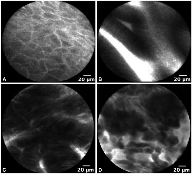
Probe-based confocal laser endomicroscopy is currently used in clinical trials as a tool for BE imaging. The Miami criteria for differentiating nondysplastic BE from dysplastic BE and adenocarcinoma was proposed in 2011.3 Normal squamous epithelium is defined as flat cells without crypts or villi, with bright vessels found within papillae (Fig. 1A). Nondysplastic BE has a uniform villiform architecture and columnar cells interspersed with dark goblet cells (Fig. 1B). High-grade dysplasia (HGD) retains a villiform structure but has dark, irregularly thickened epithelial borders, and dilated irregular vessels (Fig. 1C). In contrast, adenocarcinoma has disorganized or absent villiform structure, multiple dark columnar cells, and dilated irregular vessels (Fig. 1D). This classification was recently validated and notably, after a short formal training session, there was high interobserver agreement and accuracy between experts and recent trainees.10
Current screening and surveillance protocols rely on biopsy to differentiate BE, dysplastic BE, and adenocarcinoma. This requires endoscopists to carefully document biopsy location and, once the pathology returns dysplastic, the patient must return for endoscopic ablation. The use of pCLE theoretically will allow surveillance of a greater number of locations, reduce the total number of biopsies taken, and increase the yield of biopsy for dysplasia or cancer. Two studies have recently demonstrated the potential feasibility of this approach (Table 1). Bajbouj et al.11 reported their experience with 68 patients undergoing BE surveillance. Six hundred and seventy biopsies were performed and 11 patients (16%) had HGD or esophageal cancer (EC) found in 58 biopsies. In this study, onsite analysis and blinded review of pCLE images were performed. The specificity and negative predictive value of pCLE in excluding neoplasia was 0.97 (90% confidence interval [CI], 0.95 to 0.98) and 0.93 (90% CI, 0.91 to 0.95), which had little change in blinded evaluation with specificity of 0.95 (90% CI, 0.90 to 0.98) and a negative predictive value of 0.97 (90% CI, 0.95 to 0.98). A randomized multicenter trial using pCLE in BE demonstrated pCLE to be superior to high-definition white light (HDWL) endoscopy for detection of BE HGD. The investigators looked at the use of pCLE, HDWL, and narrow band imaging (NBI) in 101 patients, 31 of whom had EC (31%), who had a total of 874 biopsies performed. If biopsies were only taken in patients with abnormalities detected on any one of the three imaging modalities, 39% of patients could forgo biopsy without missing HGD or EC.12 These studies indicate that pCLE could decrease biopsy use, and given the high negative predictive value, provide increased reassurance for patients with negative tests.
Another area that pCLE has potential to impact management of patients with BE is in patients undergoing ablative therapy. Ablative therapy is now recommended for patients with BE with flat HGD.1 Patients often require between 2 and 10 ablation sessions to obtain remission.13,14 Probe-based CLE may play a role guiding therapy before ablation, during ablation sessions, and in the lifelong screening following these sessions. A multicenter randomized clinical trial comparing HDWL endoscopy to HDWL+pCLE in the detection of residual BE in patients who have had ablative therapy was recently performed, however was halted early due to a higher rate of residual Barrett's in both treatment arms.15 Further study is needed to determine the utility of pCLE in differentiating mucosa after ablative therapy. If feasible, given the rise in ablative therapy for BE, pCLE may develop a greater role in guiding and assessing therapy.
The potential applications for using pCLE in the stomach are similar to that of the esophagus, especially in populations with a high incidence of gastric cancer. The current method for endoscopic gastric cancer screening typically involves a careful visual examination with white light endoscopy, targeted biopsies of endoscopically visible lesions, and then random biopsies in various regions of the stomach (antrum, body, and incisura). As with Barrett's screening and surveillence, this approach is both time consuming and expensive. Furthermore, the large surface area of the stomach means this approach is even more prone to undersampling of abnormal tissue and oversampling of normal tissue. The use of chromoendoscopy, NBI, or other digital enhancement techniques has increased the accuracy of detecting gastric intestinal metaplasia (GIM) and early gastric cancer (EGC); however, these techniques are limited by significant interobserver variability.12-19 Moreover, while these modalities have increased the sensitivity and specificity of biopsy screening, no study has demonstrated the ability to accurately diagnose dysplasia or GIM-the known precursor lesions for gastric adenocarcinoma. As such, suspicious areas still require biopsy to confirm the presence of GIM or EGC and no studies have demonstrated the ability to accurately diagnose dysplasia.
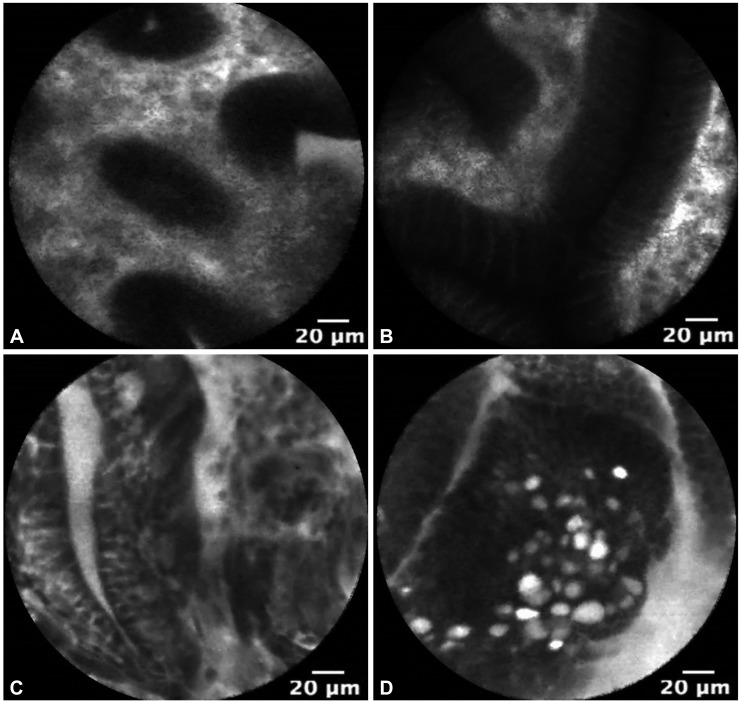
The use of pCLE is currently under investigation for the in vivo diagnosis of GIM, dysplasia, and EGC. Fig. 2 demonstrates pCLE images of normal gastric mucosa, GIM, gastric HGD, and EGC. In a recent study, pCLE performed before endoscopic resection of superficial gastric neoplasia was compared to conventional endoscopic biopsy. pCLE had significantly improved agreement over conventional biopsy for the diagnosis of adenocarcinoma (κ=0.824 vs. κ=0.617; p<0.001) and an accuracy of 91.7% compared to 85.2% for conventional biopsy (p=0.065).20 Guo et al.19 additionally demonstrated 98% sensitivity and 95% specificity for GIM using confocal microscopy. Notably, the learning curve for pCLE detection of GIM is short, typically obtaining acceptable intraobserver agreement with 1 to 2 hours of training.21 Yet to be explored is whether pCLE can improve the diagnosis of dysplasia following gastric endoscopic mucosal resection or endoscopic submucosal resection.
Similar to the applications in BE, pCLE has the potential to identify dysplasia and EGC in vivo to increase the yield of biopsies and to identify lesions that may warrant endoscopic resection. Further studies are needed to fully evaluate the potentially valuable application of pCLE for gastric malignancy.
Probe-based CLE is a promising technology with several potential applications in the management of patients with BE and GIM. One of the limitations to pCLE guided biopsy has been ensuring the visualized mucosa is sampled. Currently in development are biopsy forceps that open around the tip of the probe and extend out to directly capture the visualized mucosa. Also in development are novel contrast agents and other molecular probes for improving diagnostic accuracy.22
Studies have demonstrated the ability of pCLE to diagnose dysplasia while reducing the total number of biopsies.6 However, in order to achieve wider adoption of this technology, further studies will need to be performed to demonstrate that the strategy of targeted biopsies based on pCLE findings is as accurate as conventional biopsy and cost-effective in community practice. In addition, the role of pCLE in guiding therapy warrants further investigation.
  • 1. Kiesslich R, Burg J, Vieth M, et al. Confocal laser endoscopy for diagnosing intraepithelial neoplasias and colorectal cancer in vivo. Gastroenterology 2004;127:706–713.ArticlePubMed
  • 2. Wallace MB, Fockens P. Probe-based confocal laser endomicroscopy. Gastroenterology 2009;136:1509–1513.ArticlePubMed
  • 3. Wallace M, Lauwers GY, Chen Y, et al. Miami classification for probe-based confocal laser endomicroscopy. Endoscopy 2011;43:882–891.ArticlePubMedPDF
  • 4. Wang TD, Friedland S, Sahbaie P, et al. Functional imaging of colonic mucosa with a fibered confocal microscope for real-time in vivo pathology. Clin Gastroenterol Hepatol 2007;5:1300–1305.ArticlePubMedPMC
  • 5. Polglase AL, McLaren WJ, Skinner SA, Kiesslich R, Neurath MF, Delaney PM. A fluorescence confocal endomicroscope for in vivo microscopy of the upper- and the lower-GI tract. Gastrointest Endosc 2005;62:686–695.ArticlePubMed
  • 6. Wallace MB, Meining A, Canto MI, et al. The safety of intravenous fluorescein for confocal laser endomicroscopy in the gastrointestinal tract. Aliment Pharmacol Ther 2010;31:548–552.ArticlePubMed
  • 7. American Gastroenterological Association. Spechler SJ, Sharma P, Souza RF, Inadomi JM, Shaheen NJ. American Gastroenterological Association medical position statement on the management of Barrett's esophagus. Gastroenterology 2011;140:1084–1091.ArticlePubMedPMC
  • 8. ASGE Standards of Practice Committee. Evans JA, Early DS, et al. The role of endoscopy in Barrett's esophagus and other premalignant conditions of the esophagus. Gastrointest Endosc 2012;76:1087–1094.ArticlePubMed
  • 9. Abrams JA, Kapel RC, Lindberg GM, et al. Adherence to biopsy guidelines for Barrett's esophagus surveillance in the community setting in the United States. Clin Gastroenterol Hepatol 2009;7:736–742.ArticlePubMedPMC
  • 10. Gaddam S, Mathur SC, Singh M, et al. Novel probe-based confocal laser endomicroscopy criteria and interobserver agreement for the detection of dysplasia in Barrett's esophagus. Am J Gastroenterol 2011;106:1961–1969.ArticlePubMedPDF
  • 11. Bajbouj M, Vieth M, Rösch T, et al. Probe-based confocal laser endomicroscopy compared with standard four-quadrant biopsy for evaluation of neoplasia in Barrett's esophagus. Endoscopy 2010;42:435–440.ArticlePubMed
  • 12. Sharma P, Meining AR, Coron E, et al. Real-time increased detection of neoplastic tissue in Barrett's esophagus with probe-based confocal laser endomicroscopy: final results of an international multicenter, prospective, randomized, controlled trial. Gastrointest Endosc 2011;74:465–472.ArticlePubMedPMC
  • 13. Shaheen NJ, Overholt BF, Sampliner RE, et al. Durability of radiofrequency ablation in Barrett's esophagus with dysplasia. Gastroenterology 2011;141:460–468.ArticlePubMedPMC
  • 14. Gupta M, Iyer PG, Lutzke L, et al. Recurrence of esophageal intestinal metaplasia after endoscopic mucosal resection and radiofrequency ablation of Barrett's esophagus: results from a US Multicenter Consortium. Gastroenterology 2013;145:79–86.ArticlePubMedPMC
  • 15. Wallace MB, Crook JE, Saunders M, et al. Multicenter, randomized, controlled trial of confocal laser endomicroscopy assessment of residual metaplasia after mucosal ablation or resection of GI neoplasia in Barrett's esophagus. Gastrointest Endosc 2012;76:539–547.ArticlePubMed
  • 16. Uedo N, Iishi H, Tatsuta M, et al. Longterm outcomes after endoscopic mucosal resection for early gastric cancer. Gastric Cancer 2006;9:88–92.ArticlePubMed
  • 17. Capelle LG, Haringsma J, de Vries AC, et al. Narrow band imaging for the detection of gastric intestinal metaplasia and dysplasia during surveillance endoscopy. Dig Dis Sci 2010;55:3442–3448.ArticlePubMedPMC
  • 18. Kato M, Kaise M, Yonezawa J, et al. Magnifying endoscopy with narrow-band imaging achieves superior accuracy in the differential diagnosis of superficial gastric lesions identified with white-light endoscopy: a prospective study. Gastrointest Endosc 2010;72:523–529.ArticlePubMed
  • 19. Guo YT, Li YQ, Yu T, et al. Diagnosis of gastric intestinal metaplasia with confocal laser endomicroscopy in vivo: a prospective study. Endoscopy 2008;40:547–553.ArticlePubMed
  • 20. Bok GH, Jeon SR, Cho JY, et al. The accuracy of probe-based confocal endomicroscopy versus conventional endoscopic biopsies for the diagnosis of superficial gastric neoplasia (with videos). Gastrointest Endosc 2013;77:899–908.ArticlePubMed
  • 21. Pittayanon R, Rerknimitr R, Wisedopas N, et al. The learning curve of gastric intestinal metaplasia interpretation on the images obtained by probe-based confocal laser endomicroscopy. Diagn Ther Endosc 2012;2012:278045.ArticlePubMedPMCPDF
  • 22. Gorospe EC, Leggett CL, Sun G, et al. Diagnostic performance of two confocal endomicroscopy systems in detecting Barrett's dysplasia: a pilot study using a novel bioprobe in ex vivo tissue. Gastrointest Endosc 2012;76:933–938.ArticlePubMed
Fig. 1
Esophageal probe-based confocal microscopy images of (A) normal squamous epithelium, (B) Barrett's metaplasia without dysplasia, (C) high-grade dysplasia, and (D) adenocarcinoma.
ce-46-445-g001.jpg
Fig. 2
Gastric probe-based confocal microscopy images of (A) normal gastric epithelium from the body of the stomach, (B) gastric intestinal metaplasia without dysplasia, (C) gastric dysplasia, and (D) early gastric cancer.
ce-46-445-g002.jpg
Table 1
Studies Evaluating the Sensitivity and Specificity of Probe-Based Confocal Microscopy in Detecting Esophageal High-Grade Dysplasia or Adenocarcinoma
ce-46-445-i001.jpg

bx, biopsy; pts, patients; HGD, high-grade dysplasia; HD-WLE, high definition-white light endoscopy; NBI, narrow band imaging; pCLE, probe-based confocal microscopy.

Figure & Data

REFERENCES

    Citations

    Citations to this article as recorded by  
    • Spectrally encoded flow cytometry using few-mode fiber collection
      Reut Friedman, Dvir Yelin
      Biomedical Optics Express.2025; 16(1): 177.     CrossRef
    • Probe‐based confocal laser endomicroscopy versus biopsies in the diagnostics of oesophageal and gastric lesions: A prospective, pathologist‐blinded study
      Marek Kollar, Jana Krajciova, Lucia Prefertusova, Eva Sticova, Jana Maluskova, Zuzana Vackova, Jan Martinek
      United European Gastroenterology Journal.2020; 8(4): 436.     CrossRef
    • Overview on new progress of hereditary diffuse gastric cancer with CDH1 variants
      Mu-Ni Hu, Shu-Hui Hu, Xing-Wei Zhang, Shu-Min Xiong, Huan Deng
      Tumori Journal.2020; 106(5): 346.     CrossRef
    • Role of endoscopy in the management of hereditary diffuse gastric cancer syndrome
      Shria Kumar, Jessica M Long, Gregory G Ginsberg, Bryson W Katona
      World Journal of Gastroenterology.2019; 25(23): 2878.     CrossRef
    • Diagnosis and Management of Gastric Intestinal Metaplasia: Current Status and Future Directions
      Robert J. Huang, Alyssa Y. Choi, Camtu D. Truong, Matthew M. Yeh, Joo Ha Hwang
      Gut and Liver.2019; 13(6): 596.     CrossRef
    • Probe-based confocal laser endomicroscopy (pCLE): a preclinical investigation of the male genital tract
      Matthias Trottmann, Ronald Sroka, Herbert Stepp, Bernhard Liedl, Armin J. Becker, Christian G. Stief, Sabine Kölle
      Lasers in Medical Science.2016; 31(1): 57.     CrossRef
    • How to manage gastric polyps
      Gandhi Lanke, Atin Agarwal, Jeffrey H. Lee
      International Journal of Gastrointestinal Intervention.2016; 5(3): 159.     CrossRef
    • Prevención, escrutinio y seguimiento endoscópico de lesiones premalignas del tracto digestivo superior y medio
      Daniel Ruiz-Romero, Félix Ignacio Téllez-Ávila, Rafael Barreto-Zúñiga, Luis Eduardo Zamora-Nava.
      Endoscopia.2015; 27(3): 135.     CrossRef
    • Effect of Training in Upper Endoscopic Biopsy
      Chang Seok Bang, Gwang Ho Baik, Jong Hyeok Kim, Jin Bong Kim, Ki Tae Suk, Jai Hoon Yoon, Yeon Soo Kim, Dong Joon Kim
      The Korean Journal of Helicobacter and Upper Gastrointestinal Research.2015; 15(1): 33.     CrossRef
    • Real-time in-vivo imaging of pulmonary capillary perfusion using probe-based confocal laser scanning endomicroscopy in pigs
      Sybille Gruber, Isabella Spielauer, Stefan Böhme, David Baron, Klaus Markstaller, Roman Ullrich, Klaus Ulrich Klein
      European Journal of Anaesthesiology.2015; 32(6): 392.     CrossRef
    • Recent Advances in Molecular Imaging of Premalignant Gastrointestinal Lesions and Future Application for Early Detection of Barrett Esophagus
      Kwang Hyun Ko, Na Young Han, Chang Il Kwon, Hoo Keun Lee, Jong Min Park, Eun Hee Kim, Ki Baik Hahm
      Clinical Endoscopy.2014; 47(1): 7.     CrossRef

    • PubReader PubReader
    • ePub LinkePub Link
    • Cite
      CITE
      export Copy Download
      Close
      Download Citation
      Download a citation file in RIS format that can be imported by all major citation management software, including EndNote, ProCite, RefWorks, and Reference Manager.

      Format:
      • RIS — For EndNote, ProCite, RefWorks, and most other reference management software
      • BibTeX — For JabRef, BibDesk, and other BibTeX-specific software
      Include:
      • Citation for the content below
      Confocal Microscopy in the Esophagus and Stomach
      Clin Endosc. 2013;46(5):445-449.   Published online September 30, 2013
      Close
    • XML DownloadXML Download
    Figure
    • 0
    • 1
    Confocal Microscopy in the Esophagus and Stomach
    Image Image
    Fig. 1 Esophageal probe-based confocal microscopy images of (A) normal squamous epithelium, (B) Barrett's metaplasia without dysplasia, (C) high-grade dysplasia, and (D) adenocarcinoma.
    Fig. 2 Gastric probe-based confocal microscopy images of (A) normal gastric epithelium from the body of the stomach, (B) gastric intestinal metaplasia without dysplasia, (C) gastric dysplasia, and (D) early gastric cancer.
    Confocal Microscopy in the Esophagus and Stomach

    Studies Evaluating the Sensitivity and Specificity of Probe-Based Confocal Microscopy in Detecting Esophageal High-Grade Dysplasia or Adenocarcinoma

    bx, biopsy; pts, patients; HGD, high-grade dysplasia; HD-WLE, high definition-white light endoscopy; NBI, narrow band imaging; pCLE, probe-based confocal microscopy.

    Table 1 Studies Evaluating the Sensitivity and Specificity of Probe-Based Confocal Microscopy in Detecting Esophageal High-Grade Dysplasia or Adenocarcinoma

    bx, biopsy; pts, patients; HGD, high-grade dysplasia; HD-WLE, high definition-white light endoscopy; NBI, narrow band imaging; pCLE, probe-based confocal microscopy.


    Clin Endosc : Clinical Endoscopy Twitter Facebook
    Close layer
    TOP