Clin Endosc > Volume 46(5); 2013 > Article
Park: Tips and Tricks for Better Endoscopic Treatment of Colorectal Tumors: Usefulness of Cap and Band in Colorectal Endoscopic Mucosal Resection

Abstract

Endoscopic mucosal resection (EMR) is an endoscopic alternative to surgical resection of mucosal and submucosal neoplastic lesions. Prior to the development of knives, EMR could be performed with accessories to elevate the lesion. After the development of various knives, en bloc resection was possible without other accessories. So, recently, simple snaring without suction or endoscopic submucosal dissection using knife in the epithelial lesions such as adenoma or early mucosal cancer has been performed. However, for easy and complete resection of subepithelial lesions such as carcinoid tumor, a few accessories are needed. Complete resection of rectal carcinoid tumors is difficult to achieve with conventional endoscopic resection techniques because these tumors often extend into the submucosa. The rate of positive resection margin for tumor is lower in the group of EMR using a cap (EMR-C) or EMR with a ligation device (EMR-L) than conventional EMR group. EMR-C and EMR-L (or endoscopic submucosal resection with a ligation device) may be a superior method to conventional EMR for removing small rectal carcinoid tumors.

INTRODUCTION

Endoscopic mucosal resection (EMR) is an endoscopic alternative to surgical resection of mucosal and submucosal neoplastic lesions and intramucosal cancers. EMR offers both diagnostic and therapeutic capability. Lesions limited to the mucosa and the superficial layers of the submucosa appear to be the most amenable to endoscopic cure. The techniques for EMR can be broadly divided into two groups: suction and non-suction techniques.
This topic will provide an overview of useful accessories for colorectal EMR such as cap and band.

CAP-ASSISTED COLONOSCOPY

This technique is most commonly performed with a transparent cap (disposable distal attachment; Olympus, Tokyo, Japan) attached to the tip of the endoscope. Cap-assisted colonoscopy (CAC) uses a transparent plastic hood attached to the tip of the colonoscope to flatten the semilunar folds and improve mucosal exposure. Several studies have examined that CAC facilitated shortening of the cecal intubation time in difficult cases, and was more sensitive for detecting adenomas than was conventional colonoscopy.1,2 A meta-analysis and systemic review suggests that a transparent cap on the end of the colonoscope may give a marginally faster cecal intubation time compared with standard colonoscopy. It also suggests that there is a better polyp detection rate and less pain with the cap.3,4
CAC may reduce the time required for colonoscopic EMR of each polyp and may also improve the polyp detection rate.5

COLORECTAL EMR USING A CAP

This technique is most commonly performed using a distal attachment fitted to the distal end of the endoscope, with saline solution with a low concentration of epinephrine injected underneath the lesion. The lesion was snared and drawn into the cap using the suction function of the endoscope, and then ligated and resected using electrocautery. The most serious complication of this technique may be perforation due to the lack of submucosal saline injection and too much suction. Therefore, large volume injection, which creates a large bleb and potentially reduces the risk of perforation, is recommended.6
Moreover, EMR using a cap (EMR-C) is effective at removal of carcinoid tumor which is diagnosed increasingly each year. It is widely accepted that rectal carcinoid tumors with a diameter of 10 mm or less can be treated with local excision, including endoscopic resection.7 Complete resection of rectal carcinoid tumors, however, is difficult to achieve with conventional endoscopic resection techniques because these tumors often extend into the submucosa.
Resection via polypectomy or conventional EMR is often associated with resection margin involvement, which necessitates further intervention.8,9 The rate of positive resection margin for tumor is lower in the group of EMR-C than conventional polypectomy group.10-12
And secondary endoscopic treatment for remnant lesions of rectal carcinoid tumors after primary EMR or polypectomy is technically difficult because of fibrosis of the residual tissues. EMR-C, a method to resect the submucosal layer by suction by using a transparent cap, may be feasible as a salvage treatment. 13

COLORECTAL EMR WITH A LIGATION DEVICE (OR ENDOSCOPIC SUBMUCOSAL RESECTION WITH A LIGATION DEVICE)

A variation of the suction technique is the band and snare procedure. During the band (endoscopic ligator; Conmed, New York, NY, USA) and snare procedure, tissue is banded using an esophageal variceal banding device and then snared off in the standard fashion.14
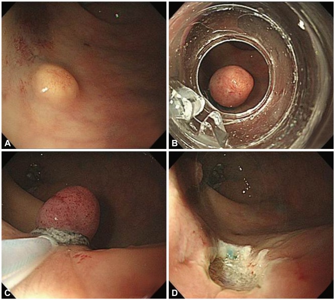
After submucosal injection beneath the lesion to elevate it away from the muscularis propria, the lesion was aspired into the ligator device and the elastic band was then deployed. Next, snare resection was performed below the band with a blended electrosurgical current (Fig. 1). The resection specimen was then removed by aspiration into the cap or by retrieving it with a grasping forceps. After resection of the specimen, the ulcer floor was endoscopically closed with clips to prevent postoperative bleeding and perforation. As with EMR-C, complete resection of rectal carcinoid tumor is important but is difficult to achieve with conventional endoscopic resection techniques because these tumors often extend into the submucosa. Resection via polypectomy or conventional EMR is often associated with resection margin involvement. Among various endoscopic resection techniques, endoscopic submucosal resection with a ligation device (ESMR-L) is known to be a useful and safe procedure.15-19

CONCLUSIONS

CAC is more sensitive for detecting adenomas and may reduce the time required for colonoscopic EMR of each polyp. EMR-C and EMR with a ligation device (or ESMR-L) may be a superior method to conventional EMR for removing small rectal carcinoids.

NOTES

The author has no financial conflicts of interest.

References

1. Kim HH, Park SJ, Park MI, Moon W, Kim SE. Transparent-cap-fitted colonoscopy shows higher performance with cecal intubation time in difficult cases. World J Gastroenterol 2012;18:1953–1958. 22563177.
crossref pmid pmc
2. Westwood DA, Alexakis N, Connor SJ. Transparent cap-assisted colonoscopy versus standard adult colonoscopy: a systematic review and meta-analysis. Dis Colon Rectum 2012;55:218–225. 22228167.
crossref pmid
3. Ng SC, Tsoi KK, Hirai HW, et al. The efficacy of cap-assisted colonoscopy in polyp detection and cecal intubation: a meta-analysis of randomized controlled trials. Am J Gastroenterol 2012;107:1165–1173. 22664471.
crossref pmid pdf
4. Morgan J, Thomas K, Lee-Robichaud H, Nelson RL, Braungart S. Transparent cap colonoscopy versus standard colonoscopy to improve caecal intubation. Cochrane Database Syst Rev 2012;12:CD008211. 23235654.
crossref pmid pmc
5. Park SY, Kim HS, Yoon KW, et al. Usefulness of cap-assisted colonoscopy during colonoscopic EMR: a randomized, controlled trial. Gastrointest Endosc 2011;74:869–875. 21824612.
crossref pmid
6. Inoue H, Kawano T, Tani M, Takeshita K, Iwai T. Endoscopic mucosal resection using a cap: techniques for use and preventing perforation. Can J Gastroenterol 1999;13:477–480. 10464347.
crossref pmid pdf
7. Kobayashi K, Katsumata T, Yoshizawa S, et al. Indications of endoscopic polypectomy for rectal carcinoid tumors and clinical usefulness of endoscopic ultrasonography. Dis Colon Rectum 2005;48:285–291. 15714250.
crossref pmid
8. Shirouzu K, Isomoto H, Kakegawa T, Morimatsu M. Treatment of rectal carcinoid tumors. Am J Surg 1990;160:262–265. 2393053.
crossref pmid
9. Matsui K, Iwase T, Kitagawa M. Small, polypoid-appearing carcinoid tumors of the rectum: clinicopathologic study of 16 cases and effectiveness of endoscopic treatment. Am J Gastroenterol 1993;88:1949–1953. 8237948.
pmid
10. Kim YJ, Lee SK, Cheon JH, et al. Efficacy of endoscopic resection for small rectal carcinoid: a retrospective study. Korean J Gastroenterol 2008;51:174–180. 18451691.
pmid
11. Oshitani N, Hamasaki N, Sawa Y, et al. Endoscopic resection of small rectal carcinoid tumours using an aspiration method with a transparent overcap. J Int Med Res 2000;28:241–246. 11092235.
crossref pmid
12. Zhao ZF, Zhang N, Ma SR, et al. A comparative study on endoscopy treatment in rectal carcinoid tumors. Surg Laparosc Endosc Percutan Tech 2012;22:260–263. 22678324.
crossref pmid
13. Jeon SM, Lee JH, Hong SP, Kim TI, Kim WH, Cheon JH. Feasibility of salvage endoscopic mucosal resection by using a cap for remnant rectal carcinoids after primary EMR. Gastrointest Endosc 2011;73:1009–1014. 21316666.
crossref pmid
14. Noda M, Kobayashi N, Kanemasa H, et al. Endoscopic mucosal resection using a partial transparent hood for lesions located tangentially to the endoscope. Gastrointest Endosc 2000;51:338–343. 10699785.
crossref pmid
15. Mashimo Y, Matsuda T, Uraoka T, et al. Endoscopic submucosal resection with a ligation device is an effective and safe treatment for carcinoid tumors in the lower rectum. J Gastroenterol Hepatol 2008;23:218–221. 18289355.
crossref pmid
16. Abe T, Kakemura T, Fujinuma S, Maetani I. Successful outcomes of EMR-L with 3D-EUS for rectal carcinoids compared with historical controls. World J Gastroenterol 2008;14:4054–4058. 18609690.
crossref pmid pmc
17. Kim HH, Park SJ, Lee SH, et al. Efficacy of endoscopic submucosal resection with a ligation device for removing small rectal carcinoid tumor compared with endoscopic mucosal resection: analysis of 100 cases. Dig Endosc 2012;24:159–163. 22507089.
crossref
18. Niimi K, Goto O, Fujishiro M, et al. Endoscopic mucosal resection with a ligation device or endoscopic submucosal dissection for rectal carcinoid tumors: an analysis of 24 consecutive cases. Dig Endosc 2012;24:443–447. 23078437.
crossref pmid
19. Lee SH, Park SJ, Kim HH, et al. Endoscopic resection for rectal carcinoid tumors: comparison of polypectomy and endoscopic submucosal resection with band ligation. Clin Endosc 2012;45:89–94. 22741138.
crossref pmid pmc
Fig. 1
(A) At 10 cm from anal verge, 6 mm sized, round, yellowish, elevated lesion with normal mucosa was noted. (B) Tissue is banded using an esophageal variceal banding device and (C) then snared off in the standard fashion. (D) The lesion was removed.
ce-46-492-g001.jpg
TOOLS
PDF Links  PDF Links
PubReader  PubReader
ePub Link  ePub Link
XML Download  XML Download
Full text via DOI  Full text via DOI
Download Citation  Download Citation
  Print
Share:      
METRICS
8
Crossref
9
Scopus
5,739
View
84
Download
Related articles
Recent Development of Techniques and Devices in Colorectal Endoscopic Submucosal Dissection  2017 November;50(6)
Proper Treatment Option for Small Rectal Neuroendocrine Tumors Using Precut Endoscopic Mucosal Resection  2017 November;50(6)
Editorial Office
Korean Society of Gastrointestinal Endoscopy
#817, 156 Yanghwa-ro (LG Palace, Donggyo-dong), Mapo-gu, Seoul, 04050, Korea
TEL: +82-2-335-1552   FAX: +82-2-335-2690    E-mail: CE@gie.or.kr
Copyright © Korean Society of Gastrointestinal Endoscopy.                 Developed in M2PI
Close layer