Skip Navigation
Skip to contents

Clin Endosc : Clinical Endoscopy

OPEN ACCESS

Articles

Page Path
HOME > Clin Endosc > Volume 46(5); 2013 > Article
Special Issue Article of IDEN 2013
Review
Endoscopic Treatments of Endoscopic Retrograde Cholangiopancreatography-Related Duodenal Perforations
Tae Hoon Lee1, Joung-Ho Han2, Sang-Heum Park1
Clinical Endoscopy 2013;46(5):522-528.
DOI: https://doi.org/10.5946/ce.2013.46.5.522
Published online: September 30, 2013

1Division of Gastroenterology, Department of Internal Medicine, Soonchunhyang University Cheonan Hospital, Soonchunhyang University College of Medicine, Cheonan, Korea.

2Division of Gastroenterology, Department of Internal Medicine, Chungbuk National University College of Medicine, Cheongju, Korea.

Correspondence: Tae Hoon Lee. Division of Gastroenterology, Department of Internal Medicine, Soonchunhyang University Cheonan Hospital, Soonchunhyang University College of Medicine, 31 Suncheonhyang 6-gil, Dongnam-gu, Cheonan 330-930, Korea. Tel: +82-41-570-3662, Fax: +82-41-574-5762, thlee9@schmc.ac.kr
• Received: June 7, 2013   • Accepted: June 25, 2013

Copyright © 2013 Korean Society of Gastrointestinal Endoscopy

This is an Open Access article distributed under the terms of the Creative Commons Attribution Non-Commercial License (http://creativecommons.org/licenses/by-nc/3.0/) which permits unrestricted non-commercial use, distribution, and reproduction in any medium, provided the original work is properly cited.

  • 14,798 Views
  • 191 Download
  • 27 Crossref
  • 37 Scopus
prev next
  • Iatrogenic duodenal perforation associated with endoscopic retrograde cholangiopancreatography (ERCP) is a very uncommon complication that is often lethal. Perforations during ERCP are caused by endoscopic sphincterotomy, placement of biliary or duodenal stents, guidewire-related causes, and endoscopy itself. In particular, perforation of the medial or lateral duodenal wall usually requires prompt diagnosis and surgical management. Perforation can follow various clinical courses, and management depends on the cause of the perforation. Cases resulting from sphincterotomy or guidewire-induced perforation can be managed by conservative treatment and biliary diversion. The current standard treatment for perforation of the duodenal free wall is early surgical repair. However, several reports of primary endoscopic closure techniques using endoclip, endoloop, or newly developed endoscopic devices have recently been described, even for use in direct perforation of the duodenal wall.
Gut perforation during endoscopic retrograde cholangiopancreatography (ERCP) and stenting or due to migration of stents is a very rare complication, with an incidence of well below 2%. Moreover, direct duodenoscope-induced perforation of the lateral or medial duodenal wall is even less common, accounting for 0.1% of patients who undergo ERCP, but the perforation tends to be large and further away from the ampulla.1-6 Duodenal perforations from the superior duodenal angle to the descending duodenum may be complicated by leakage of digestive enzymes from the pancreas; this may severely damage other abdominal organs. Therefore, duodenal wall perforation is one of the most serious complications of ERCP.
Traditionally, the standard treatment for traumatic or iatrogenic duodenal perforation has been early surgical closure because of a relatively high mortality rate of 16% to 18%.1-4 However, although ERCP-related perforations of the lateral or medial duodenal wall usually require immediate surgery, some cases are inoperable because of the patient's advanced age and comorbidities that prohibit surgical treatment. For nonoperative management of a large perforation of the lateral or medial duodenal wall, early endoscopic closure of the perforation is essential.
Recently of endoscopic trials on perforation treatment have been increased, and successful primary repairs of duo denal perforation using the endoscope itself have been reported.1-5 However, the best therapeutic option for these compli cations remains a matter of debate.
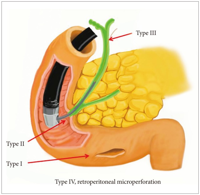
Several researchers have classified ERCP-related duodenal perforations according to the location or mechanism of injury and have recommended various treatments (Table 1, Fig. 1). Stapfer et al.1 classified perforations into four types, in decreasing order of severity, with implications for management. Type I perforations are perforations of the lateral or medial wall of the duodenum; these perforations involve large rents, are remote from the ampulla, and are caused by the endoscope itself or by the stent. These perforations cause considerable spillage, either retroperitoneally or intraperitoneally, necessitating aggressive surgical intervention. Type II perforations are perforations of the medial wall of the duodenum; these perforations are peri-Vaterian, are generally retroperitoneal, and occur during sphincterotomies. Type II perforations tend to be treated with conservative or minimally invasive therapies. Type III injuries are distal bile duct injuries caused by instrumentation and/or stenting in the proximity of an obstruction. These perforations are small and amenable to conservative management. Type IV perforations are tiny retroperitoneal perforations caused by the use of compressed air during endoscopy and can be managed conservatively. Howard et al.2 classified perforations into three types according to the mechanism of injury. Group I refers to guidewire-induced perforations; group II, periampullary perforations; and group III, duodenal perforations. Group III perforations require immediate surgery. Enns et al.3 suggested classification into three categories. Esophageal, gastric, and duodenal perforations require surgical management. Sphincterotomy-related perforation or guidewire-related perforation usually requires conservative management.
Known risk factors for ERCP-related duodenal perforation include old age, suspected dysfunction of the sphincter of Oddi, dilated bile duct, papillary stenosis, Billroth-II reconstruction, precut sphincterotomy, and long procedure duration.7-9 The classic presentation of duodenal perforation, with severe epigastric pain, vomiting, and epigastric tenderness progressing to generalized board-like rigidity, is seen only in a minority of cases. The symptoms and signs of ERCP-related perforations are often mild when this complication is recognized early.1,10-12 Therefore, the initial clinical presentation of patients with perforation is nonspecific. Duodenal perforation secondary to placement of a biliary endoprosthesis should be considered in all patients presenting with abdominal pain after such a placement. If a perforation is not recognized or suspected during ERCP, early diagnosis is difficult. Moreover, diagnosis is likely to be delayed if patients have concurrent elevated amylase level and the pain is attributed to post-ERCP pancreatitis. Clinical suspicion and diagnosis of a procedure-related perforation can be facilitated greatly by clinical findings and particularly by radiographic imaging with contrast studies, computed tomography (CT), and even magnetic resonance imaging. A multi-slice CT scan can provide an exact diagnosis of this complication.
Duodenal wall perforation
Type I (perforation of lateral or medial duodenal wall; Stapfer et al.1) or group III (duodenal perforation remote from the papilla; Howard et al.2) injuries are usually large and traditionally require immediate surgery for repair after prompt diagnosis. In a study by Stapfer et al.,1 surgery was recommended for patients with the following criteria: large contrast extravasation on ERCP; contrast-enhanced CT scans showing intraperitoneal or retroperitoneal fluid collection; massive subcutaneous emphysema or suspected perforation in association with retained material (i.e., stones or ERCP wire/basket). In cases of peri-Vaterian injuries, they suggested conservative management with serial radiographic examination. Howard et al.2 also suggested the use of endoscopic drainage to divert the bile, pancreatic, and/or duodenal fluids away from the perforation, and showed that the endoscopic approach reduced the rates of surgery and mortality, and the length of hospital stay. Unlike more common spontaneous perforations resulting from peptic ulcer disease, endoscopic therapy-related iatrogenic perforations have a relatively lower chance of bacterial contamination in a fasting state; therefore, these patients can occasionally be managed using nonsurgical means such as endoscopic therapies. A small amount of bacterial contamination may be controlled by conservative managements, including nil by mouth, nasogastric or nasoduodenal drainage, intensive fluid therapy, and antibiotics.13
Trials of endoscopic management have recently been performed and endorsed. There have been sporadic reports on the use of an endoscopic clipping device for the closure of iatrogenic perforation during endoscopic mucosal resection (EMR), sphincterotomy, or stent migration in the esophagus, stomach, and duodenum.14-19 Although surgery remains the standard treatment for duodenal perforations caused by the endoscope itself, the outcomes from case reports support the beneficial role of endoclips in the closure of these defects.5,20-22 In particular, some reports have indicated that nonsurgical treatment is possible for perforation of the upper gastrointestinal tract when peritonitis remains localized. The clinician's familiarity with endoclips and the immediate availability and proper use of endoclips may help to avoid surgery for a selected group of patients with a high surgical risk.5
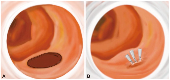
Typical primary endoscopic closure by endoclips may be difficult because of the position of the lateral wall and the complexity of aligning the perforation with the endoscope. To approximate the perforated hole and adherent hemoclips, glue injection and spraying may be successfully performed under cap-fitted endoscopy (Figs. 2, 3).18,20 Glue injection may cause multiple clips to adhere to each other and provide a cushion effect below the base of the perforation. In addition, since clipping with a duodenoscopy can be technically difficult, a cap-assisted, forward-view endoscopy can be used effectively. The cap-assisted endoscopy method under direct visualization through a transparent hood may be helpful in reducing the manipulation time of the procedure by allowing a good visual field and ensuring the safety margin during clipping.5
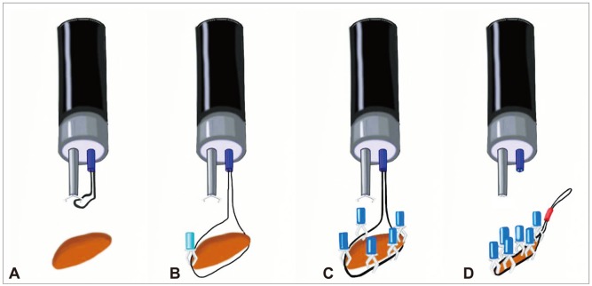
However, endoscopic repair using endoclips can be limited in large perforations or in those with tangential angles. A wide perforation is difficult to close because of slippage of the perforation edge from the clip while the clip is maneuvered across the defect to grasp the opposite edge of the perforation. Everted perforation edges also make it impossible to grasp the tissue with endoclips. As a modified method without additional glue injection, endoscopic closure using endoloops with endoclips has provided good clinical results, even though it has been described only in case reports.23-25 Clinical trials have been performed using an endoloop and multiple hemoclips to cover large mucosal defects after endoscopic submucosal dissection or ERCP-induced perforation.23-28 After placement of the endoloop around the perforated area through 1- or 2-channel endoscopy, multiple clips are attached with the endoloop to the perforated area; finally, the endoloop is tightened, and this closes the perforated area (Figs. 4, 5).29,30
Technically, endoscopic band ligation (EBL) is a simple procedure to treat bleeding from esophageal varices. The simplicity of the technique and low complication rates compared with those of sclerotherapy have contributed to its growing popularity.5 EBL has also been widely used in the management of non-variceal hemorrhage from Dieulafoy's ulcer, gastric angiodysplasia, and polypectomy-induced bleeding. In addition, several reports have described the use of EBL in rectal and duodenal perforations during EMR. Theoretically, EBL can readily approximate both edges of the perforation. Therefore, complete sutures to the remaining wall by additional bands or endoclips may be simple to achieve, even for a large perforation (Fig. 6). EBL can also reduce the procedure time in comparison with that required for clipping. Immediate closure can prevent the need for surgery or the development of serious peritonitis caused by leakage of gastric contents. Finally, the use of additional clips to suture the perforation after EBL might not be necessary.17 Thus, EBL may be also adopted for duodenoscope-induced duodenal wall perforation. However, more experimental studies are needed to confirm its suitability.
Finally, recent studies have reported high technical success rates for primary closure of an acute iatrogenic perforation with newly developed clips or endoscopic suturing devices developed for NOTES.5,13-16 These devices include through the scope (TTS) clips, such as the QuickClip 2 (Olympus Inc., Center Valley, PA, USA), the Resolution clip (Boston Scientific Inc., Natick, MA, USA), and the Tri-Clip and Instinct clip (Cook Medical, Winston-Salem, NC, USA); the over the scope clip (OTSC) system (Ovesco Endoscopy AG, Tubingen, Germany); and endoscopic suturing devices such as T tags (Ethicon Endo-Surgery, Cincinnati, OH, USA) and the flexible Endo Stitch (Covidien, Mansfield, MA, USA). Closure of luminal perforations >20 mm in size may be difficult. For larger gastric defects, TTS clips can be placed around the circumference of the perforation and lassoed together with a detachable plastic snare (Endo-loop; Olympus Inc.).17 Among the newly developed devices, OTSC has been used for the closure of perforation <20 mm in size, and ex vivo studies have shown that colon defects measuring 10 to 30 mm can be closed with a single OTSC.16 However, although some techniques have been developed to correct deficits in clip placement, they are not commonly practiced. Some of these devices may prove suitable for the closure of defects throughout the intestinal tract, but their use is limited by the endoscopist's experience, device availability, and cost. Currently, no one particular technique has demonstrated proven efficacy or greater reliability over other closure modalities.
Stent-induced perforation
Gut perforation has been reported following migration of all types of stents, irrespective of material, make, mode of placement, caliber, or size. The flexibility of the stent might play a role in influencing stent migration.10-12,31-36 None of the stents in current use have been implicated in particular. However, a straight-type stent with low flexibility may increase migration, especially in sphincterotomy state. For duodenal perforation secondary to placement of a biliary endoprosthesis, the treatment of choice is endoscopic removal of the endoprosthesis in the case of a localized process, and then the perforation can be su tured by a simple closure with hemoclips or hemoclips with glue injection.18 However, surgery should be reserved for patie nts with peritonitis or extensive retroperitoneal phlegmon.35,36
Sphincterotomy-induced perforation
The management of perforations after therapeutic periampullary endoscopic procedures has remained controversial. If perforation is suspected, prompt diagnosis and initiation of systemic support with broad-spectrum antibiotics and intravenous resuscitation is mandatory. Asymptomatic patients with evidence of retroperitoneal free air alone can normally be treated with conservative management. When perforation is confirmed, external/internal biliary, duodenal, and/or pancreatic drainage may be indicated.26 Some patients can be managed by diversion or decompression of contents from the duodenum by placing a nasoduodenal tube alone or with internal pancreaticobiliary drainage. Another option is diversion: a fully covered self-expandable metal stent can be used for the treatment of biliary sphincterotomy-induced perforation.27,28 However, inappropriate biliary drainage may cause bile infiltration or fluid leakage into the perforated site, thus increasing morbidity.1 If patients develop abdominal pain or fever or show clinical signs of toxicity, surgical consultation should be obtained, and operative exploration for effective repair or drainage should be considered, especially in elderly people or otherwise chronically ill patients less able to withstand physiologic stress.
Guidewire-induced perforation
Guidewire-induced perforation is defined as the entrance of the guidewire into the retroperitoneal space or through the bile duct during cannulation attempts or attempts to pass a stricture. These perforations may be also associated with injection of contrast into the retroperitoneal space.37 They are often very small in size and usually have no clinical consequences. Therefore, most cases of guidewire-induced perforation can be treated with conservative management, as mentioned above for sphincterotomy-induced perforations, or temporary diversion of the bile duct by a plastic or fully covered metallic stent.
In summary, for the successful management of ERCP-related duodenal perforations, prompt recognition and rapid management, including endoscopic closure or surgery, are the key determinants for lessening the significant morbidity and mortality. Delayed diagnosis and surgery are associated with a high mortality rate. Until recently, immediate surgical closure has been the standard treatment for traumatic or iatrogenic duodenal wall perforation, with the exception of guidewire-induced perforations or peri-Vaterian injuries. Recent endoscopic trials using clips, endoloops, glue injection, and newly developed devices such as OTSCs for the management of duodenal perforation have reported successful outcomes, although there is no clear consensus for primary repair because of the limited number of cases. Although nonsurgical suturing therapies are not widely accepted yet as the primary treatment for ERCP-related duodenal wall perforation, endos copic indications may be broadened in selected cases with the de velopment of endoscopic skills and devices. In addition, with im provement in endoscopic devices, endoscopic treatment may replace surgical management in the near future.
  • 1. Stapfer M, Selby RR, Stain SC, et al. Management of duodenal perforation after endoscopic retrograde cholangiopancreatography and sphin cterotomy. Ann Surg 2000;232:191–198. 10903596.ArticlePubMedPMC
  • 2. Howard TJ, Tan T, Lehman GA, et al. Classification and management of perforations complicating endoscopic sphincterotomy. Surgery 1999;126:658–663. 10520912.ArticlePubMed
  • 3. Enns R, Eloubeidi MA, Mergener K, et al. ERCP-related perforations: risk factors and management. Endoscopy 2002;34:293–298. 11932784.ArticlePubMedPDF
  • 4. Assalia A, Suissa A, Ilivitzki A, et al. Validity of clinical criteria in the management of endoscopic retrograde cholangiopancreatography related duodenal perforations. Arch Surg 2007;142:1059–1064. 18025334.ArticlePubMed
  • 5. Lee TH, Bang BW, Jeong JI, et al. Primary endoscopic approximation suture under cap-assisted endoscopy of an ERCP-induced duodenal perforation. World J Gastroenterol 2010;16:2305–2310. 20458771.ArticlePubMedPMC
  • 6. Vandervoort J, Soetikno RM, Tham TC, et al. Risk factors for complica tions after performance of ERCP. Gastrointest Endosc 2002;56:652–656. 12397271.ArticlePubMed
  • 7. Freeman ML. Complications of endoscopic biliary sphincterotomy: a review. Endoscopy 1997;29:288–297. 9255535.ArticlePubMedPDF
  • 8. Krims PE, Cotton PB. Papillotomy and functional disorders of the sphincter of Oddi. Endoscopy 1988;20(Suppl 1):203–206. 3049058.ArticlePubMedPDF
  • 9. Wu HM, Dixon E, May GR, Sutherland FR. Management of perforation after endoscopic retrograde cholangiopancreatography (ERCP): a population-based review. HPB 2006;8:393–399. 18333093.ArticlePubMedPMC
  • 10. Miller G, Yim D, Macari M, Harris M, Shamamian P. Retroperitoneal perforation of the duodenum from biliary stent erosion. Curr Surg 2005;62:512–515. 16125609.ArticlePubMed
  • 11. Melita G, Currò G, Iapichino G, Princiotta S, Cucinotta E. Duodenal per foration secondary to biliary stent dislocation: a case report and review of the literature. Chir Ital 2005;57:385–388. 16231831.PubMed
  • 12. Sánchez-Tembleque MD, Naranjo Rodrìguez A, Ruiz Morales R, Hervás Molina AJ, Calero Ayala B, de Dios Vega JF. Duodenal perforation due to an endoscopic biliary prosthesis. Gastroenterol Hepatol 2005;28:225–227. 15811264.ArticlePubMed
  • 13. Kaneko T, Akamatsu T, Shimodaira K, et al. Nonsurgical treatment of duodenal perforation by endoscopic repair using a clipping device. Gastrointest Endosc 1999;50:410–413. 10462667.ArticlePubMed
  • 14. Katsinelos P, Paroutoglou G, Papaziogas B, Beltsis A, Dimiropoulos S, Atmatzidis K. Treatment of a duodenal perforation secondary to an endoscopic sphincterotomy with clips. World J Gastroenterol 2005;11:6232–6234. 16273659.ArticlePubMedPMC
  • 15. Baron TH, Gostout CJ, Herman L. Hemoclip repair of a sphincterotomy-induced duodenal perforation. Gastrointest Endosc 2000;52:566–568. 11023583.ArticlePubMed
  • 16. Baron TH, Wong Kee, Zielinski MD, Emura F, Fotoohi M, Kozarek RA. A comprehensive approach to the management of acute endoscopic perforations (with videos). Gastrointest Endosc 2012;76:838–859. 22831858.ArticlePubMed
  • 17. Han JH, Lee TH, Jung Y, et al. Rescue endoscopic band ligation of iatrogenic gastric perforations following failed endoclip closure. World J Gastroenterol 2013;19:955–959. 23429885.ArticlePubMedPMC
  • 18. Kim KW, Lee TH, Park SH, et al. a primary repair with hemoclips and fibrin glue injection in biliary stent induced duodenal perforation. Korean J Gastrointest Endosc 2011;42:437–441.
  • 19. Marano BJ Jr, Bonanno CA. Metallic biliary endoprosthesis causing duodenal perforation and acute upper gastrointestinal bleeding. Gastrointest Endosc 1994;40(2 Pt 1):257–258. 8013842.ArticlePubMed
  • 20. Mutignani M, Iacopini F, Dokas S, et al. Successful endoscopic closure of a lateral duodenal perforation at ERCP with fibrin glue. Gastrointest Endosc 2006;63:725–727. 16564890.ArticlePubMed
  • 21. Seibert DG. Use of an endoscopic clipping device to repair a duodenal perforation. Endoscopy 2003;35:189. 12561015.ArticlePubMedPDF
  • 22. Sebastian S, Byrne AT, Torreggiani WC, Buckley M. Endoscopic closure of iatrogenic duodenal perforation during endoscopic ultrasound. Endoscopy 2004;36:245. 14986227.ArticlePubMed
  • 23. Nakagawa Y, Nagai T, Soma W, et al. Endoscopic closure of a large ERCP-related lateral duodenal perforation by using endoloops and endoclips. Gastrointest Endosc 2010;72:216–217. 20304402.ArticlePubMed
  • 24. Endo M, Inomata M, Terui T, et al. New endoscopic technique to close large mucosal defects after endoscopic mucosal resection in patients with gastric mucosal tumors. Dig Endosc 2004;16:372–375.Article
  • 25. Matsuda T, Fujii T, Emura F, et al. Complete closure of a large defect after EMR of a lateral spreading colorectal tumor when using a two-channel colonoscope. Gastrointest Endosc 2004;60:836–838. 15557972.ArticlePubMed
  • 26. Cotton PB, Lehman G, Vennes J, et al. Endoscopic sphincterotomy com plications and their management: an attempt at consensus. Gastrointest Endosc 1991;37:383–393. 2070995.ArticlePubMed
  • 27. Vezakis A, Fragulidis G, Nastos C, Yiallourou A, Polydorou A, Voros D. Closure of a persistent sphincterotomy-related duodenal perforation by placement of a covered self-expandable metallic biliary stent. World J Gastroenterol 2011;17:4539–4541. 22110286.ArticlePubMedPMC
  • 28. Park WY, Cho KB, Kim ES, Park KS. A case of ampullary perforation treated with a temporally covered metal stent. Clin Endosc 2012;45:177–180. 22866262.ArticlePubMedPMC
  • 29. Kwon CI, Song SH, Hahm KB, Ko KH. Unusual complications related to endoscopic retrograde cholangiopancreatography and its endoscopic treatment. Clin Endosc 2013;46:251–259. 23767036.ArticlePubMedPMC
  • 30. Shi Q, Chen T, Zhong YS, et al. Complete closure of large gastric defects after endoscopic full-thickness resection, using endoloop and metallic clip interrupted suture. Endoscopy 2013;45:329–334. 23468195.ArticlePubMedPDF
  • 31. Aliperti G. Complications related to diagnostic and therapeutic endoscopic retrograde cholangiopancreatography. Gastrointest Endosc Clin N Am 1996;6:379–407. 8673333.ArticlePubMed
  • 32. Saranga Bharathi R, Rao P, Ghosh K. Iatrogenic duodenal perforations caused by endoscopic biliary stenting and stent migration: an update. Endoscopy 2006;38:1271–1274. 17163332.ArticlePubMedPDF
  • 33. Esterl RM Jr, St Laurent M, Bay MK, Speeg KV, Halff GA. Endoscopic biliary stent migration with small bowel perforation in a liver transplant recipient. J Clin Gastroenterol 1997;24:106–110. 9077729.ArticlePubMed
  • 34. Mastorakos DP, Milman PJ, Cohen R, Goldenberg SP. An unusual complication of a biliary stent-small bowel perforation of an incarcerated hernia sac. Am J Gastroenterol 1998;93:2533–2535. 9860420.ArticlePubMed
  • 35. Coppola R, Masetti R, Riccioni ME, et al. Early retroduodenal perforation following endoscopic internal biliary drainage. Endoscopy 1993;25:255–256. 8519251.ArticlePubMedPDF
  • 36. Humar A, Barron PT, Sekar AS, Lum A. Pancreatitis and duodenal perforation as complications of an endoscopically placed biliary stent. Gastrointest Endosc 1994;40:365–366. 8056245.ArticlePubMed
  • 37. Lee TH, Park SH, Son BS, et al. Four cases of guidewire induced periampullary perforation during endoscopic retrograde cholangiopancreatography. Korean J Gastrointest Endosc 2011;42:334–340.
Fig. 1
Classical types of iatrogenic duodenal perforations: type I, endoscope-related lateral or medial duodenal wall perforation; type II, sphincterotomy-related periampullary perforations; type III, ductal or duodenal perforations due to endoscopic instruments such as a guidewire; type IV, retroperitoneal micoperforation caused by compressed air during endoscopy.
ce-46-522-g001.jpg
Fig. 2
Primary endoscopic closure using endoclips through a cap-fitted endoscopy. (A) Direct duodenoscope-induced duodenal wall perforation. (B) Simple endoscopic closure using multiple hemoclips via cap-fitted endoscopy.
ce-46-522-g002.jpg
Fig. 3
Primary endoscopic closure using endoclips with glue injection in stent-induced duodenal wall perforation. (A) Cap-fitted endoscopy shows a circular perforated hole after removal of the stent. (B) The therapeutic attempt to generate primary closure using hemoclips and glue injection and spraying around the closed hole of the perforation.
ce-46-522-g003.jpg
Fig. 4
Endoscopic closure of a large defect using an endoloop with multiple endoclips through 2-channel endoscopy. (A) Placement of endoscope and endoloop-containing catheter around the perforated area. (B) The tip of the endoloop is caught by the endoclip from the distal margin. (C) Multiple clips are attached with the endoloop to the perforated area. (D) The endoloop is tightened, thus closing the perforated area completely.
ce-46-522-g004.jpg
Fig. 5
Modified endoscopic closure using multiple endoloops with endoclips. (A) An endoloop was anchored with a clip to normal mucosa near the proximal resection margin. (B) The maneuver was repeated to anchor the same endoloop at the distal resection margin. (C) The endoloop was tightened slightly, resulting in approximation of the borders of the defect. (D) Another endoloop was required to bring the margins together. Additional clips can be placed to obtain complete wound closure if necessary.
ce-46-522-g005.jpg
Fig. 6
Endoscopic band ligation (EBL) in an iatrogenic intestinal wall perforation. (A) Iatrogenic intestinal wall perforation after endoscopic mucosal resection or dissection. (B) Primary EBL was performed successfully in technical difficulty with endoclip closure. (C) Additional clips were applied around the band and surrounding mucosa.
ce-46-522-g006.jpg
Table 1
Classification of Iatrogenic Duodenal Perforations during Endoscopic Retrograde Cholangiopancreatography
ce-46-522-i001.jpg

Figure & Data

REFERENCES

    Citations

    Citations to this article as recorded by  
    • Endoscopic nasobiliary drainage tube placement through a periampullary perforation for management of intestinal leak and necrotizing pancreatitis
      Mitsuru Okuno, Keisuke Iwata, Tsuyoshi Mukai, Yuhei Iwasa, Tomio Ogiso, Yoshiyuki Sasaki, Eiichi Tomita
      VideoGIE.2023; 8(2): 75.     CrossRef
    • Post-endoscopic Retrograde Cholangiopancreatography Complications: A Case of Duodenal Perforation and Literature Review
      Rabira R Dufera, Tamiru B Berake, Benedict Maliakkal
      Cureus.2023;[Epub]     CrossRef
    • Causes and Management of Endoscopic Retrograde Cholangiopancreatography-Related Perforation: A Retrospective Study
      Tetsuya Shimizu, Masato Yoshioka, Akira Matsushita, Keiko Kaneko, Junji Ueda, Mampei Kawashima, Toshiyuki Irie, Takashi Ono, Hiroyasu Furuki, Tomohiro Kanda, Yoshiaki Mizuguchi, Yoichi Kawano, Yoshiharu Nakamura, Hiroshi Yoshida
      Journal of Nippon Medical School.2023; 90(4): 316.     CrossRef
    • Endoscopic management of upper gastrointestinal perforations, leakage and fistula
      Hee Seok Moon
      Journal of Innovative Medical Technology.2023; 1(1): 15.     CrossRef
    • Endoscopic clipping to prevent papillary obstruction when closing a duodenal perforation with an over-the-scope clip
      Kazuya Kanaya, Haruka Toyonaga, Tsuyoshi Hayashi, Kuniyuki Takahashi, Akio Katanuma
      VideoGIE.2021; 6(6): 266.     CrossRef
    • Endoscopic Closure of an Acute Duodenal Perforation Occurring during Endoscopic Ultrasound Using Endoclips and Polyglycolic Acid Sheets with Fibrin Glue
      Mana Matsuoka, Katsumasa Kobayashi, Yukito Okura, Tomohiro Mochida, Takahito Nozaka, Ayako Sato, Masato Yauchi, Taichi Matsumoto, Yohei Furumoto, Takao Horiuchi, Toru Asano
      Case Reports in Gastroenterology.2021; 15(1): 253.     CrossRef
    • ERCP-related perforation: an analysis of operative outcomes in a large series over 12 years
      Nilesh Sadashiv Patil, Nisha Solanki, Pramod Kumar Mishra, Barjesh Chander Sharma, Sundeep Singh Saluja
      Surgical Endoscopy.2020; 34(1): 77.     CrossRef
    • Computed tomography classification of endoscopic retrograde cholangiopancreatography‐related perforation
      Jhong‐Han Wu, Hong‐Ming Tsai, Chiung‐Yu Chen, Yao‐Sheng Wang
      The Kaohsiung Journal of Medical Sciences.2020; 36(2): 129.     CrossRef
    • Retracted: Endoscopic Treatment of Endoscopic Retrograde Cholangiopancreatography-Related Duodenal Perforations
      Ding Shi, Jian feng Yang, Yong pan Liu
      Journal of Laparoendoscopic & Advanced Surgical Techniques.2019; 29(3): 385.     CrossRef
    • Duodenal perforation after the cutting an ENPD tube in a patient with pancreatic cancer and acute suppurative pancreatic ductitis
      Akihisa OHNO, Nao FUJIMORI, Kazuhide MATSUMOTO, Katsuhito TERAMATSU, Yu TAKAMATSU, Masami MIKI, Takehiro TAKAOKA, Takamasa OONO
      Suizo.2019; 34(1): 30.     CrossRef
    • Endoscopic management of iatrogenic gastrointestinal perforations
      Kan Wang, Jihao Shi, Linna Ye
      Laparoscopic, Endoscopic and Robotic Surgery.2019; 2(2): 41.     CrossRef
    • Clinical outcomes of iatrogenic upper gastrointestinal endoscopic perforation: a 10-year study
      Dae Hwan Kang, Dae Gon Ryu, Cheol Woong Choi, Hyung Wook Kim, Su Bum Park, Su Jin Kim, Hyeong Seok Nam
      BMC Gastroenterology.2019;[Epub]     CrossRef
    • Outcome and management of endoscopic retrograde cholangiopancreatography perforations: A community perspective
      Mallory S. Bray, Andrew J. Borgert, Milan E. Folkers, Shanu N. Kothari
      The American Journal of Surgery.2017; 214(1): 69.     CrossRef
    • Algorithm for the management of ERCP-related perforations
      Vivek Kumbhari, Amitasha Sinha, Aditi Reddy, Elham Afghani, Deanna Cotsalas, Yuval A. Patel, Andrew C. Storm, Mouen A. Khashab, Anthony N. Kalloo, Vikesh K. Singh
      Gastrointestinal Endoscopy.2016; 83(5): 934.     CrossRef
    • Recent Advanced Endoscopic Management of Endoscopic Retrograde Cholangiopancreatography Related Duodenal Perforations
      Seon Mee Park
      Clinical Endoscopy.2016; 49(4): 376.     CrossRef
    • Closure of echoendoscope-related duodenal free wall perforation by placement of a covered metallic stent
      Selçuk Disibeyaz, Erkin Öztaş, UfukBarış Kuzu, Mustafa Özdemir
      Endoscopic Ultrasound.2016; 5(6): 399.     CrossRef
    • Retroperitoneales Emphysem nach endoskopischer retrograder Cholangiopankreatikographie
      T. Vowinkel, N. Senninger
      Der Chirurg.2015; 86(5): 462.     CrossRef
    • Tratamiento conservador de la perforación duodenal tras realización de colangiopancreatografía retrógrada endoscópica terapéutica
      Antonio Palomeque Jiménez, Cristina González Puga, Beatriz Pérez Cabrera, José Antonio Jiménez Ríos
      Gastroenterología y Hepatología.2015; 38(4): 285.     CrossRef
    • Efficacy and Safety of Endoscopic Papillary Balloon Dilation Using Cap-Fitted Forward-Viewing Endoscope in Patients Who Underwent Billroth II Gastrectomy
      Jong Soon Jang, Seungho Lee, Hee Seung Lee, Myeong Ho Yeon, Joung-Ho Han, Soon Man Yoon, Hee Bok Chae, Sei Jin Youn, Seon Mee Park
      Clinical Endoscopy.2015; 48(5): 421.     CrossRef
    • Fatal pneumoperitoneum following endoscopic retrograde cholangiopancreatography confirmed by post-mortem computed tomography
      Ann Sophie Schröder, Axel Heinemann, Hideyuki Nushida, Jan Peter Sperhake
      Forensic Science, Medicine, and Pathology.2015; 11(3): 460.     CrossRef
    • Endoscopic Retrograde Cholangiopancreatography–Related Adverse Events
      Tarun Rustagi, Priya A. Jamidar
      Gastrointestinal Endoscopy Clinics of North America.2015; 25(1): 97.     CrossRef
    • Closure of transmural defects in the gastrointestinal tract by methods other than clips and sutures
      Juliana Yang, David Lee, Deepak Agrawal
      Techniques in Gastrointestinal Endoscopy.2015; 17(3): 141.     CrossRef
    • Duodenal perforation following esophagogastroduodenoscopy (EGD) with cautery and epinephrine injection for peptic ulcer disease: An interesting case of nonoperative management in the medical intensive care unit (MICU)
      Jason Chertoff, Vikas Khullar, Lucas Burke
      International Journal of Surgery Case Reports.2015; 10: 121.     CrossRef
    • Endoscopic Retrograde Cholangiopancreatography-Related Large Jejunal Perforation: Operate or Apply Over-the-Scope Clip Device?
      George Tribonias, Evangelos Voudoukis, Emmanouil Vardas, Angeliki Theodoropoulou, Manola Margarita-Eleni, Gregorios Paspatis
      Clinical Endoscopy.2014; 47(3): 281.     CrossRef
    • The Management of Endoscopic Retrograde Cholangiopancreatography-Related Duodenal Perforation
      Kwang Bum Cho
      Clinical Endoscopy.2014; 47(4): 341.     CrossRef
    • Colic and gastric over-the-scope clip (Ovesco) for the treatment of a large duodenal perforation during endoscopic retrograde cholangiopancreatography
      Gianfranco Donatelli, Jean-Loup Dumont, Bertrand Marie Vergeau, Renaud Chiche, Jean-Jacques Quioc, Thierry Tuszynski, Bruno Meduri
      Therapeutic Advances in Gastroenterology.2014; 7(6): 282.     CrossRef
    • Value of temporary stents for the management of perivaterian perforation during endoscopic retrograde cholangiopancreatography
      Sang Min Lee
      World Journal of Clinical Cases.2014; 2(11): 689.     CrossRef

    • PubReader PubReader
    • ePub LinkePub Link
    • Cite
      CITE
      export Copy Download
      Close
      Download Citation
      Download a citation file in RIS format that can be imported by all major citation management software, including EndNote, ProCite, RefWorks, and Reference Manager.

      Format:
      • RIS — For EndNote, ProCite, RefWorks, and most other reference management software
      • BibTeX — For JabRef, BibDesk, and other BibTeX-specific software
      Include:
      • Citation for the content below
      Endoscopic Treatments of Endoscopic Retrograde Cholangiopancreatography-Related Duodenal Perforations
      Clin Endosc. 2013;46(5):522-528.   Published online September 30, 2013
      Close
    • XML DownloadXML Download
    Figure
    • 0
    • 1
    • 2
    • 3
    • 4
    • 5
    Related articles
    Endoscopic Treatments of Endoscopic Retrograde Cholangiopancreatography-Related Duodenal Perforations
    Image Image Image Image Image Image
    Fig. 1 Classical types of iatrogenic duodenal perforations: type I, endoscope-related lateral or medial duodenal wall perforation; type II, sphincterotomy-related periampullary perforations; type III, ductal or duodenal perforations due to endoscopic instruments such as a guidewire; type IV, retroperitoneal micoperforation caused by compressed air during endoscopy.
    Fig. 2 Primary endoscopic closure using endoclips through a cap-fitted endoscopy. (A) Direct duodenoscope-induced duodenal wall perforation. (B) Simple endoscopic closure using multiple hemoclips via cap-fitted endoscopy.
    Fig. 3 Primary endoscopic closure using endoclips with glue injection in stent-induced duodenal wall perforation. (A) Cap-fitted endoscopy shows a circular perforated hole after removal of the stent. (B) The therapeutic attempt to generate primary closure using hemoclips and glue injection and spraying around the closed hole of the perforation.
    Fig. 4 Endoscopic closure of a large defect using an endoloop with multiple endoclips through 2-channel endoscopy. (A) Placement of endoscope and endoloop-containing catheter around the perforated area. (B) The tip of the endoloop is caught by the endoclip from the distal margin. (C) Multiple clips are attached with the endoloop to the perforated area. (D) The endoloop is tightened, thus closing the perforated area completely.
    Fig. 5 Modified endoscopic closure using multiple endoloops with endoclips. (A) An endoloop was anchored with a clip to normal mucosa near the proximal resection margin. (B) The maneuver was repeated to anchor the same endoloop at the distal resection margin. (C) The endoloop was tightened slightly, resulting in approximation of the borders of the defect. (D) Another endoloop was required to bring the margins together. Additional clips can be placed to obtain complete wound closure if necessary.
    Fig. 6 Endoscopic band ligation (EBL) in an iatrogenic intestinal wall perforation. (A) Iatrogenic intestinal wall perforation after endoscopic mucosal resection or dissection. (B) Primary EBL was performed successfully in technical difficulty with endoclip closure. (C) Additional clips were applied around the band and surrounding mucosa.
    Endoscopic Treatments of Endoscopic Retrograde Cholangiopancreatography-Related Duodenal Perforations

    Classification of Iatrogenic Duodenal Perforations during Endoscopic Retrograde Cholangiopancreatography

    Table 1 Classification of Iatrogenic Duodenal Perforations during Endoscopic Retrograde Cholangiopancreatography


    Clin Endosc : Clinical Endoscopy Twitter Facebook
    Close layer
    TOP