Skip Navigation
Skip to contents

Clin Endosc : Clinical Endoscopy

OPEN ACCESS

Articles

Page Path
HOME > Clin Endosc > Volume 46(1); 2013 > Article
Case Report A Case of Spontaneous Esophagopleural Fistula Successfully Treated by Endoscopic Stent Insertion
Gu Hyum Kang, Beom Yong Yoon, Beom Hee Kim, Hee Seok Moon, Hyun Yong Jeong, Jae Kyu Sung, Eaum Seok Lee
Clinical Endoscopy 2013;46(1):91-94.
DOI: https://doi.org/10.5946/ce.2013.46.1.91
Published online: January 31, 2013

Department of Internal Medicine, Chungnam National University School of Medicine, Daejeon, Korea.

Correspondence: Hee Seok Moon. Department of Internal Medicine, Chungnam National University Hospital, Chungnam National University School of Medicine, 282 Munhwa-ro, Junggu, Daejeon 301-721, Korea. Tel: +82-42-280-8049, Fax: +82-42-257-5753, mhs1357@cnuh.co.kr
• Received: March 6, 2012   • Revised: April 22, 2012   • Accepted: April 26, 2012

Copyright © 2013 Korean Society of Gastrointestinal Endoscopy

This is an Open Access article distributed under the terms of the Creative Commons Attribution Non-Commercial License (http://creativecommons.org/licenses/by-nc/3.0/) which permits unrestricted non-commercial use, distribution, and reproduction in any medium, provided the original work is properly cited.

  • 6,543 Views
  • 46 Download
  • 9 Web of Science
  • 4 Crossref
  • 6 Scopus
prev next
  • The most common cause of esophagorespiratory fistulas (ERFs) is associated with malignancy. The use of self-expandable metal stents is effective for the treatment of malignant ERFs, but benign ERF is rare, which is why its optimal treatment is not defined yet. There have been few reports describing benign esophagopleural fistula and its treatments in South Korea. Here, we report a rare case of spontaneous esophagopleural fistula, which was successfully treated by endoscopic placement of a membrane covered metal stent.
Esophagorespiratory fistula (ERF) is an uncommon condition, despite of the anatomical proximity of these structures. Malignancy of esophagus, lung, or mediastinum is recognized as the most common cause. Causes of ERF development include direct tumor invasion and subsequent perforation or after radiation, laser therapy, chemotherapy, or pre-existing stents.1 Benign ERF is rare and may be due to trauma or infection.2 However, there have been few reports describing benign ERF and its treatments in South Korea. Here we report a case of nonmalignant esophagopleural fistula successfully treated by covered self-expanding metallic stents.
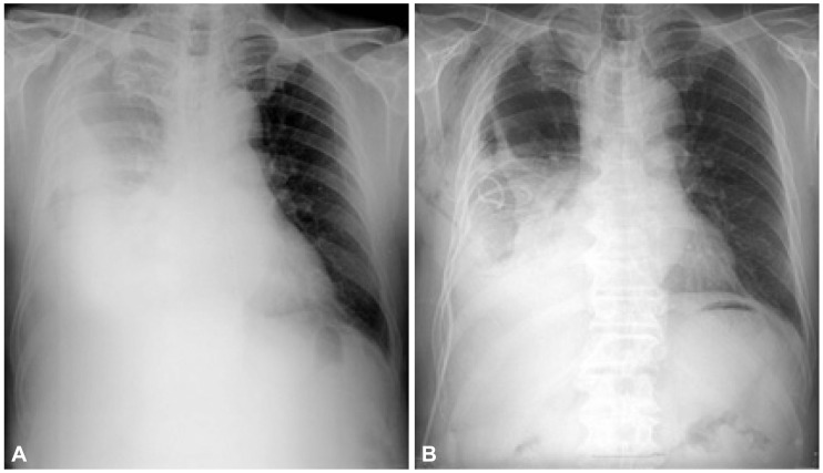
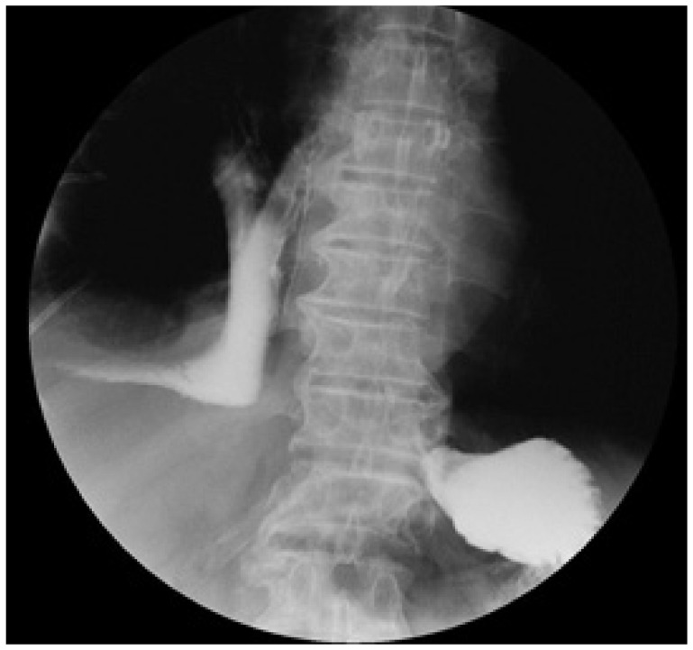
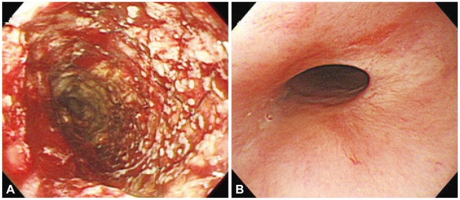
A 73-year-old man was admitted to Chungnam National University Hospital complaining right chest discomfort. He had 20 pack-years smoking history but had neither an operation history nor a medical history such as diabetes, hypertension, pulmonary tuberculosis, or hepatitis. Physical examination was unremarkable except for diminished respiratory sounds in the whole right lung field. The results of laboratory studies showed a white blood cell (WBC) count of 8,500/mm2; hemoglobin of 12.5 g/dL; and platelet count of 164,000/mm2. The serum biochemistry presented Na+ of 140 mEq/L; K+ 4.3 mEq/L; Cl- 107 mEq/L; aspartate aminotransferase 19.5 IU/L; alanine aminotransferase 12.4 IU/L; total bilirubin 0.9 mg/dL; and lactate dehydrogenase (LDH) 246 IU/L (all within normal ranges) with hypoalbuminemia (2.8 g/dL) and elevated serum creatinine level (1.65 mg/dL). Chest X-ray revealed right pleural effusion (Fig. 1A). Pleural fluid analysis revealed pH of 7.0; red blood cell 3,200/mm3; WBC 4,800/mm3; total protein 4.0 g/dL; and LDH 2,246 IU/L compatible with exudative parapneumonic effusion, for which an antibiotic therapy was initiated with chest tube catheter insertion (Fig. 1B). Thereafter food material was drained into the chest catheter. Esophagogram using gastrografin documented extravasation into the pleural cavity (Fig. 2). Finally, upper endoscopy confirmed a large fistulous tract measuring 12 mm (Fig. 3A). Stent insertion was considered as the first treatment due to the large fistula and pleuritis. A 14-cm long, 18-mm diameter covered Choo stent (MITech, Seoul, Korea) was deployed under direct vision (Fig. 3B). A repeat endoscopic and radiologic study demonstrated neither extravasation nor migration. Three months after the stent insertion, the stent was easily removed by pulling removal snare with an alligator forceps (Fig. 4A). Stent removal was uncomplicated. The patient remained asymptomatic (Fig. 4B) at the subsequent follow-up 6 months after the stent removal.
ERF is used to describe all fistulas located between the esophagus and the airway tree; however, esophagotracheal and esophagobronchial fistulas mostly account for the remaining ERF cases (3% to 11%) of esophagopulmonary fistulas.3,4 Accordingly, there have been very few previous reports concerning benign esophagopleural fistula and its treatment. Recent reports5 regarding malignant esophagopulmonary fistula other than esophagotracheal and esophagobronchial fistulas showed technically successful stent placement outcome in 14 patients and complete fistula sealing in 12 patients (86%).
Known causes of acquired benign ERF include trauma and infection.6-8 The former lesions may be due to the increase in fistula caused by surgical procedures, blunt chest trauma, and prolonged ventilator assistance. The most common infectious cause is granulomatous disease, particularly tuberculosis. Other reported infectious causes include syphilis, mycotic disease, and Crohn's disease. In our case, the cause of the esophagopleural fistula was unclear. Evidence of malignancy was nearly undetectable from radiologic and tumor marker studies. Also a specific infectious agent was not isolated at the time of diagnosis and treatment.
Esophagopleural fistulas rarely heal spontaneously. Leaks of the esophagus are associated with a high mortality rate and need to be treated as soon as possible. Therapeutic options include surgical repair or resection or conservative management with antibiotic therapy and cessation of oral intake. Surgical correction requires aggressive and technically demanding procedures, results for which are uncertain. Endoscopic techniques to overcome this issue have been reported with obliterating agents, such as fibrin glue or cyan acrylic glue,9-11 endoscopic clip application,12,13 and endoscopic suturing device.14,15 Recently several types of covered and uncovered stents have been used in the treatment of ERFs.
Self-expanding metal stents (SEMSs) have been considered a safe and effective treatment for malignant dysphagia stricture, and fistula in inoperable patients.16 Primarily SEMS has been used to palliate malignant state but are less commonly used in benign state due to concerns about their nonremovability and high complication rate. Recent technical improvement overcame these limitations. To evaluate fistula closure and stent patency or migration, repeat esophagogram at 1 week, then every 1 to 2 months after the procedure is suggested.
In our case, the cause of the esophagopleural fistula was unclear. However, esophagopleural fistula was successfully treated by an endoscopic stent insertion. Therefore, it appears that implantation of membrane-covered metal stents is an effective alternative for the treatment of esophagopleural fistula.
  • 1. Shin JH, Song HY, Ko GY, Lim JO, Yoon HK, Sung KB. Esophagorespiratory fistula: long-term results of palliative treatment with covered expandable metallic stents in 61 patients. Radiology 2004;232:252–259. 15166325.ArticlePubMed
  • 2. Wychulis AR, Ellis FH Jr, Andersen HA. Acquired nonmalignant esophagotracheobronchial fistula: report of 36 cases. JAMA 1966;196:117–122. 5952106.ArticlePubMed
  • 3. Duranceau A, Jamieson GG. Malignant tracheoesophageal fistula. Ann Thorac Surg 1984;37:346–354. 6201144.ArticlePubMed
  • 4. Angorn IB. Intubation in the treatment of carcinoma of the esophagus. World J Surg 1981;5:535–541. 6172904.ArticlePubMed
  • 5. Kim KR, Shin JH, Song HY, et al. Palliative treatment of malignant esophagopulmonary fistulas with covered expandable metallic stents. AJR Am J Roentgenol 2009;193:W278–W282. 19770295.ArticlePubMed
  • 6. Coleman FP. Acquired non-malignant esophagorespiratory fistula. Am J Surg 1957;93:321–328. 13394811.ArticlePubMed
  • 7. Wesselhoeft CW Jr, Keshishian JM. Acquired nonmalignant esophagotracheal and esophagobronchial fistulas. Ann Thorac Surg 1968;6:187–195. 4918289.ArticlePubMed
  • 8. Spalding AR, Burney DP, Richie RE. Acquired benign bronchoesophageal fistulas in the adult. Ann Thorac Surg 1979;28:378–383. 507984.ArticlePubMed
  • 9. Vandenplas Y, Helven R, Derop H, et al. Endoscopic obliteration of recurrent tracheoesophageal fistula. Dig Dis Sci 1993;38:374–377. 8425452.ArticlePubMed
  • 10. Lopes MF, Pires J, Nogueria Brandão A, Reis A, Morais Leitão L. Endoscopic obliteration of a recurrent tracheoesophageal fistula with enbucrilate and polidocanol in a child. Surg Endosc 2003;17:657. 12582778.Article
  • 11. Truong S, Böhm G, Klinge U, Stumpf M, Schumpelick V. Results after endoscopic treatment of postoperative upper gastrointestinal fistulas and leaks using combined Vicryl plug and fibrin glue. Surg Endosc 2004;18:1105–1108. 15156390.ArticlePubMed
  • 12. van Bodegraven AA, Kuipers EJ, Bonenkamp HJ, Meuwissen SG. Esophagopleural fistula treated endoscopically with argon beam electrocoagulation and clips. Gastrointest Endosc 1999;50:407–409. 10462666.ArticlePubMed
  • 13. Raymer GS, Sadana A, Campbell DB, Rowe WA. Endoscopic clip application as an adjunct to closure of mature esophageal perforation with fistulae. Clin Gastroenterol Hepatol 2003;1:44–50. 15017516.ArticlePubMed
  • 14. Adler DG, McAfee M, Gostout CJ. Closure of an esophagopleural fistula by using fistula tract coagulation and an endoscopic suturing device. Gastrointest Endosc 2001;54:652–653. 11677492.ArticlePubMed
  • 15. Rocca R, Fracchia M, Daperno M, et al. Treatment of tracheoesophageal fistula in a child by using an endoscopic suturing device. Gastrointest Endosc 2007;65:932–935. 17208241.ArticlePubMed
  • 16. Al-Haddad M, Craig CA, Odell J, Pajaro O, Wallace MB. The use of self-expandable plastic stents for non-malignant esophago-pleural fistulas. Dis Esophagus 2007;20:538–541. 17958732.ArticlePubMedPDF
Fig. 1
Chest X-ray showing (A) a huge left pleural effusion and (B) inserted chest tube catheter.
ce-46-91-g001.jpg
Fig. 2
Esophagogram showing apparent leakage from above the esophagogastric junction to the left pleural cavity.
ce-46-91-g002.jpg
Fig. 3
Endoscopy images showing (A) a purulent fistular tract opening in the lower esophagus and (B) fully expanded stent.
ce-46-91-g003.jpg
Fig. 4
Endoscopy images showing (A) the lower esophagus after stent removal and (B) complete closure of the esophagopleural fistula 6 months later.
ce-46-91-g004.jpg

Figure & Data

REFERENCES

    Citations

    Citations to this article as recorded by  
    • Delayed esophagopleural fistula after endoscopic injection sclerotherapy for esophageal varices
      Mingliang Sui, Weibing Tang, Changjiang Wu, Jinhu Yang, Huiping Liu, Chaofa Huang, Xianzhu Hu, Damei Xia, Yadi Yang
      Medicine.2020; 99(3): e18806.     CrossRef
    • Unusual Presentation of an Esophagopleural Fistula in a Patient With Severe Esophagitis: Blunt Trauma or Iatrogenic?
      Yanting Wang, Oscar A. Rivas Chicas, Anupam Basu
      ACG Case Reports Journal.2019; 6(10): e00240.     CrossRef
    • Tubercular esophago-pleural fistula—a rare case report and review of literature
      Manoj Kumar Pattnaik, Sameer Kumar Panigrahy, Sarada Prasanna Sahoo, Naba Kumar Pattnaik
      Indian Journal of Thoracic and Cardiovascular Surgery.2015; 31(4): 316.     CrossRef
    • Treatment of Esophagopleural Fistulas Using Covered Retrievable Expandable Metallic Stents
      Tae-Hyung Kim, Ji Hoon Shin, Kyung Rae Kim, Jung-Hoon Park, Jin Hyoung Kim, Ho-Young Song
      Journal of Vascular and Interventional Radiology.2014; 25(4): 623.     CrossRef

    • PubReader PubReader
    • ePub LinkePub Link
    • Cite
      CITE
      export Copy Download
      Close
      Download Citation
      Download a citation file in RIS format that can be imported by all major citation management software, including EndNote, ProCite, RefWorks, and Reference Manager.

      Format:
      • RIS — For EndNote, ProCite, RefWorks, and most other reference management software
      • BibTeX — For JabRef, BibDesk, and other BibTeX-specific software
      Include:
      • Citation for the content below
      A Case of Spontaneous Esophagopleural Fistula Successfully Treated by Endoscopic Stent Insertion
      Clin Endosc. 2013;46(1):91-94.   Published online January 31, 2013
      Close
    • XML DownloadXML Download
    Figure
    • 0
    • 1
    • 2
    • 3
    Related articles
    A Case of Spontaneous Esophagopleural Fistula Successfully Treated by Endoscopic Stent Insertion
    Image Image Image Image
    Fig. 1 Chest X-ray showing (A) a huge left pleural effusion and (B) inserted chest tube catheter.
    Fig. 2 Esophagogram showing apparent leakage from above the esophagogastric junction to the left pleural cavity.
    Fig. 3 Endoscopy images showing (A) a purulent fistular tract opening in the lower esophagus and (B) fully expanded stent.
    Fig. 4 Endoscopy images showing (A) the lower esophagus after stent removal and (B) complete closure of the esophagopleural fistula 6 months later.
    A Case of Spontaneous Esophagopleural Fistula Successfully Treated by Endoscopic Stent Insertion

    Clin Endosc : Clinical Endoscopy Twitter Facebook
    Close layer
    TOP