Skip Navigation
Skip to contents

Clin Endosc : Clinical Endoscopy

OPEN ACCESS

Articles

Page Path
HOME > Clin Endosc > Volume 47(1); 2014 > Article
Case Report A Case of Esophageal Fibrovascular Polyp That Induced Asphyxia during Sleep
Jin-Seok Park1, Byoung Wook Bang1, Junyoung Shin1, Kye Sook Kwon1, Hyung Gil Kim1, Yong Woon Shin1, Suk Jin Choi2
Clinical Endoscopy 2014;47(1):101-103.
DOI: https://doi.org/10.5946/ce.2014.47.1.101
Published online: January 24, 2014

1Division of Gastroenterology, Department of Internal Medicine, Inha University School of Medicine, Incheon, Korea.

2Department of Pathology, Inha University School of Medicine, Incheon, Korea.

Correspondence: Byoung Wook Bang. Division of Gastroenterology, Department of Internal Medicine, Inha University Hospital, Inha University School of Medicine, 27 Inhang-ro, Jung-gu, Incheon 400-711, Korea. Tel: +82-32-890-2548, Fax: +82-32-890-2549, bangbu@inha.ac.kr
• Received: February 19, 2013   • Revised: March 10, 2013   • Accepted: March 20, 2013

Copyright © 2014 Korean Society of Gastrointestinal Endoscopy

This is an Open Access article distributed under the terms of the Creative Commons Attribution Non-Commercial License (http://creativecommons.org/licenses/by-nc/3.0/) which permits unrestricted non-commercial use, distribution, and reproduction in any medium, provided the original work is properly cited.

  • 8,351 Views
  • 86 Download
  • 18 Web of Science
  • 15 Crossref
  • 17 Scopus
prev next
  • Esophageal fibrovascular polyps are rare, benign, submucosal tumors of the upper digestive tract that usually have an indolent course until the lesion attains a very large size. The most frequent complaints associated with these tumors include dysphagia and foreign body sensation. However, a long pedunculated polyp can regurgitate into the pharynx or oral cavity and cause asphyxia and sudden death if the larynx is occluded. We describe the case of a 51-year-old man who experienced snoring and occasional asphyxia during sleep. Upper endoscopy was performed, which indicated the presence of a pedunculated esophageal polyp that regurgitated into the vocal cords. The polyp was removed using a polypectomy snare and was confirmed to be a fibrovascular polyp based on pathologic examination findings. Three months after the excision of the polyp, the patient was found to be doing well without any further occurrence of asphyxia or sleep disturbances.
Fibrovascular polyps (FVPs) of the esophagus are rare, benign intraluminal tumor-like lesions arising from the submucosal layer of the cervical esophagus.1 These masses are composed of loose or dense fibrous tissue, vascular structures, and adipose tissue covered by normal squamous epithelium.2 FVPs are the most common intraluminal, nonepithelial neoplasms of the esophagus, accounting for 0.5% to 1% of all benign esophageal tumors.3 They are most typically observed in middle-aged and elderly men.4 Because of an initial lack of symptoms, the tumor attains a very large size inside the lumen of the esophagus.5 Thus, FVPs may cause dysphagia and sudden death from asphyxiation when the polyp regurgitates and occludes the larynx.6 In the present report, we describe the case of a patient with an FVP who suffered from snoring and asphyxia during sleep. This individual was successfully treated endoscopically.
A 51-year-old man visited our institution for routine endoscopy. He had a long history of snoring and obstructive sleep apnea. His wife frequently heard his labored breathing while he slept and would wake him up. However, the patient did not undergo a complete medical examination because he regarded his condition as simple snoring. Moreover, he did not have a history of regurgitation, pain during swallowing, weight loss, or signs and symptoms of upper gastrointestinal bleeding. His medical and surgical history was unremarkable, and the physical examination findings were normal. Laboratory results were also unremarkable.
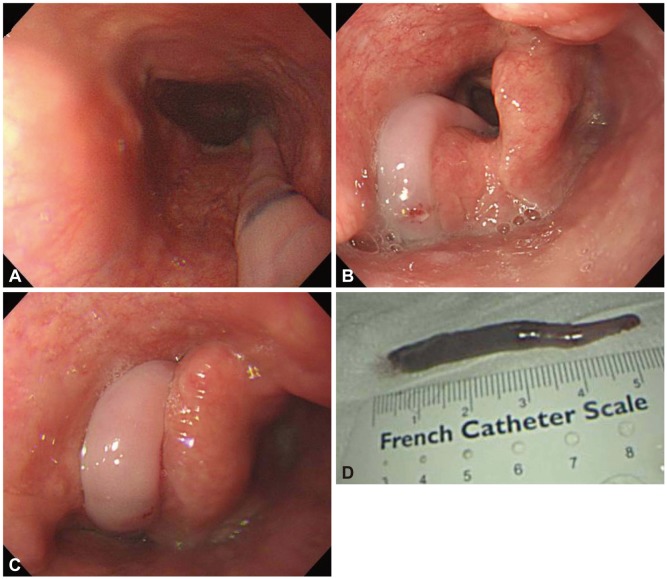
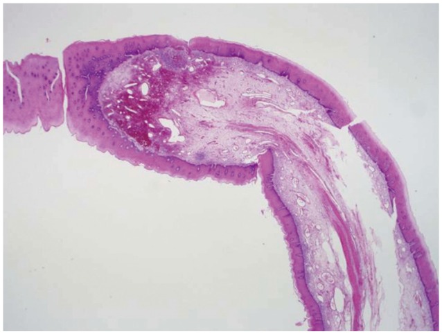
Endoscopy revealed a pedunculated polyp, >5 cm in length, that was detected immediately after inserting the endoscope into the patient's throat (Fig. 1A). The polyp arose from just below the pyriform sinus and regurgitated into the oral cavity during endoscopy. Occasionally, the polyp passed through the vocal cords (Fig. 1B) and obstructed the airway (Fig. 1C). We removed the polyp endoscopically using a polypectomy snare (Fig. 1D) because we believed that the polyp could induce asphyxia during sleep. Histopathological examination indicated that the polypoid lesion contained vascularized loose fibrous shafts covered by squamous epithelium with acanthosis, which were indicative of an FVP (Fig. 2). The patient was discharged on the next day without any complications. At the 3-month follow-up examination, the patient was found to be doing well without any further occurrence of snoring or sleep disturbances.
FVPs usually remain asymptomatic for years until reaching significant and occasionally enormous sizes because of their slow growth. The most frequently reported symptoms of FVPs are progressive dysphagia and the sensation of a mass. The severity of these symptoms is reported to be related to increases in polyp size.7 The other complaints include retrosternal or epigastric discomfort, odynophagia, vomiting, bleeding, weight loss, and respiratory symptoms such as persisting cough or difficulty in breathing.8
Because they originate from the cervical esophagus and due to their loose, elongated features, FVPs easily regurgitate into the mouth or pharynx. However, although asphyxia due to a regurgitating FVP is well known, no endoscopic imaging evidence indicating that regurgitating FVPs can be aspirated into the airway and cause asphyxia is available. In the present case, we obtained high quality pictures of the regurgitating polyp that was obstructing the airway during endoscopy. In addition, we observed that the patient suffered from breathing difficulties and coughing when the FVP passed through his airway.
Similar to the present case, relatively small regurgitating FVPs induce snoring and sleep disturbances. However, extremely large FVPs can cause potentially life-threatening conditions if the regurgitating polyps completely obstruct the airway.9 Due to the rarity of FVPs in the esophagus, there are no data on the mortality rate due to FVP-associated asphyxia. However, there have been seven reported cases of death due to aspiration of regurgitating polyps,7 and up to 30% of patients may die without a correct diagnosis.10 Therefore, FVPs of the upper esophagus should be removed regardless of the symptoms.
The definitive treatment of an FVP is excision, which can be performed either endoscopically or surgically. Appropriate treatment depends on the accurate assessment of the origin, diameter, and vascularity of the pedicle along with tumor size.11 Giant polyps or ones with a thick and richly vascularized pedicle are removed by surgical excision, which enables controlling of the bleeding.12 The pedicle should be completely excised because incomplete excision may result in recurrence.13 Endoscopic resection is also an option but is not preferable for treating giant FVPs because of the risk of hemorrhage from vessels in the polyp stalk. In general, endoscopic resection is recommended for polyps less than 2 cm in diameter and with a thin pedicle.14 Endoscopic ultrasonography can be helpful for identifying the presence of feeding vessels prior to attempting endoscopic resection.15 In the present case, we successfully removed the FVP endoscopically, even though it was more than 5 cm in length, because the pedicle was relatively thin. No complications, such as bleeding or recurrence, occurred during the 3 months after endoscopic resection.
In summary, FVPs of the esophagus are benign tumors. Despite their slow growth, these lesions should be removed because of potentially serious complications such as asphyxia or sudden death. Appropriate treatment should be based on FVP size and origin as well as on vascularity of the pedicle.
This study was supported by an Inha University Research Grant.
  • 1. Sargent RL, Hood IC. Asphyxiation caused by giant fibrovascular polyp of the esophagus. Arch Pathol Lab Med 2006;130:725–727.ArticlePubMedPDF
  • 2. LiVolsi VA, Perzin KH. Inflammatory pseudotumors (inflammatory fibrous polyps) of the esophagus. A clinicopathologic study. Am J Dig Dis 1975;20:475–481.ArticlePubMed
  • 3. Ginai AZ, Halfhide BC, Dees J, Zondervan PE, Klooswijk AI, Knegt PP. Giant esophageal polyp: a clinical and radiological entity with variable histology. Eur Radiol 1998;8:264–269.ArticlePubMed
  • 4. Fries MR, Galindo RL, Flint PW, Abraham SC. Giant fibrovascular polyp of the esophagus. A lesion causing upper airway obstruction and syncope. Arch Pathol Lab Med 2003;127:485–487.PubMed
  • 5. Avezzano EA, Fleischer DE, Merida MA, Anderson DL. Giant fibrovascular polyps of the esophagus. Am J Gastroenterol 1990;85:299–302.PubMed
  • 6. Levine MS, Buck JL, Pantongrag-Brown L, Buetow PC, Hallman JR, Sobin LH. Fibrovascular polyps of the esophagus: clinical, radiographic, and pathologic findings in 16 patients. AJR Am J Roentgenol 1996;166:781–787.ArticlePubMed
  • 7. Caceres M, Steeb G, Wilks SM, Garrett HE Jr. Large pedunculated polyps originating in the esophagus and hypopharynx. Ann Thorac Surg 2006;81:393–396.ArticlePubMed
  • 8. Drenth J, Wobbes T, Bonenkamp JJ, Nagengast FM. Recurrent esophageal fibrovascular polyps: case history and review of the literature. Dig Dis Sci 2002;47:2598–2604.ArticlePubMed
  • 9. Carrick C, Collins KA, Lee CJ, Prahlow JA, Barnard JJ. Sudden death due to asphyxia by esophageal polyp: two case reports and review of asphyxial deaths. Am J Forensic Med Pathol 2005;26:275–281.ArticlePubMed
  • 10. Timmons B, Sedwitz JL, Oller DW. Benign fibrovascular polyp of the esophagus. South Med J 1991;84:1370–1372.ArticlePubMed
  • 11. Chourmouzi D, Drevelegas A. Giant fibrovascular polyp of the oesophagus: a case report and review of the literature. J Med Case Rep 2008;2:337.ArticlePubMedPMCPDF
  • 12. van Lanschot JJ, Poublon RM, Zonderland HM, van Houten H. Benign pedunculated tumor of the esophagus. Neth J Surg 1987;39:83–85.PubMed
  • 13. Lee SY, Chan WH, Sivanandan R, Lim DT, Wong WK. Recurrent giant fibrovascular polyp of the esophagus. World J Gastroenterol 2009;15:3697–3700.ArticlePubMedPMC
  • 14. Tasaka Y, Makimoto K, Yamauchi M, Haebara H. Benign pedunculated intraluminal tumor of the esophagus. J Otolaryngol 1982;11:111–115.PubMed
  • 15. Eberlein TJ, Hannan R, Josa M, Sugarbaker DJ. Benign schwannoma of the esophagus presenting as a giant fibrovascular polyp. Ann Thorac Surg 1992;53:343–345.ArticlePubMed
Fig. 1
Endoscopic and gross images of a fibrovascular polyp of the esophagus. (A) Elongated polyp originating from the cervical esophagus. (B) Regurgitating polyp aspirated into the vocal cords. (C) Regurgitated polyp obstructing the airway. (D) Resected esophageal polyp measuring greater than 5 cm in length.
ce-47-101-g001.jpg
Fig. 2
Histopathological features of the fibrovascular polyp. The core of the polyp is composed of dilated blood vessels and fibrous connective tissue covered by squamous epithelium with acanthosis (H&E stain, ×40).
ce-47-101-g002.jpg

Figure & Data

REFERENCES

    Citations

    Citations to this article as recorded by  
    • Hybrid laparo-endoscopic access: New approach to surgical treatment for giant fibrovascular polyp of esophagus: A case report and review of literature
      Seda Dzhantukhanova, Lyudmila Grigori Avetisyan, Amina Badakhova, Yury Starkov, Andrey Glotov
      World Journal of Gastrointestinal Endoscopy.2023; 15(11): 666.     CrossRef
    • Esophageal Lipoma and Liposarcoma: A Systematic Review
      Davide Ferrari, Daniele Bernardi, Stefano Siboni, Veronica Lazzari, Emanuele Asti, Luigi Bonavina
      World Journal of Surgery.2021; 45(1): 225.     CrossRef
    • Recent developments in gastroesophageal mesenchymal tumours
      David J. Papke, Jason L. Hornick
      Histopathology.2021; 78(1): 171.     CrossRef
    • Deep Learning-Based Automatic Detection of Rectal Polyps Using Abdominal CT Images Guided by Cold Snare Polypectomy
      Haijun Lin, Qi Chen, Caijuan Li, Aifen Zheng, Lei Yang, Jiemin Hong, Hanqing Chen, Xuni He, Wuna Feng, Gustavo Ramirez
      Scientific Programming.2021; 2021: 1.     CrossRef
    • Difficult endoscopic resection of a giant esophageal fibrovascular polyp: case report and literature review
      Dan Nie, Ye Zong, Jielin Li
      Journal of International Medical Research.2021;[Epub]     CrossRef
    • Endoscopic resection for giant oesophageal fibrovascular polyp
      N Acar, T Acar, F Cengiz, B Şuataman, C Tavusbay, M Haciyanli
      The Annals of The Royal College of Surgeons of England.2020; 102(4): e89.     CrossRef
    • Well-Differentiated Liposarcoma (Atypical Lipomatous Tumor) Presenting as an Esophageal Polyp
      Aoife J. McCarthy, Paul Carroll, Rajkumar Vajpeyi, Gail Darling, Runjan Chetty
      Journal of Gastrointestinal Cancer.2019; 50(3): 589.     CrossRef
    • First Characterization with Ultrasound Contrast Agent of a Fibrovascular Polyp Before Its Endoscopic Resection: A Case Report (with Videos)
      Nicolas Williet, Radwan Kassir, Francois Casteillo, Violaine Yvorel, Cyril Habougit, Xavier Roblin, Jean-Marc Phelip
      Clinical Endoscopy.2019; 52(2): 186.     CrossRef
    • Asphyxia Caused by a Giant Fibrovascular Polyp of the Esophagus
      Santiago A. Endara, Gerardo A. Dávalos, Ramiro J. Yepez, Diego F. Luna, Fabián B. Corral, Gabriel A. Molina, W. Javier Cisneros
      ACG Case Reports Journal.2019; 6(7): e00126.     CrossRef
    • Polypoid fibroadipose tumors of the esophagus: ‘giant fibrovascular polyp’ or liposarcoma? A clinicopathological and molecular cytogenetic study of 13 cases
      Rondell P Graham, Saba Yasir, Karen J Fritchie, Michelle D Reid, Patricia T Greipp, Andrew L Folpe
      Modern Pathology.2018; 31(2): 337.     CrossRef
    • Presentation and Management of Giant Fibrovascular Polyps of the Hypopharynx and Esophagus
      Julina Ongkasuwan, C. Lane Anzalone, Esperanza Salazar, Donald T. Donovan
      Annals of Otology, Rhinology & Laryngology.2017; 126(1): 29.     CrossRef
    • Giant Esophageal Fibrovascular Polyp: A Rare Cause of Dysphagia
      Joseph Cano, Clark Hair, Robert Jay Sealock
      Clinical Gastroenterology and Hepatology.2017; 15(5): e93.     CrossRef
    • Gastroscopic removal of a giant fibrovascular polyp from the esophagus
      Jie Li, Hua Yu, Renfu Pu, Zhongsheng Lu
      Thoracic Cancer.2016; 7(3): 363.     CrossRef
    • A Case of Giant Fibrovascular Polyp of the Esophagus, Treated Successfully by Endoscopic Resection
      Jong Wook Lee, Gwang Ha Kim, Joong Keun Kim, Chul Hong Park, Byeong Gu Song, Dong Hun Shin, Dong Woo Ha, Geun Am Song
      The Korean Journal of Gastroenterology.2016; 67(5): 253.     CrossRef
    • Highlights from the 50th Seminar of the Korean Society of Gastrointestinal Endoscopy
      Eun Young Kim, Il Ju Choi, Kwang An Kwon, Ji Kon Ryu, Seok Ho Dong, Ki Baik Hahm
      Clinical Endoscopy.2014; 47(4): 285.     CrossRef

    • PubReader PubReader
    • ePub LinkePub Link
    • Cite
      CITE
      export Copy Download
      Close
      Download Citation
      Download a citation file in RIS format that can be imported by all major citation management software, including EndNote, ProCite, RefWorks, and Reference Manager.

      Format:
      • RIS — For EndNote, ProCite, RefWorks, and most other reference management software
      • BibTeX — For JabRef, BibDesk, and other BibTeX-specific software
      Include:
      • Citation for the content below
      A Case of Esophageal Fibrovascular Polyp That Induced Asphyxia during Sleep
      Clin Endosc. 2014;47(1):101-103.   Published online January 24, 2014
      Close
    • XML DownloadXML Download
    Figure
    • 0
    • 1
    A Case of Esophageal Fibrovascular Polyp That Induced Asphyxia during Sleep
    Image Image
    Fig. 1 Endoscopic and gross images of a fibrovascular polyp of the esophagus. (A) Elongated polyp originating from the cervical esophagus. (B) Regurgitating polyp aspirated into the vocal cords. (C) Regurgitated polyp obstructing the airway. (D) Resected esophageal polyp measuring greater than 5 cm in length.
    Fig. 2 Histopathological features of the fibrovascular polyp. The core of the polyp is composed of dilated blood vessels and fibrous connective tissue covered by squamous epithelium with acanthosis (H&E stain, ×40).
    A Case of Esophageal Fibrovascular Polyp That Induced Asphyxia during Sleep

    Clin Endosc : Clinical Endoscopy Twitter Facebook
    Close layer
    TOP