Skip Navigation
Skip to contents

Clin Endosc : Clinical Endoscopy

OPEN ACCESS

Articles

Page Path
HOME > Clin Endosc > Volume 47(6); 2014 > Article
Focused Review Series: Endoscopic Screening and Surveillance for Gastrointestinal Cancer Prevention Strategies for Gastric Cancer: A Global Perspective
Jin Young Park1, Lawrence von Karsa2, Rolando Herrero1
Clinical Endoscopy 2014;47(6):478-489.
DOI: https://doi.org/10.5946/ce.2014.47.6.478
Published online: November 30, 2014

1Prevention and Implementation Group, Section of Early Detection and Prevention (EDP), International Agency for Research on Cancer (IARC), Lyon, France.

2Quality Assurance Group, Section of Early Detection and Prevention (EDP), International Agency for Research on Cancer (IARC), Lyon, France.

Correspondence: Rolando Herrero. Prevention and Implementation Group, Section of Early Detection and Prevention, International Agency for Research on Cancer, 150 Cours Albert Thomas, 69372 Lyon Cedex 08, France. Tel: +33-4-7273-8683, Fax: +33-4-7273-8518, herreror@iarc.fr
• Received: November 3, 2014   • Revised: November 11, 2014   • Accepted: November 11, 2014

Copyright © 2014 Korean Society of Gastrointestinal Endoscopy

This is an Open Access article distributed under the terms of the Creative Commons Attribution Non-Commercial License (http://creativecommons.org/licenses/by-nc/3.0/) which permits unrestricted non-commercial use, distribution, and reproduction in any medium, provided the original work is properly cited.

  • 13,737 Views
  • 198 Download
  • 110 Web of Science
  • 94 Crossref
  • 117 Scopus
prev next
  • Despite the substantial burden of gastric cancer worldwide, population strategies for primary prevention have not been introduced in any country. Recognizing the causal role of Helicobacter pylori infection, there is increasing interest in population-based programs to eradicate the infection to prevent gastric cancer. Nonetheless, the paucity of available evidence on feasibility and effectiveness has prevented implementation of this approach. There are very few secondary prevention programs based on screening with endoscopy or radiography, notably in the Republic of Korea and Japan, two of the countries with the highest incidence rates of gastric cancer. In Korea, where the organized screening program is in place, survival rate of gastric cancer is as high as 67%. More research is needed to quantify the specific contribution of the screening program to observed declines in mortality rates. Gastric cancer screening is unlikely to be feasible in many Low-Middle Income Countries where the gastric cancer burden is high. Prevention strategies are still under development and the optimal approach may differ depending on local conditions and societal values. The present review gives an overview of the etiology and burden of the disease, and possible prevention strategies for countries and regions confronted with a significant burden of disease.
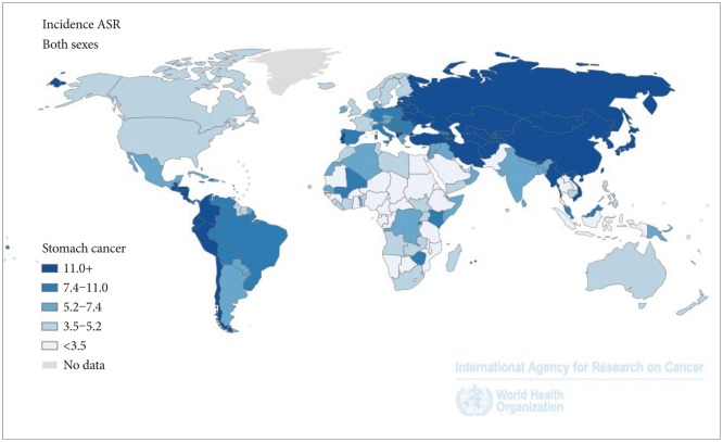
In spite of declining incidence in recent years, gastric cancer is still one of the most frequently occurring cancers. The most recent estimates from the International Agency for Research on Cancer (IARC) GLOBOCAN 2012 indicate that nearly one million new gastric cancer cases and more than 700,000 deaths occurred globally in 2012.1 The disease accounted for 7% of the total new cancer cases and 9% of the total cancer deaths. Gastric cancer is now the fifth most common cancer in the world, after lung, breast, colorectal and prostate cancer and the third leading cause of cancer death.1 There is a 10-fold variation in gastric cancer incidence internationally, with high rates seen in many countries of Eastern Asia (age standardized rate, 42.4/100,000 in 2008), Central and Eastern Europe (21.9/100,000), and Central (12.7/100,000) and South America (17.3/100,000), and much lower rates reported from North America (5.8/100,000), and Africa (4.7/100,000) (Fig. 1). In addition, rates in men are approximately double those observed in women.1,2
Over the past four decades, the age-standardized incidence as well as mortality rates from gastric cancer have steadily declined in nearly all populations, independently from their background risk of gastric cancer or sex (Fig. 2).3 Allowing for the estimated annual percentage change of -2%, the annual number of new gastric cancer cases (both sexes) would still slightly increase or remain stable in the year 2030 due to ageing of the population.1,4 The relative 5-year survival rates for gastric cancer are low in most countries with less than 30% except for the Republic of Korea and Japan with around 70% where screening programmes that lead to early detection are in place. The case fatality rate is lower in countries with high levels of human development (overall mortality-to-incidence ratio=0.65) than in countries at low or medium levels of human development (0.83).2
Currently, the incidence rates of gastric cancer among men in the Republic of Korea is the highest in the world.4 It has been estimated that the age-standardized rates were 63.3 in men and 25.1 per 100,000 in women, respectively, with approximately 32,000 new gastric cases being diagnosed in 2011.5 The rates have remained stable for many years (67.2 in 2001 and 63.3 in 2011 for men; 26.2 in 2001 and 25.1 in 2011 for women) without geographical variation across the country.6 Gastric cancer was the most frequently occurring cancer in men, accounting for 19.6% of all cases and the third most frequently diagnosed cancer in women, accounting for 10.3% of all cases occurred in Koreans in 2012.7 Recent mortality data in Korea showed that gastric cancer was the third leading cause of cancer death in men, and the second leading cause of cancer death in women.5 The mortality rate for gastric cancer has decreased continuously both in men and women since 1997. The 5-year survival rate for gastric cancer in Korea have been much improved over the last two decades, showing an increase from 43% in 1993 to 1995 to 67% in 2006 to 2010.7
Gastric cancer represents a biologically and genetically heterogeneous group of tumours with multifactorial aetiologies, both environmental and genetic.2,8 Gastric cancers arising from the most proximal (cardia) or distal (non-cardia) region are likely to have different aetiologies. The majority of gastric cancers are noncardia cancers, while cardia cancers have been most commonly reported in North American and European populations.2
In most cases of noncardia gastric cancer, the cancer develops from the epithelial cells of the gastric mucosa with a transition from dysplasia to confirmed cancer. The successive steps in the sequence of gastric carcinogenesis, in relation to Helicobacter pylori infection of the mucosa, have been described by Correa.9 Those steps include: (1) chronic atrophic gastritis of the mucosa up to the lamina propria by the inflammation preceded by H. pylori bacterial infection; (2) intestinal metaplasia in large areas of the epithelium; (3) premalignant adenomatous lesion with succession of low and high grade dysplasia; and (4) intramucosal cancer.
Histological classification of gastric cancer
One of the most commonly used histological classifications of gastric cancer is the Laurén classification, where diffuse and intestinal types are identified as the two major subtypes.10 Mixed and indeterminate types are relatively uncommon.10 Diffuse type consists of poorly cohesive cells with little or no gland formation while intestinal type forms glands with various degrees of differentiation.8
The World Health Organization (WHO) classification is another widely used classification and it recognizes five major types: (1) tubular; (2) papillary; (3) mucinous; (4) poorly cohesive, including signet ring cell carcinoma and other variants; and (5) mixed carcinomas. This is a descriptive scheme and does not take into account histogenesis, differentiation or epidemiological data.8 Tubular and papillary types in the WHO classification roughly correspond to the intestinal type in the Laurén classification, and poorly cohesive cancers correspond to the diffuse type.2
Classification of dysplasia/intraepithelial neoplasia
The well-known discrepancies between Japanese and European/North American pathologists in categorizing gastric dysplasia/intraepithelial neoplasia11,12 resulted in attempts to standardize the terminology of the morphological spectrum of lesions ranging from non-neoplastic changes to early invasive cancer.8,13 The Vienna classification of gastrointestinal epithelial neoplasia is one of them and the categories are designed to be associated with different recommendations for clinical management. The Vienna Classification has been revised and it identifies five categories: (1) negative for neoplasia/dysplasia; (2) indefinite for neoplasia/dysplasia; (3) mucosal low grade neoplasia (low grade adenoma; low grade dysplasia); (4) mucosal high grade neoplasia which is subdivided into: 4.1) high grade adenoma/dysplasia; 4.2) noninvasive carcinoma (carcinoma in situ); 4.3) suspicious for invasive carcinoma; 4.4) intramucosal carcinoma; and (5) submucosal invasion by carcinoma. This revised Vienna classification aims to be more closely related to patient management, with more clinical usefulness.12
For premalignant lesions, the current 2010 WHO classification considers: (1) negative for intraepithelial neoplasia (dysplasia); (2) indefinite for intraepithelial neoplasia (dysplasia); (3) intraepithelial neoplasia (dysplasia) which is further subdivided into two grades: 3.1) low-grade intraepithelial neoplasia (dysplasia) and 3.2) high-grade intraepithelial neoplasia (dysplasia); and (4) intramucosal invasive neoplasia/intramucosal carcinoma.8 The WHO classification acknowledges that both dysplasia and intraepithelial neoplasia are acceptable terms.
Despite these attempts to lessen disagreements between Japanese and European/North American, lesions diagnosed as intramucosal carcinoma in Japan are still classified as high-grade adenoma/dysplasia in Europe/North America and often not diagnosed as cancer.14 In many cases neither Western nor Japanese pathologists could distinguish between "high grade adenoma/dysplasia," "noninvasive carcinoma (carcinoma in situ)," and "suspicion of invasive carcinoma" in a reproducible way.15
Endoscopic classification
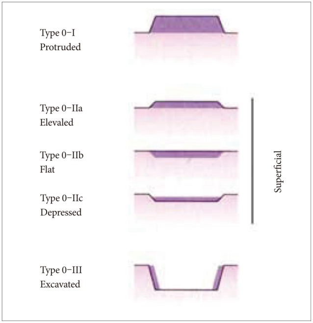
Gastric cancers are classified endoscopically according to growth pattern.8 Early gastric cancer is an invasive carcinoma that is limited to the mucosa, or the mucosa and submucosa, regardless of nodal status. Early gastric cancers are divided into three types (Fig. 3): (1) protruded (type 0-I); (2) superficial (type 0-II); and (3) excavated (type 0-III). The superficial type accounts for the majority of early gastric cancers and is further divided into type 0-IIa (elevated type), type 0-IIb (flat type), and type 0-IIc (depressed type).8,14
The Paris endoscopic classification of superficial neoplastic lesions of the stomach defines "superficial" at endoscopy when the endoscopic appearance suggests that the depth of penetration in the digestive wall is not more than into the submucosa, i.e., there is no infiltration of the muscularis propria.16 This classification considers the tumour grossly and endoscopically.17
In contrast to early gastric cancer, the gross appearance of advanced carcinoma is described macroscopically according to the Borrmann classification which includes: (1) polypoid type; (2) fungating type; (3) ulcerated type; and (4) infiltrative type, in which fungating and ulcerated types are common.8 With advanced gastric cancer invading to the proper muscle layer or beyond, 5-year survival rate was around 60% or less18 while early gastric carcinoma has a favourable prognosis, with a 5-year survival rate as high as 90%19,20 or higher even in elderly patients.21 A recent study of 598 Korean patients and 159 United States patients showed 5-year probability of death due to early-stage node-negative (T1N0) gastric cancer was 1.6 % in Korean patients and 3.2% in the United States patients between 1995 to 2005.22
Precursor lesions: gastritis
One of the recognized precursor lesions of the intestinal type, distal gastric cancer is gastritis. Despite the general consensus on the morphological aspect of the lesions, there have been controversies over the different types and pattern of gastritis, which led to considerable confusion among pathologists.23 In an attempt to establish an agreed terminology of gastritis and to identify, define and resolve some of the problems associated with the Sydney System, the Updated Sydney System was proposed as one of the most representative histological methods for gastritis assessment. The Updated Sydney system recommends an itemized description of various microscopic findings including lymphoplasma cell infiltrates, neutrophilic infiltrates, intestinal metaplasia, mucosal atrophy, and H. pylori. Severity/intensity of each factor is quantitatively expressed using a four-point grading system of the histological lesions, ranging from 0 (absence) to 3 (marked).23
Although the Updated Sydney System has been widely used with useful recommendations, it does not provide a clinically meaningful link between the presence of gastritis and risk of developing gastric cancer with no immediate prognostic or therapeutic information to clinicians.24 Recognizing the consistent evidence of the association of the extent of atrophy with the risk of gastric cancer, the Operative Link for Gastritis Assessment (OLGA) staging system was proposed.24 The OLGA staging system ranks gastritis-associated cancer risk according to both the topography and the extent of gastric mucosal atrophy.25 It stages corpus and antrum from stage 0 (no atrophy) to stage IV (severe atrophy) where the risk to develop gastric cancer is substantially high.24 A modification of the OLGA staging system, the Operative Link on Gastric Intestinal Metaplasia (OLGIM) assessment replaces the atrophy score with an assessment of intestinal metaplasia alone but is still based on the OLGA frame.26 Since the OLGIM proposal replaces the global atrophy score with a semiquantitative assessment of intestinal metaplasia both for extent and site, it has a higher inter-observer agreement compared with the histopathological diagnosis of atrophic gastritis.26 However, OLGIM staging alone is less sensitive than OLGA staging in the identification of patients at high risk of gastric cancer because it primarily focuses on intestinal metaplasia.26 Nonetheless, both OLGA and OLGIM gastritis staging systems have contributed to provide prognostically important information on the gastritis-associated cancer risk.
Only a very small proportion of gastric cancer cases is linked to germline mutation in E-cadherin/CDH1, which has been found in approximately 25% to 40% of families with hereditary diffuse gastric cancer.27,28 The penetrance of CDH1 gene mutations is high, with an estimated risk of >80% by the age of 80 years with poor prognosis and late presentation with a highly invasive diffuse type tumour.29
Two major autosomal dominant forms of heritable colorectal cancer, familial adenomatous polyposis and hereditary nonpolyposis colorectal cancer (Lynch syndrome), are also linked to increased risk of other cancers including gastric cancer. Lynch syndrome is caused by germline mutations in four mismatch repair genes (MLH1, MSH2, MSH6, and PMS2), resulting in development of a spectrum of different tumours and Lynch syndrome mutation carriers have shown a substantial risk for developing gastric cancer.30 The risk of gastric cancer is also increased in Li-Fraumeni syndrome with germline mutation of TP53.31
Peutz-Jeghers syndrome is an inherited multiple gastrointestinal polyposis associated with mucocutaneous pigmentation.32 Germline mutation in the LKB1 gene that encodes serine/threonine-protein kinase that acts as a tumour suppressor results in the loss of LKB1 function which can lead to gastric cancer carcinogenesis.33
While hereditary gastric cancer is uncommon, genetic alterations in sporadic gastric cancer cases are frequently reported. However, no major high-penetrance genes have been discovered so far,29 although genetic factors might play an important role in gastric carcinogenesis by possibly influencing immune and inflammatory responses especially to H. pylori infection and thereby altering susceptibility to gastric cancer.34
Polymorphisms of the interleukin 1β gene (IL-1β), which contributes to initiation and amplification of inflammatory response, and of the interleukin-1 receptor antagonist (IL-1RN) gene, which modulates inflammation, have been associated with gastric cancer risk. However, a systematic review of 35 studies did not find an overall association of polymorphisms in these inflammatory genes with risk of gastric cancer, but there was significant heterogeneity among study results.35 Results from very large genome-wide association studies (GWAS) showed reproducible associations between single-nucleotide polymorphisms located at prostate stem cell antigen gene (PSCA), PLCE1, and Mucin 1, cell surface-associated gene (MUC1) genes and different subtypes of gastric cancer risk. These results from GWAS are mainly from Chinese, Korean and Japanese populations and the clear biological mechanism behind these polymorphisms is not yet clearly known.29
The main cause of gastric cancer is chronic infection with H. pylori.36,37 The IARC classified H. pylori as a class I carcinogen in 1994 and reconfirmed this classification in 2009 after reviewing newer evidence.38 According to the most recent estimates using a more sensitive method to detect H. pylori, approximately 89% (770,000 cases) of distal gastric cancer cases worldwide have been estimated to be attributable to chronic H. pylori infection.39 H. pylori infection is present throughout the world and causes most cases of peptic ulcer disease and mucosa-associated lymphoid tissue lymphoma, as well as gastric cancer. The prevalence of infection in adults varies between 20% to 50% in industrialized countries and over 80% in many developing countries.40 H. pylori infection is strongly associated with socioeconomic conditions,41 and transmission appears to occur through close personal contact, particularly within the family38 and typically in early childhood. Once established, infection usually persists throughout life unless treated. The prevalence of H. pylori infection has decreased in recent decades, particularly among children in developed countries, probably reflecting improvements in hygiene.42,43 However, there is only limited information on prevalence trends among children from most countries,41,44 and H. pylori still infects an estimated 50% of the world's population.
Factors associated with colonization and pathogenicity of H. pylori include virulence factors like cytotoxin-associated gene A (cagA) in the cag pathogenicity island and the vacuolating cytotoxin A (vacA). cagA is a gene that is part of the cag pathogenicity island and colonization with strains that possess cagA is associated with increased risk of developing both intense tissue responses and premalignant and malignant lesions in the distal stomach through secreting a functional cytotoxin which induces more severe gastric injury and further augment the risk.45,46 CagA has been shown to associate with the apoptosis-stimulating protein of p53, inhibiting the apoptotic function of p53, in a manner similar to human papillomavirus (HPV) and other oncogenic DNA viruses.47 vacA encodes a secreted bacterial toxin (VacA) and induces multiple structural and functional alterations in cells, the most prominent of which is the formation of large intracellular vacuoles.48 Primarily due to variations in vacA gene structure, vacA positive strains vary considerably in production of cytotoxin activity, despite its universal presence in all H. pylori strains examined.45
Another infectious agent that has been associated with gastric cancer is the Epstein-Barr virus (EBV). The virus is a ubiquitous infectious agent with a prevalence of over 90% in adults and has been causally linked to the development of several malignancies, including Burkitt's lymphoma, immunosuppression-related lymphoma, Hodgkin's disease, and nasopharyngeal carcinoma.49 Approximately 8% of gastric cancers have been estimated to harbour EBV,50 but there is insufficient epidemiological evidence of a clear etiological role for EBV in gastric carcinogenesis.38 The EBV genome is present in the tumour cell in a monoclonal form, and transforming EBV proteins are expressed in the tumour cell. A meta-analysis reported that the prevalence of EBV-positivity in gastric cancer varied jointly by age, sex and anatomic subsite, i.e., EBV-positivity decreased with age among men, more steeply for tumours localised to the antrum.50 In addition, EBV-positivity has been associated with longer survival,51 suggesting that EBV-positive gastric cancer may have distinct clinical and genetic features, and therefore may be a separate clinical entity.
Various other risk factors including older age, male sex, cigarette smoking, low socioeconomic status, low level of physical activity, and radiation exposure have been linked to increased risk of gastric cancer in general while gastroesophageal reflux disease and obesity have been associated with increased risk of gastric cardia cancer specifically.29 Nonsteroidal anti-inflammatory drugs and statin intake have shown inverse associations with gastric cancer risk.29
Dietary factors have been investigated in many studies in relation to gastric cancer risk, since food carcinogens can potentially interact directly with the epithelial cells that line the stomach. A comprehensive systematic review of dietary factors and cancer prevention published by the World Cancer Research Fund/American Institute for Cancer Research (WCRF/AICR) concluded that nonstarchy vegetables, including specifically allium vegetables, as well as fruits probably protect against gastric cancer while salt, and also salt-preserved foods, are probably causes of this cancer.52 A high concentration of sodium chloride has been shown to cause damage to the gastric mucosa, cell death and consequent regenerative cell proliferation in animal models,53,54 which may lead to inflammation and damage such as diffuse erosion and degeneration.55 Studies have found that a high salt diet was associated with colonization of H. pylori56 and exacerbated H. pylori-induced inflammation,57 which in consequence was responsible for promotion of gastric carcinogenesis in a dose-dependent fashion.58 This was also confirmed in other studies showing that salt worked synergistically with H. pylori infection to enhance the expression of proinflammatory enzymes and cytokines such as inducible nitric oxide synthase and cyclooxygenase-2 in the gastric mucosa.59 In addition, high salt concentrations have been shown to stimulate increased expression of H. pylori CagA, which in turn leads to an increased amount of CagA translocated into gastric epithelial cells and an enhanced ability of H. pylori to alter gastric epithelial cell function.60
Another dietary factor that might play a role in gastric carcinogenesis is N-nitroso compounds.52 Nitrates, produced endogenously in gastric acid, and added as preservatives to processed meats may contribute to N-nitroso compound production and exposure that are suspected mutagens and carcinogens.52 Previous studies have shown a significant increase of gastrointestinal cancer risk associated with dietary61 or endogenous exposure to N-nitroso compounds,62 especially with noncardia gastric cancer rather than cardia gastric cancer.62 Many processed meats contain high levels of salt and nitrite. Haem found in red meat promotes the formation of N-nitroso compounds through direct reaction between nitric oxide and haemoglobin and myoglobin.61 Red meat also contains iron, which can lead to the production of free radicals. Vitamin C may be protective against H. pylori-associated gastric carcinogenesis by enhancing mucosal immune response, neutralizing free radicals, reducing the formation of gastric N-nitroso compounds, inhibiting cell proliferation and directly affecting H. pylori growth.62 The gastric juice vitamin C levels were also shown to be lower with infection with CagA positive H. pylori strains compared with CagA negative H. pylori strains.63,64 Even with the biologically plausible mechanisms, epidemiological evidence suggesting that dietary nitrate and nitrite or other N-nitroso compounds are the causes of gastric cancer is limited. The WCRF/AICR report assessed other dietary factors and concluded that the available evidence suggesting that pulses (legumes) and foods containing selenium protect against gastric cancer and that chilli, processed meat, smoked foods, and grilled (broiled) and barbecued (charbroiled) animal foods are causes of stomach cancer is also limited.52
The two major primary prevention strategies for gastric cancer at a population level might include improvement in diet and reduction in the prevalence of H. pylori, the main cause of gastric cancer.2 Prevention through dietary intervention would be with increased fruit and vegetable intakes and decreased consumption of salt or salt-preserved foods. Lifestyle modification such as increased physical activity and smoking cessation may also help reduce risk of getting the disease. Interventions for primary prevention at the population level may potentially involve very large numbers of people that require convincing evidence of effectiveness. However, interventions based on these dietary strategies have not yet been evaluated in large-scale randomized clinical trials examining their impact on gastric cancer incidence or mortality.
Another strategy to prevent gastric cancer would be through H. pylori eradication. Recognition of the causal role of chronic H. pylori infection in gastric cancer led some authors by 2005 to call for broad-scale programmes to eradicate the infection as a way to prevent the disease.65 However, no countries with high burden of gastric cancer have yet implemented a population-based H. pylori eradication programme. Some of this inaction may reflect doubts about the effectiveness of H. pylori eradication in preventing gastric cancer as there are still scant data available from randomized clinical trials.66,67 Recently, a new meta-analysis of all six reported randomized trials among asymptomatic adults estimated a benefit (relative risk=0.66, 95% confidence interval [CI], 0.46 to 0.95).68 In 2012, a trial in a general population of adults in China reported a statistically significant reduction in gastric cancer risk following treatment after 15 years of follow-up (4.6% in control group, 3.0% in treated group; odds ratio, 0.61; 95% CI, 0.38 to 0.96).69 Two trials have assessed the effect of H. pylori treatment after endoscopic mucosal resection of early gastric cancer.70,71 One of the trials, in Japan, reported a statistically significant reduction in risk of metachronous gastric cancer.71 Risk also appeared lower in a study conducted in the Republic of Korea, but the result was not statistically significant.70 Although these results of randomized trials indicate that H. pylori treatment may lower gastric cancer incidence by 30% to 40%, there are important limitations to the available data. Firstly, the meta-analysis finding is based on relatively few trials, many of which had too few cancer endpoints to contribute much weight to the analyses. Secondly, because of the relative paucity of endpoint data, the estimated size of the overall beneficial effect of treatment is likely to be imprecise. Thirdly, the larger trials were all conducted among middle-aged adults in Asia, notably in China, and the generalizability of their results to other populations is not certain. Lastly, the trials do not provide direct data on the feasibility, costs, effectiveness, and adverse effects of treatment programmes as they are applied in community settings.72
A recently published report of a working group of the IARC of the WHO concluded that-randomized clinical trials have found that H. pylori treatment is effective in preventing gastric cancer, and models indicate that H. pylori screening and treatment strategies would be cost-effective. However, uncertainties remain about the generalizability of results and about the cost-effectiveness and possible adverse consequences of programmes applied in community settings. The Working Group therefore recommends that countries explore the possibility of introducing population-based H. pylori screening and treatment programmes, but cautions that decisions as to whether and how to implement H. pylori testing and treatment must hinge on local considerations of disease burden, other health priorities, and cost-effectiveness analyses. Moreover, these programmes should be implemented in conjunction with a scientifically valid assessment of programme processes, feasibility, effectiveness, and possible adverse consequences."73,74
Early detection efforts
Early detection and treatment of cancer requires accessible, high-quality health services with adequate human, financial, and technical resources. Screening methods that are based on rigorous evidence and are implemented in national or regional cancer screening programmes currently include: mammography for breast cancer; cytology, HPV testing and visual inspection of the cervix for cervical cancer; and faecal occult blood testing or faecal immunochemical testing, flexible sigmoidoscopy, and colonoscopy for colorectal cancer.2 With the exception of the republic of Korea (see below), gastric cancer screening is generally not included in national cancer prevention strategies, even in countries with a high burden of the disease. This may reflect not only high cost, decreasing incidence worldwide and the paucity of data to answer the questions, "how, who, and when to screen." It may also result from the complexity of the screening process and the substantial knowledge, skill, and effort needed to assure the quality of the screening services.2,75 Given the wide variation in gastric cancer burden around the world and the substantial numbers of cases and deaths projected in the coming years, further improvement in early diagnosis and treatment, and development and validation of effective methods for population-based screening in countries with a significant projected burden of disease should be a priority on the public health agenda.
Different types of tests have been proposed and used in a few countries for gastric cancer screening. Barium-meal photofluorography (or barium swallow) has been used for mass screening in Japan since 196076 and upper endoscopy has been used for nationally organized gastric cancer screening in the Republic of Korea since 1999.77 Active intervention in a population usually requires randomized controlled clinical trial data showing that the intervention is effective.2 Photofluorography has been used for screening in the absence of evidence for reducing gastric cancer mortality in randomized controlled clinical trials.75 Current guidelines for gastric cancer screening in Japan that recommended use of photofluorography for both population-based and opportunistic screening, are largely based on results of case-control studies which suggested a 40% to 60% decrease in gastric cancer mortality with photofluorography.78 Prospective cohort studies were limited in numbers and inconsistent with the results in demonstrating the reduction in gastric cancer mortality. The sensitivity of photofluorography ranged from 60% to 80%, whereas the specificity ranged from 80% to 90%.78
Upper gastrointestinal endoscopy has been regarded as the gold standard for the diagnosis of gastric cancer.29 It is also used for minimally invasive treatment of early gastric cancer through endoscopic mucosal resection and endoscopic submucosal dissection. Widespread use of the latter method has resulted in a dramatic increase in the number of early gastric cancer cases treated endoscopically in Japan and Korea.14 In high risk populations it may also play a role in secondary prevention (screening) as the examination used for primary testing for gastric cancer or its precursors. The technique is highly dependent on the skills of the endoscopists; however, and the feasibility of endoscopy-based screening depends on capacity that is limited in most countries. Endoscopic screening was shown to be cost-effective in areas with high gastric cancer burden,79 although further studies are needed to determine the suitability of this technique for recommending its broad use as a primary screening test.80
The effectiveness of endoscopic screening to reduce gastric cancer mortality has not yet been confirmed in randomized controlled trials. A study conducted retrospectively in a community in an isolated island in Japan found that in the district where endoscopic screening was introduced, the odds ratio of death from gastric cancer among the participants versus non-participants of endoscopic screening was 0.12 (1 case out of 16 screened vs. 8 cases out of 22 nonscreened), while it was 0.09 (2 out of 34 screened vs. 48 out of 114 nonscreened) in the radiographic screening district, after 6 years of follow-up on average, suggesting that both radiographic and endoscopic screening may prevent death from gastric cancer.81 However, very small numbers analysed in the study and its retrospective nature limit interpretation of the study.
In a cohort study carried out in Linqu County, China with a high incidence of gastric cancer, the number of observed deaths from gastric cancer (n=37) during 11 years of follow-up with repeated endoscopic screening was close to the number expected (n=36.8), resulting in a standardized mortality ratio of 1.01 (95% CI, 0.72 to 1.37) for men and women combined.82 The most recent evidence comes from a community-based case-control study conducted in Japan, which suggested a 30% reduction in gastric cancer mortality by endoscopic screening compared with no screening within 36 months before the date of diagnosis of gastric cancer.83 The study results must be interpreted cautiously; however, due to various methodological limitations. For example, symptomatic individuals were not excluded and no other background information was collected which might have explained the results.
In Korea, the performance of upper endoscopy has been intensively studied within the setting of the National Cancer Screening Programme (NCSP) especially in comparison with upper gastrointestinal series (UGIS), which is another modality that participants can choose for gastric cancer screening. The detection rates of gastric cancer were 0.68 and 2.61 per 1,000 UGIS and endoscopy screenings, respectively.80 The sensitivities of UGIS and endoscopy screening to detect gastric cancer were 36.7% and 69.0%, respectively, and their specificities were 96.1% and 96.0%, respectively.80 The detection rate of gastric cancer by endoscopic screening is 2.7- to 4.6-fold higher than that by gastric radiography.80,84,85 Overall, endoscopy performed better than UGIS in the NCSP for gastric cancer. The unit costs of screening using UGIS and upper endoscopy were US $32.67 and $34.89, respectively. In 2008, the estimated cost of identifying one case of gastric cancer was US $53,094 using UGIS and $16,900 using upper endoscopy.84 In a study of more than 18,000 participants who underwent endoscopy within NCSP between 2001 and 2007, a total of 81 gastric cancer cases were found, in which 81% were early gastric cancers.86 The study reported a 50% decreased incidence of gastric cancer in the repeated endoscopic screening group within 2 years compared the group with infrequent screening. In addition, gastric cancers detected by a repeated endoscopy within 2 years were found to be mostly early gastric cancers that can be treated by endoscopic resection.86
Pepsinogen for atrophic gastritis detection
Plasma biomarkers tests may identify patients at increased risk of developing gastric cancer, especially with atrophic gastritis, who would then be eligible for endoscopic surveillance for cancer risk.87 The method that has been investigated most extensively is pepsinogen levels.
Pepsinogens I and II are produced by cells from the gastric mucosa, whose production can be altered by atrophic gastritis.87 These pepsinogen alterations have been shown to be a marker for atrophy in the gastric mucosa. Since atrophic gastritis is the highest known independent risk factor for distal, noncardia gastric cancer,87 the test for pepsinogen levels has long been considered for screening to identify a high risk group. A pooled analysis of Japanese studies in a total of about 300,000 individuals, pepsinogen test resulted in a sensitivity of 77% and a specificity of 73% for atrophic gastritis, suggesting the test is reliable as a marker for the identification of individuals at risk of progression to intestinal gastric cancer.88
Both Chinese and Japanese studies89,90,91 have shown a synergistic effect of both H. pylori and pepsinogens, i.e., the highest risk of developing gastric cancer shown in the group positive for pepsinogen test but negative for H. pylori, suggesting that the utility of the pepsinogen test might be improved if the test is combined with serology for H. pylori infection. Outside Asia, however, evidence for the utility of pepsinogen test has been less convincing, especially in Latin America, requiring further studies to establish the performance of the test and define cutoff values for the population.92 Moreover, it should be noted that the pepsinogen test is not useful for screening of diffuse gastric cancer, and performance would vary depending on the proportion of diffuse gastric cancer in the population.92 Considering that the pepsinogen test is still too expensive to be applied in community-based screening programmes, particularly in low-middle income countries in Latin America and Asia that present the highest mortality rates, the possible population-based application needs to be further evaluated.92
More recently, amidated gastrin-17 (G-17) has been suggested to characterize atrophy in the antral part of the stomach. G-17 is secreted exclusively by the G cells in antrum, and G-17 levels have been evaluated as a serological marker to distinguish antral atrophic gastritis from corpus atrophic gastritis. In particular, patients with antral atrophic gastritis present with low levels of circulating G-17.87,93,94 However, the concentrations of G-17 in the circulation are influenced by several factors, and as a result the sensitivity for diagnosing atrophy in the antral part of the stomach is not satisfactory (15.4% in a fasting state and 30.8% after stimulation),95 possibly due also to inadequate performance of available commercial kits.92 In a recent study that evaluated associations of temporal changes in multiple serological markers including gastrin-17 with risk for progression of gastric precancerous lesions during 14 years of follow-up, changes in gastrin-17 were not statistically significantly associated with progression.96 Given that there are very few epidemiological studies of G-17 in relation to gastric cancer, the value of G-17 for gastric cancer screening is unclear.
Gastric cancer remains the third leading cause of cancer death worldwide and the burden of disease, which for unknown reasons affects twice more men than women and varies widely from place to place, is expected to remain high for many decades despite declining trends. The disease is usually detected late and survival is poor in most settings. With few exceptions, there are no public health programmes aimed at reducing gastric cancer burden.
Primary prevention interventions, including dietary modifications and eradication of H. pylori need to be considered as potentially valuable tools in the control of this disease. H. pylori can be treated with antibiotics and in clinical trials, its eradication has been shown to reduce gastric cancer incidence between 30% and 40%. However, given the high prevalence of infection in high risk areas and limited information about feasibility, acceptability and potential adverse consequences of the intervention, it has not been broadly implemented. Several trials are underway that should provide additional data, but demonstrations projects with proper evaluation of the programme were recently recommended by an IARC working group of international experts.
More rapid impact on the burden of disease might be forthcoming from secondary prevention through early detection and prompt treatment of precursor lesions and early cancer. Encouraging progress has been achieved in Japan where opportunistic screening is widespread, and in the Republic of Korea where screening is conducted in an organized programme. However, the methods used in these countries require infrastructure and human resources with ample expertise which are not available in less developed areas. In addition, the effectiveness of the screening methods has not been validated in randomized controlled clinical trials.
Endoscopic detection and treatment is only one important facet of the complex process of gastric cancer screening. To achieve an appropriate benefit, screening should only be considered in regions with a significant burden of disease.97,98,99 In addition, screening should be conducted in organized, population-based programmes to promote equal access through personal invitation and to facilitate comprehensive quality assurance systems. Before gastric cancer screening can be recommended as a public health policy in other countries, information is required on expected detection rates, participation rates, acceptability, economic costs, as well as any complications or adverse effects associated with screening in different populations. The highly successfully organized screening programme in the Republic of Korea provides a unique opportunity to conduct population-based studies within the framework of the programme. This will not only help maintain and further improve the quality of the programme, but also provide valuable evidence for other countries considering the potential of gastric cancer screening to reduce the burden of disease in their populations.
We acknowledge Dr René Lambert for his contribution to this review.
  • 1. Ferlay J, Soerjomataram I, Ervik M, et al. GLOBOCAN 2012 v1.0, Cancer incidence and mortality worldwide: IARC CancerBase No. 11 [Internet]. Lyon: International Agency for Research on Cancer; 2013. cited 2014 Apr 25]. Available from: http://globocan.iarc.fr
  • 2. Carneiro F. Stomach cancer. In: Steward BW, Wild CP, editors. World Cancer Report 2014. Lyon: International Agency for Research on Cancer; 2014. p. 383–391.
  • 3. Ferlay J, Bray F, Steliarova-Foucher E, Forman D. Cancer incidence in five continents, CI5plus: IARC CancerBase No. 9 [Internet]. Lyon: International Agency for Research on Cancer; 2014. cited 2014 Apr 25]. Available from: http://ci5.iarc.fr
  • 4. Forman D, Sierra MS. Introduction: the current and projected global burden of gastric cancer. IARC Helicobacter pylori Working Group. Helicobacter pylori Eradication as a Strategy for Preventing Gastric Cancer [Internet]. Lyon: International Agency for Research on Cancer (IARC Working Group Reports, No. 8); 2014. cited 2014 Apr 25]. p. 5-15. Available from:http://www.iarc.fr/en/publications/pdfs-online/wrk/wrk8/index.php
  • 5. Jung KW, Won YJ, Kong HJ, Oh CM, Lee DH, Lee JS. Cancer statistics in Korea: incidence, mortality, survival, and prevalence in 2011. Cancer Res Treat 2014;46:109–123.ArticlePubMedPMCPDF
  • 6. Choi IJ, Park JY, Herrero R. Effect of Helicobacter pylori eradication on gastric cancer prevention in the Republic of Korea: a randomized controlled clinical trial. IARC Helicobacter pylori Working Group. Helicobacter pylori Eradication as a Strategy for Preventing Gastric Cancer [Internet]. Lyon: International Agency for Research on Cancer (IARC Working Group Reports, No. 8); 2014. cited 2014 Apr 25]. p. 154-160. Available from: http://www.iarc.fr/en/publications/pdfs-online/wrk/wrk8/index.php
  • 7. National Cancer Center. Cancer Facts & Figures 2013 in the Republic of Korea. Goyang: President of National Cancer Center, Minister for Health and Welfare; 2013. p. 8.
  • 8. Lauwers GY, Carneiro F, Graham DY, et al. Gastric carcinoma. In: Bosman FT, Carneiro F, Hruban RH, Theise ND, editors. WHO Classification of Tumours of the Digestive System. Lyon: International Agency for Research on Cancer; 2010. p. 48–58.
  • 9. Correa P. A human model of gastric carcinogenesis. Cancer Res 1988;48:3554–3560.PubMed
  • 10. Lauren P. The two histological main types of gastric carcinoma: diffuse and so-called intestinal-type carcinoma. An attempt at a histo-clinical classification. Acta Pathol Microbiol Scand 1965;64:31–49.ArticlePubMed
  • 11. Lauwers GY, Shimizu M, Correa P, et al. Evaluation of gastric biopsies for neoplasia: differences between Japanese and Western pathologists. Am J Surg Pathol 1999;23:511–518.ArticlePubMed
  • 12. Dixon MF. Gastrointestinal epithelial neoplasia: Vienna revisited. Gut 2002;51:130–131.ArticlePubMedPMC
  • 13. Dinis-Ribeiro M, Areia M, de Vries AC, et al. Management of precancerous conditions and lesions in the stomach (MAPS): guideline from the European Society of Gastrointestinal Endoscopy (ESGE), European Helicobacter Study Group (EHSG), European Society of Pathology (ESP), and the Sociedade Portuguesa de Endoscopia Digestiva (SPED). Endoscopy 2012;44:74–94.ArticlePubMedPMCPDF
  • 14. Yada T, Yokoi C, Uemura N. The current state of diagnosis and treatment for early gastric cancer. Diagn Ther Endosc 2013;2013:241320.ArticlePubMedPMCPDF
  • 15. Schlemper RJ, Riddell RH, Kato Y, et al. The Vienna classification of gastrointestinal epithelial neoplasia. Gut 2000;47:251–255.ArticlePubMedPMC
  • 16. Participants in the Paris Workshop. The Paris endoscopic classification of superficial neoplastic lesions: esophagus, stomach, and colon: November 30 to December 1, 2002. Gastrointest Endosc 2003;58(6 Suppl):S3–S43.PubMed
  • 17. Hu B, El Hajj N, Sittler S, Lammert N, Barnes R, Meloni-Ehrig A. Gastric cancer: classification, histology and application of molecular pathology. J Gastrointest Oncol 2012;3:251–261.PubMedPMC
  • 18. Yoshikawa K, Maruyama K. Characteristics of gastric cancer invading to the proper muscle layer: with special reference to mortality and cause of death. Jpn J Clin Oncol 1985;15:499–503.PubMed
  • 19. Everett SM, Axon AT. Early gastric cancer in Europe. Gut 1997;41:142–150.ArticlePubMedPMC
  • 20. Yokota T, Kunii Y, Teshima S, et al. Significant prognostic factors in patients with early gastric cancer. Int Surg 2000;85:286–290.PubMed
  • 21. Kim DY, Joo JK, Ryu SY, Park YK, Kim YJ, Kim SK. Clinicopathologic characteristics of gastric carcinoma in elderly patients: a comparison with young patients. World J Gastroenterol 2005;11:22–26.ArticlePubMedPMC
  • 22. Strong VE, Song KY, Park CH, et al. Comparison of disease-specific survival in the United States and Korea after resection for early-stage node-negative gastric carcinoma. J Surg Oncol 2013;107:634–640.ArticlePubMedPMC
  • 23. Dixon MF, Genta RM, Yardley JH, Correa P. Classification and grading of gastritis. The updated Sydney System. International workshop on the histopathology of gastritis, Houston 1994. Am J Surg Pathol 1996;20:1161–1181.ArticlePubMed
  • 24. Rugge M, Meggio A, Pennelli G, et al. Gastritis staging in clinical practice: the OLGA staging system. Gut 2007;56:631–636.ArticlePubMedPMC
  • 25. Rugge M, Correa P, Di Mario F, et al. OLGA staging for gastritis: a tutorial. Dig Liver Dis 2008;40:650–658.ArticlePubMed
  • 26. Capelle LG, de Vries AC, Haringsma J, et al. The staging of gastritis with the OLGA system by using intestinal metaplasia as an accurate alternative for atrophic gastritis. Gastrointest Endosc 2010;71:1150–1158.ArticlePubMed
  • 27. Carneiro F, Huntsman DG, Smyrk TC, et al. Model of the early development of diffuse gastric cancer in E-cadherin mutation carriers and its implications for patient screening. J Pathol 2004;203:681–687.ArticlePubMed
  • 28. Carneiro F, Oliveira C, Suriano G, Seruca R. Molecular pathology of familial gastric cancer, with an emphasis on hereditary diffuse gastric cancer. J Clin Pathol 2008;61:25–30.ArticlePubMed
  • 29. Karimi P, Islami F, Anandasabapathy S, Freedman ND, Kamangar F. Gastric cancer: descriptive epidemiology, risk factors, screening, and prevention. Cancer Epidemiol Biomarkers Prev 2014;23:700–713.ArticlePubMedPMC
  • 30. Capelle LG, Van Grieken NC, Lingsma HF, et al. Risk and epidemiological time trends of gastric cancer in Lynch syndrome carriers in the Netherlands. Gastroenterology 2010;138:487–492.ArticlePubMed
  • 31. Masciari S, Dewanwala A, Stoffel EM, et al. Gastric cancer in individuals with Li-Fraumeni syndrome. Genet Med 2011;13:651–657.ArticlePubMedPMC
  • 32. Song SH, Kim KW, Kim WH, et al. Gastrointestinal cancers in a peutz-jeghers syndrome family: a case report. Clin Endosc 2013;46:572–575.ArticlePubMedPMC
  • 33. Takahashi M, Sakayori M, Takahashi S, et al. A novel germline mutation of the LKB1 gene in a patient with Peutz-Jeghers syndrome with early-onset gastric cancer. J Gastroenterol 2004;39:1210–1214.ArticlePubMed
  • 34. El-Omar EM, Carrington M, Chow WH, et al. Interleukin-1 polymorphisms associated with increased risk of gastric cancer. Nature 2000;404:398–402.ArticlePubMedPDF
  • 35. Kamangar F, Cheng C, Abnet CC, Rabkin CS. Interleukin-1B polymorphisms and gastric cancer risk: a meta-analysis. Cancer Epidemiol Biomarkers Prev 2006;15:1920–1928.ArticlePubMed
  • 36. González CA, Megraud F, Buissonniere A, et al. Helicobacter pylori infection assessed by ELISA and by immunoblot and noncardia gastric cancer risk in a prospective study: the Eurgast-EPIC project. Ann Oncol 2012;23:1320–1324.ArticlePubMedPDF
  • 37. Lochhead P, El-Omar EM. Helicobacter pylori infection and gastric cancer. Best Pract Res Clin Gastroenterol 2007;21:281–297.ArticlePubMed
  • 38. IARC Working Group. Helicobacter pylori. IARC Working Group. IARC Monographs on the Evaluation of Carcinogenic Risks to Humans. Vol. 100B. Biological Agents. Lyon: International Agency for Research on Cancer; 2012. p. 385–436.
  • 39. Plummer M, Franceschi S, Vignat J, Forman D, de Martel C. Global burden of gastric cancer attributable to pylori. Int J Cancer 2015;136:487–490.ArticlePubMed
  • 40. Suerbaum S, Michetti P. Helicobacter pylori infection. N Engl J Med 2002;347:1175–1186.ArticlePubMed
  • 41. Porras C, Nodora J, Sexton R, et al. Epidemiology of Helicobacter pylori infection in six Latin American countries (SWOG Trial S0701). Cancer Causes Control 2013;24:209–215.ArticlePubMedPMC
  • 42. Oona M, Utt M, Nilsson I, Uibo O, Vorobjova T, Maaroos HI. Helicobacter pylori infection in children in Estonia: decreasing seroprevalence during the 11-year period of profound socioeconomic changes. Helicobacter 2004;9:233–241.ArticlePubMed
  • 43. Yim JY, Kim N, Choi SH, et al. Seroprevalence of Helicobacter pylori in South Korea. Helicobacter 2007;12:333–340.ArticlePubMed
  • 44. Leja M, Cine E, Rudzite D, et al. Prevalence of Helicobacter pylori infection and atrophic gastritis in Latvia. Eur J Gastroenterol Hepatol 2012;24:1410–1417.ArticlePubMed
  • 45. Peek RM Jr, Crabtree JE. Helicobacter infection and gastric neoplasia. J Pathol 2006;208:233–248.ArticlePubMed
  • 46. Peek RM Jr, Vaezi MF, Falk GW, et al. Role of Helicobacter pylori cagA(+) strains and specific host immune responses on the development of premalignant and malignant lesions in the gastric cardia. Int J Cancer 1999;82:520–524.ArticlePubMed
  • 47. Buti L, Spooner E, Van der Veen AG, Rappuoli R, Covacci A, Ploegh HL. Helicobacter pylori cytotoxin-associated gene A (CagA) subverts the apoptosis-stimulating protein of p53 (ASPP2) tumor suppressor pathway of the host. Proc Natl Acad Sci U S A 2011;108:9238–9243.ArticlePubMedPMC
  • 48. Gebert B, Fischer W, Weiss E, Hoffmann R, Haas R. Helicobacter pylori vacuolating cytotoxin inhibits T lymphocyte activation. Science 2003;301:1099–1102.ArticlePubMed
  • 49. IARC Working Group. Epstein-Barr virus. IARC Working Group. IARC Monographs on the Evaluation of Carcinogenic Risks to Humans. Vol. 70. Epstein-Barr Virus and Kaposi's Sarcoma Herpesvirus/Human Herpesvirus 8. Lyon: International Agency for Research on Cancer; 1997. p. 47–373.
  • 50. Camargo MC, Murphy G, Koriyama C, et al. Determinants of Epstein-Barr virus-positive gastric cancer: an international pooled analysis. Br J Cancer 2011;105:38–43.ArticlePubMedPMCPDF
  • 51. Camargo MC, Kim WH, Chiaravalli AM, et al. Improved survival of gastric cancer with tumour Epstein-Barr virus positivity: an international pooled analysis. Gut 2014;63:236–243.ArticlePubMedPMC
  • 52. World Cancer Research Fund. American Institute for Cancer Research. Stomach. World Cancer Research Fund; American Institute for Cancer Research. Food, Nutrition, Physical Activity, and the Prevention of Cancer: a Global Perspective. Washington DC: WCRF/AICR; 2007. p. 265–270.
  • 53. Cohen AJ, Roe FJ. Evaluation of the aetiological role of dietary salt exposure in gastric and other cancers in humans. Food Chem Toxicol 1997;35:271–293.ArticlePubMed
  • 54. Furihata C, Ohta H, Katsuyama T. Cause and effect between concentration-dependent tissue damage and temporary cell proliferation in rat stomach mucosa by NaCl, a stomach tumor promoter. Carcinogenesis 1996;17:401–406.ArticlePubMedPDF
  • 55. Tsugane S, Sasazuki S, Kobayashi M, Sasaki S. Salt and salted food intake and subsequent risk of gastric cancer among middle-aged Japanese men and women. Br J Cancer 2004;90:128–134.ArticlePubMedPMCPDF
  • 56. Fox JG, Dangler CA, Taylor NS, King A, Koh TJ, Wang TC. High-salt diet induces gastric epithelial hyperplasia and parietal cell loss, and enhances Helicobacter pylori colonization in C57BL/6 mice. Cancer Res 1999;59:4823–4828.PubMed
  • 57. Gaddy JA, Radin JN, Loh JT, et al. High dietary salt intake exacerbates Helicobacter pylori-induced gastric carcinogenesis. Infect Immun 2013;81:2258–2267.ArticlePubMedPMC
  • 58. Kato S, Tsukamoto T, Mizoshita T, et al. High salt diets dose-dependently promote gastric chemical carcinogenesis in Helicobacter pylori-infected Mongolian gerbils associated with a shift in mucin production from glandular to surface mucous cells. Int J Cancer 2006;119:1558–1566.ArticlePubMed
  • 59. Toyoda T, Tsukamoto T, Hirano N, et al. Synergistic upregulation of inducible nitric oxide synthase and cyclooxygenase-2 in gastric mucosa of Mongolian gerbils by a high-salt diet and Helicobacter pylori infection. Histol Histopathol 2008;23:593–599.PubMed
  • 60. Loh JT, Torres VJ, Cover TL. Regulation of Helicobacter pylori cagA expression in response to salt. Cancer Res 2007;67:4709–4715.ArticlePubMed
  • 61. Loh YH, Jakszyn P, Luben RN, Mulligan AA, Mitrou PN, Khaw KT. N-Nitroso compounds and cancer incidence: the European Prospective Investigation into Cancer and Nutrition (EPIC)-Norfolk Study. Am J Clin Nutr 2011;93:1053–1061.ArticlePubMedPDF
  • 62. Jakszyn P, Bingham S, Pera G, et al. Endogenous versus exogenous exposure to N-nitroso compounds and gastric cancer risk in the European Prospective Investigation into Cancer and Nutrition (EPIC-EURGAST) study. Carcinogenesis 2006;27:1497–1501.ArticlePubMedPDF
  • 63. Rokkas T, Liatsos C, Petridou E, et al. Relationship of Helicobacter pylori CagA(+) status to gastric juice vitamin C levels. Eur J Clin Invest 1999;29:56–62.ArticlePubMed
  • 64. Zhang ZW, Patchett SE, Perrett D, Katelaris PH, Domizio P, Farthing MJ. The relation between gastric vitamin C concentrations, mucosal histology, and CagA seropositivity in the human stomach. Gut 1998;43:322–326.ArticlePubMedPMC
  • 65. Graham DY, Shiotani A. The time to eradicate gastric cancer is now. Gut 2005;54:735–738.ArticlePubMedPMC
  • 66. Park JY, Forman D, Greenberg ER, Herrero R. Helicobacter pylori eradication in the prevention of gastric cancer: are more trials needed? Curr Oncol Rep 2013;15:517–525.ArticlePubMed
  • 67. Greenberg ER, Park JY. Effectiveness of Helicobacter pylori eradication. IARC Helicobacter pylori Working Group. Helicobacter pylori Eradication as a Strategy for Preventing Gastric Cancer [Internet]. Lyon: International Agency for Research on Cancer (IARC Working Group Reports, No. 8); 2014. cited 2014 Apr 25]. p. 64-71. Available from:http://www.iarc.fr/en/publications/pdfs-online/wrk/wrk8/index.php
  • 68. Ford AC, Forman D, Hunt RH, Yuan Y, Moayyedi P. Helicobacter pylori eradication therapy to prevent gastric cancer in healthy asymptomatic infected individuals: systematic review and meta-analysis of randomised controlled trials. BMJ 2014;348:g3174.ArticlePubMedPMC
  • 69. Ma JL, Zhang L, Brown LM, et al. Fifteen-year effects of Helicobacter pylori, garlic, and vitamin treatments on gastric cancer incidence and mortality. J Natl Cancer Inst 2012;104:488–492.ArticlePubMedPMCPDF
  • 70. Choi J, Kim SG, Yoon H, et al. Eradication of Helicobacter pylori after endoscopic resection of gastric tumors does not reduce incidence of metachronous gastric carcinoma. Clin Gastroenterol Hepatol 2014;12:793–800.ArticlePubMed
  • 71. Fukase K, Kato M, Kikuchi S, et al. Effect of eradication of Helicobacter pylori on incidence of metachronous gastric carcinoma after endoscopic resection of early gastric cancer: an open-label, randomised controlled trial. Lancet 2008;372:392–397.ArticlePubMed
  • 72. Herrero R, Parsonnet J, Greenberg ER. Prevention of gastric cancer. JAMA 2014;312:1197–1198.ArticlePubMed
  • 73. IARC Helicobacter pylori Working Group. Helicobacter pylori Eradication as a Strategy for Preventing Gastric Cancer [Internet]. Lyon: International Agency for Research on Cancer (IARC Working Group Reports, No. 8); 2014. cited 2014 Apr 25]. Available from: http://www.iarc.fr/en/publications/pdfs-online/wrk/wrk8/index.php
  • 74. Park JY, Greenberg ER, Parsonnet J, Wild CP, Forman D, Herrero R. Summary of IARC Working Group Meeting on Helicobacter pylori eradication as a strategy for preventing gastric cance. IARC Helicobacter pylori Working Group. Helicobacter pylori Eradication as a Strategy for Preventing Gastric Cancer [Internet]. Lyon: International Agency for Research on Cancer (IARC Working Group Reports, No. 8); 2014. cited 2014 Apr 25]. p. 1-4. Available from: http://www.iarc.fr/en/publications/pdfs-online/wrk/wrk8/index.php
  • 75. Leung WK, Wu MS, Kakugawa Y, et al. Screening for gastric cancer in Asia: current evidence and practice. Lancet Oncol 2008;9:279–287.ArticlePubMed
  • 76. Tsubono Y, Hisamichi S. Screening for gastric cancer in Japan. Gastric Cancer 2000;3:9–18.ArticlePubMed
  • 77. Choi KS, Jun JK, Lee HY, et al. Performance of gastric cancer screening by endoscopy testing through the National Cancer Screening Program of Korea. Cancer Sci 2011;102:1559–1564.ArticlePubMed
  • 78. Hamashima C, Shibuya D, Yamazaki H, et al. The Japanese guidelines for gastric cancer screening. Jpn J Clin Oncol 2008;38:259–267.ArticlePubMedPDF
  • 79. Areia M, Carvalho R, Cadime AT, Rocha Goncalves F, Dinis-Ribeiro M. Screening for gastric cancer and surveillance of premalignant lesions: a systematic review of cost-effectiveness studies. Helicobacter 2013;18:325–337.ArticlePubMed
  • 80. Choi KS, Jun JK, Park EC, et al. Performance of different gastric cancer screening methods in Korea: a population-based study. PLoS One 2012;7:e50041.ArticlePubMedPMC
  • 81. Matsumoto S, Ishikawa S, Yoshida Y. Reduction of gastric cancer mortality by endoscopic and radiographic screening in an isolated island: A retrospective cohort study. Aust J Rural Health 2013;21:319–324.ArticlePubMed
  • 82. Riecken B, Pfeiffer R, Ma JL, et al. No impact of repeated endoscopic screens on gastric cancer mortality in a prospectively followed Chinese population at high risk. Prev Med 2002;34:22–28.ArticlePubMed
  • 83. Hamashima C, Ogoshi K, Okamoto M, Shabana M, Kishimoto T, Fukao A. A community-based, case-control study evaluating mortality reduction from gastric cancer by endoscopic screening in Japan. PLoS One 2013;8:e79088.ArticlePubMedPMC
  • 84. Lee HY, Park EC, Jun JK, Choi KS, Hahm MI. Comparing upper gastrointestinal X-ray and endoscopy for gastric cancer diagnosis in Korea. World J Gastroenterol 2010;16:245–250.ArticlePubMedPMC
  • 85. Tashiro A, Sano M, Kinameri K, Fujita K, Takeuchi Y. Comparing mass screening techniques for gastric cancer in Japan. World J Gastroenterol 2006;12:4873–4874.PubMedPMC
  • 86. Nam SY, Choi IJ, Park KW, et al. Effect of repeated endoscopic screening on the incidence and treatment of gastric cancer in health screenees. Eur J Gastroenterol Hepatol 2009;21:855–860.ArticlePubMed
  • 87. Agreus L, Kuipers EJ, Kupcinskas L, et al. Rationale in diagnosis and screening of atrophic gastritis with stomach-specific plasma biomarkers. Scand J Gastroenterol 2012;47:136–147.ArticlePubMedPMC
  • 88. Miki K. Gastric cancer screening using the serum pepsinogen test method. Gastric Cancer 2006;9:245–253.ArticlePubMed
  • 89. Mizuno S, Miki I, Ishida T, et al. Prescreening of a high-risk group for gastric cancer by serologically determined Helicobacter pylori infection and atrophic gastritis. Dig Dis Sci 2010;55:3132–3137.ArticlePubMed
  • 90. Ohata H, Kitauchi S, Yoshimura N, et al. Progression of chronic atrophic gastritis associated with Helicobacter pylori infection increases risk of gastric cancer. Int J Cancer 2004;109:138–143.ArticlePubMed
  • 91. Zhang X, Xue L, Xing L, et al. Low serum pepsinogen I and pepsinogen I/II ratio and Helicobacter pylori infection are associated with increased risk of gastric cancer: 14-year follow up result in a rural Chinese community. Int J Cancer 2012;130:1614–1619.ArticlePubMed
  • 92. Torres J. The role of biomarkers of gastric cancer risk to target interventions. IARC Helicobacter pylori Working Group. Helicobacter pylori Eradication as a Strategy for Preventing Gastric Cancer [Internet]. Lyon: International Agency for Research on Cancer (IARC Working Group Reports, No. 8); 2014. cited 2014 Apr 25]. p. 122-135. Available from: http://www.iarc.fr/en/publications/pdfs-online/wrk/wrk8/index.php
  • 93. Sipponen P, Ranta P, Helske T, et al. Serum levels of amidated gastrin-17 and pepsinogen I in atrophic gastritis: an observational case-control study. Scand J Gastroenterol 2002;37:785–791.ArticlePubMed
  • 94. Väänänen H, Vauhkonen M, Helske T, et al. Non-endoscopic diagnosis of atrophic gastritis with a blood test. Correlation between gastric histology and serum levels of gastrin-17 and pepsinogen I: a multicentre study. Eur J Gastroenterol Hepatol 2003;15:885–891.ArticlePubMed
  • 95. Leja M, Kupcinskas L, Funka K, et al. Value of gastrin-17 in detecting antral atrophy. Adv Med Sci 2011;56:145–150.ArticlePubMedPDF
  • 96. Tu H, Sun L, Dong X, et al. Temporal changes in serum biomarkers and risk for progression of gastric precancerous lesions: a longitudinal study. Int J Cancer 2014;136:425–434.ArticlePubMedPMC
  • 97. von Karsa L, Qiao YL, Ramadas K, Keita N, Arrossi S, Dean PB. Screening: implementation. In: Stewart BW, Wild CP, editors. World Cancer Report 2014. Lyon: International Agency for Research on Cancer; 2014. p. 330–336.
  • 98. Karsa LV, Lignini TA, Patnick J, Lambert R, Sauvaget C. The dimensions of the CRC problem. Best Pract Res Clin Gastroenterol 2010;24:381–396.ArticlePubMed
  • 99. von Karsa L, Dean PB, Arrossi S, Sankaranarayanan R. Screening: principles. In: Stewart BW, Wild CP, editors. World Cancer Report 2014. Lyon: International Agency for Research on Cancer; 2014. p. 322–329.
Fig. 1
Estimated age-standardized incidence rates of gastric cancer (ASR-World) per 100,000, IARC GLOBOCAN 2012. Adapted from Ferlay et al. GLOBOCAN 2012 v1.0.1
ce-47-478-g001.jpg
Fig. 2
Trends of mortality from gastric cancer in selected countries with high or low burden in men (A) and women (B). Adapted from Ferlay et al. Cancer incidence in five continents.3
ce-47-478-g002.jpg
Fig. 3
Growth patterns of early gastric carcinoma. Adapted from Lauwers et al. WHO Classification of Tumours of the Digestive System, with permission from with permission from Dr D.Y. Graham and the International Agency for Research on Cancer International Agency for Research on Cancer.8
ce-47-478-g003.jpg

Figure & Data

REFERENCES

    Citations

    Citations to this article as recorded by  
    • Birth cohort and age-specific trends in global Helicobacter pylori seroprevalence: a scoping review
      Chelsea S. Taylor, Mercedes V. McMahon, Zachary J. Ward, Fernando Alarid-Escudero, M. Constanza Camargo, Monika Laszkowska, Jorge Roa, Jennifer M. Yeh
      The Lancet Regional Health - Americas.2025; 41: 100877.     CrossRef
    • A meta-analysis of randomized controlled trials examining the efficacy and safety of elemene in combination with chemotherapy for the treatment of gastric cancer
      Yige Liu, Feng Du, Bo Chen, Sanjay Rastogi, Yanhua Huang
      EXPLORE.2025; 21(1): 103076.     CrossRef
    • Burden of Stomach Cancer Incidence, Mortality, Disability-Adjusted Life Years, and Risk Factors in 204 Countries, 1990–2019: An Examination of Global Burden of Disease 2019
      Rajesh Sharma
      Journal of Gastrointestinal Cancer.2024; 55(2): 787.     CrossRef
    • S-1 plus oxaloplatin (S-1OX) versus capecitabine plus oxaloplatin (CAPOX) for advanced gastric cancer: A systematic review and meta-analysis
      S.M.Zeeshan Qadar, Zhiyong Dong, Sheikh Mohammed Shariful Islam, Jianxue Wang, Xiling Xu, Fakhsheena Anjum, Sana Shamim, Bafreen Sherif, Sundas Ali
      Computer Methods and Programs in Biomedicine Update.2024; 5: 100151.     CrossRef
    • Gastric Cancer: A Comprehensive Literature Review
      Reda H Mithany, M Hasaan Shahid, Mina Manasseh, Muhammad Talha Saeed, Samana Aslam, Mohamed S Mohamed, Nesma Daniel
      Cureus.2024;[Epub]     CrossRef
    • Circulating long noncoding RNA, Zfpm2-As1, and XIST based on medical data analysis are potential plasma biomarkers for gastric cancer diagnosis
      Han Liang, Hao Li, Nan Xia, Jingjing Chen, Linlin Gao, Hao Liu, Ping Lyu, Xiaolin Guo, Ziwei Yang, Chi Lin, Chang Wu Yu, Ning Wang
      Technology and Health Care.2024; 32(6): 4919.     CrossRef
    • Gastric Cancer: An Up-to-Date Review with New Insights into Early-Onset Gastric Cancer
      Marek Mazurek, Monika Szewc, Monika Z. Sitarz, Ewa Dudzińska, Robert Sitarz
      Cancers.2024; 16(18): 3163.     CrossRef
    • Investigating the causal association between serum uric acid levels and gastric cancer risk: a Mendelian randomization study
      Xiaokun Zhao, Na Ding
      Scientific Reports.2024;[Epub]     CrossRef
    • PHF5A facilitates the development and progression of gastric cancer through SKP2-mediated stabilization of FOS
      Zhandong Zhang, Liangqun Peng, Wei Yang, Baodong Li, Yawei Hua, Suxia Luo
      Journal of Translational Medicine.2023;[Epub]     CrossRef
    • Microbiota Effect on Trimethylamine N-Oxide Production: From Cancer to Fitness—A Practical Preventing Recommendation and Therapies
      Edoardo Tacconi, Giuseppe Palma, Davide De Biase, Antonio Luciano, Massimiliano Barbieri, Filomena de Nigris, Francesca Bruzzese
      Nutrients.2023; 15(3): 563.     CrossRef
    • Dual-Regulated Mechanism of EZH2 and KDM6A on SALL4 Modulates Tumor Progression via Wnt/β-Catenin Pathway in Gastric Cancer
      Lei Ren, Hong Deng, Yu Jiang, Chunfeng Liu
      Digestive Diseases and Sciences.2023; 68(4): 1292.     CrossRef
    • Investigation of The Antiproliferative Effect of Colchicine on SNU-1 Gastric Cancer Cells
      Fatih Yulak
      Cumhuriyet Science Journal.2023; 44(2): 263.     CrossRef
    • Incidence and Patterns of Digestive Organ Cancer in Georgia: Insights from a Population-Based Registry Study in 2021
      Miranda Nonikashvili, Maia Kereselidze, Otar Toidze, Tina Beruchashvili
      Journal of Personalized Medicine.2023; 13(7): 1121.     CrossRef
    • Role of microorganisms in the developmental, prognostic, and therapeutics of gastric cancer
      Sadaf Wajahat
      Iranian Journal of Blood and Cancer.2023; 15(4): 272.     CrossRef
    • M-CSF secreted by gastric cancer cells exacerbates the progression of gastric cancer by increasing the expression of SHP2 in tumor-associated macrophages
      Shaohua Zhang, Dongfei Ren, Huiyu Hou, Li Yao, Hufang Yuan
      Aging.2023; 15(24): 15525.     CrossRef
    • Sijunzi Decoction Inhibits Stemness by Suppressing β-Catenin Transcriptional Activity in Gastric Cancer Cells
      Yue-jun Li, Lin-li Liao, Pei Liu, Ping Tang, Hong Wang, Qing-hua Peng
      Chinese Journal of Integrative Medicine.2022; 28(8): 702.     CrossRef
    • The Effect of Palliative Invasive Interventions on Overall Survival in the Last 3 Months of Life in Metastatic Gastric Cancer
      Ferhat Ekinci, Atike Pınar Erdoğan
      Journal of Gastrointestinal Cancer.2022; 53(2): 496.     CrossRef
    • Prediction Models for Gastric Cancer Risk in the General Population: A Systematic Review
      Jianhua Gu, Ru Chen, Shao-Ming Wang, Minjuan Li, Zhiyuan Fan, Xinqing Li, Jiachen Zhou, Kexin Sun, Wenqiang Wei
      Cancer Prevention Research.2022; 15(5): 309.     CrossRef
    • Prevalence of Helicobacter pylori in Non-Cardia Gastric Cancer in China: A Systematic Review and Meta-Analysis
      Yong Lu, Fei Xiao, Yuan Wang, Zhenyu Wang, Di Liu, Feng Hong
      Frontiers in Oncology.2022;[Epub]     CrossRef
    • Evaluation of serum tRF-23-Q99P9P9NDD as a potential biomarker for the clinical diagnosis of gastric cancer
      Yu Zhang, Xinliang Gu, Xinyue Qin, Yuejiao Huang, Shaoqing Ju
      Molecular Medicine.2022;[Epub]     CrossRef
    • Molecular and Circulating Biomarkers of Gastric Cancer
      Wojciech Jelski, Barbara Mroczko
      International Journal of Molecular Sciences.2022; 23(14): 7588.     CrossRef
    • Diagnostic value of long noncoding RNA LINC01060 in gastric cancer
      Junhui Huang, Junrong Wu, Zuojian Hu, Cuiju Mo, Huaping Chen, Liuyi Lu, Mingxing Chen, Xiamei Huang, Xue Qin
      Journal of Laboratory Medicine.2022; 46(5): 361.     CrossRef
    • Nadir Görülen Malign Mide Tümörlerinde Cerrahi Tedavi Sonuçlarımız
      Ramazan GÜNDOĞDU, Hakan YABANOĞLU, Serkan ERKAN, Murat KUŞ
      Muğla Sıtkı Koçman Üniversitesi Tıp Dergisi.2022; 9(2): 137.     CrossRef
    • Global burden of major gastrointestinal cancers and its association with socioeconomics, 1990–2019
      Mei-Zhu Hong, Jing-Mao Li, Zhi-Jian Chen, Xiao-Yun Lin, Jin-Shui Pan, Li-Li Gong
      Frontiers in Oncology.2022;[Epub]     CrossRef
    • GCdiscrimination: identification of gastric cancer based on a milliliter of blood
      Jiangpeng Wu, Yifan Yang, Long Cheng, Jing Wu, Lili Xi, Ying Ma, Pengyi Zhang, Xiaoying Xu, Dekui Zhang, Shuyan Li
      Briefings in Bioinformatics.2021; 22(1): 536.     CrossRef
    • Association of Helicobacter pylori Infection and Host Cytokine Gene Polymorphism with Gastric Cancer
      Md. Zeyaullah, Abdullah M. AlShahrani, Irfan Ahmad, Giovanni Marasco
      Canadian Journal of Gastroenterology and Hepatology.2021; 2021: 1.     CrossRef
    • Silencing lncRNA XIST exhibits antiproliferative and proapoptotic effects on gastric cancer cells by up-regulating microRNA-132 and down-regulating PXN
      Ping Li, Liuhua Wang, Pengfei Li, Fangyong Hu, Yi Cao, Dong Tang, Gang Ye, Hongbo Li, Daorong Wang
      Aging.2021; 13(10): 14469.     CrossRef
    • Knockdown of PYCR1 suppressed the malignant phenotype of human hepatocellular carcinoma cells via inhibiting the AKT pathway activation
      Jiang Guo, Xiaoyan Cheng, Yanjie Tian, Baoming Li, Xiaoli Zhang, Xuesong Gao, Yunhe An
      Reproductive Biology.2021; 21(3): 100534.     CrossRef
    • Identification and epidemiological evaluation of gastric cancer risk factors: based on a field synopsis and meta-analysis in Chinese population
      Fujiao Duan, Chunhua Song, Jiachen Shi, Peng Wang, Hua Ye, Liping Dai, Jianying Zhang, Kaijuan Wang
      Aging.2021; 13(17): 21451.     CrossRef
    • Evaluation of the Methylation of MIR129-2 Gene in Gastric Cancer
      Nazila Alizadeh, Milad Asadi, Dariush Shanehbandi, Venus Zafari, Navid Shomali, Touraj Asvadi, Bita Sepehri
      Journal of Gastrointestinal Cancer.2020; 51(1): 267.     CrossRef
    • miR‐449c‐5p availability is antagonized by circ‐NOTCH1 for MYC‐induced NOTCH1 upregulation as well as tumor metastasis and stemness in gastric cancer
      Xiaoyong Zhao, Qinggui Zhong, Xueyan Cheng, Shang Wang, Ruiqing Wu, Xinling Leng, Lei Shao
      Journal of Cellular Biochemistry.2020; 121(10): 4052.     CrossRef
    • Human adipose-derived mesenchymal stem cells inhibit proliferation and induce apoptosis of human gastric cancer HGC-27 cells
      Jianhong Zhao, Zilong Zhang, Qingfeng Cui, Lina Zhao, Yongjun Hu, Subin Zhao
      3 Biotech.2020;[Epub]     CrossRef
    • Exosomal Long Non-Coding RNA CEBPA-AS1 Inhibits Tumor Apoptosis and Functions as a Non-Invasive Biomarker for Diagnosis of Gastric Cancer


      Hai-yan Piao, Shuai Guo, Yue Wang, Jun Zhang
      OncoTargets and Therapy.2020; Volume 13: 1365.     CrossRef
    • Plasma Exosomal Long Noncoding RNA lnc-SLC2A12-10:1 as a Novel Diagnostic Biomarker for Gastric Cancer


      Peiming Zheng, Haoliang Zhang, Huijie Gao, Jingfang Sun, Junmeng Li, Xiulei Zhang, Lan Gao, Ping Ma, Shibao Li
      OncoTargets and Therapy.2020; Volume 13: 4009.     CrossRef
    • Gastric Cancer: Epidemiology, Risk Factors, Classification, Genomic Characteristics and Treatment Strategies
      Julita Machlowska, Jacek Baj, Monika Sitarz, Ryszard Maciejewski, Robert Sitarz
      International Journal of Molecular Sciences.2020; 21(11): 4012.     CrossRef
    • Mining topoisomerase isoforms in gastric cancer
      Guoxin Hou, Jingjing Deng, Xin You, Jing Chen, Yiming Jiang, Tingting Qian, Yanyu Bi, Binbin Song, Yufen Xu, Xinmei Yang
      Gene.2020; 754: 144859.     CrossRef
    • Circulating exosomal long non-coding RNA H19 as a potential novel diagnostic and prognostic biomarker for gastric cancer
      Hui Zhou, Weifeng Shen, Hongxing Zou, Qingshan Lv, Pingyang Shao
      Journal of International Medical Research.2020;[Epub]     CrossRef
    • Immune suppressed tumor microenvironment by exosomes derived from gastric cancer cells via modulating immune functions
      Juan Liu, Shaoxian Wu, Xiao Zheng, Panpan Zheng, Yuanyuan Fu, Changping Wu, Binfeng Lu, Jingfang Ju, Jingting Jiang
      Scientific Reports.2020;[Epub]     CrossRef
    • Comparison of prognostic factors in different age groups and prognostic significance of neutrophil-lymphocyte ratio in patients with gastric cancer
      Qiong Li, Lin-Yi Huang, Hui-Ping Xue
      World Journal of Gastrointestinal Oncology.2020; 12(10): 1146.     CrossRef
    • Gastric cancer: factors affecting survival
      Suleyman Orman, Haci Murat Cayci
      Acta Chirurgica Belgica.2019; 119(1): 24.     CrossRef
    • Evaluation of serum markers for gastric cancer and its precursor diseases among high incidence and mortality rate of gastric cancer area
      Boldbaatar Gantuya, Khasag Oyuntsetseg, Dashdorj Bolor, Yansan Erdene-Ochir, Ruvjir Sanduijav, Duger Davaadorj, Tegshee Tserentogtokh, Tomohisa Uchida, Yoshio Yamaoka
      Gastric Cancer.2019; 22(1): 104.     CrossRef
    • Hsa_circ_0000467 promotes cancer progression and serves as a diagnostic and prognostic biomarker for gastric cancer
      Jun Lu, Peng‐yang Zhang, Jian‐wei Xie, Jia‐bin Wang, Jian‐xian Lin, Qi‐yue Chen, Long‐long Cao, Chang‐ming Huang, Ping Li, Chao‐hui Zheng
      Journal of Clinical Laboratory Analysis.2019;[Epub]     CrossRef
    • A robust qualitative transcriptional signature for the correct pathological diagnosis of gastric cancer
      Haidan Yan, Meifeng Li, Longlong Cao, Haifeng Chen, Hungming Lai, Qingzhou Guan, Huxing Chen, Wenbin Zhou, Baotong Zheng, Zheng Guo, Chaohui Zheng
      Journal of Translational Medicine.2019;[Epub]     CrossRef
    • The effect of Jianpi Yangzheng Xiaozheng Decoction and its components on gastric cancer
      Jian Wu, Xing-Xing Zhang, Xi Zou, Min Wang, Hong-xing Wang, Yao-hui Wang, Chang-yin Li, Lin-gang Zhao, Min Chen, Li-xia Pei, Shen-Lin Liu, Qing-Min Sun
      Journal of Ethnopharmacology.2019; 235: 56.     CrossRef
    • CD36 mediates palmitate acid-induced metastasis of gastric cancer via AKT/GSK-3β/β-catenin pathway
      Jiaomeng Pan, Zhiyuan Fan, Zhenqiang Wang, Qingqiang Dai, Zhen Xiang, Fei Yuan, Min Yan, Zhenggang Zhu, Bingya Liu, Chen Li
      Journal of Experimental & Clinical Cancer Research.2019;[Epub]     CrossRef
    • Frequency of CDH1 germline variants and contribution of dietary habits in early age onset gastric cancer patients in Brazil
      Rodrigo Santa Cruz Guindalini, Marina Cândido Visontai Cormedi, Simone Maistro, Fátima Solange Pasini, Priscila Cristina Abduch Adas Branas, Liliane dos Santos, Gláucia Fernanda de Lima Pereira, Geertruida Hendrika de Bock, Daniela Marques Saccaro, Maria
      Gastric Cancer.2019; 22(5): 920.     CrossRef
    • Palmitate acid promotes gastric cancer metastasis via FABP5/SP1/UCA1 pathway
      Jiaomeng Pan, Qingqiang Dai, Tianqi Zhang, Chen Li
      Cancer Cell International.2019;[Epub]     CrossRef
    • Prognostic values of signal transducers activators of transcription in gastric cancer
      Yujie Zhang, Chaoran Yu
      Bioscience Reports.2019;[Epub]     CrossRef
    • Downregulation of IQGAP1 inhibits epithelial‐mesenchymal transition via the HIF1α/VEGF‐A signaling pathway in gastric cancer
      Junqiang Liu, Xiufan Ni, Yafang Li, Mengjiao Chen, Wei Chen, Ying Wu, Baoding Chen, Yan Wu, Min Xu
      Journal of Cellular Biochemistry.2019; 120(9): 15790.     CrossRef
    • Efficacy and safety of intensity-modulated radiation therapy versus three-dimensional conformal radiation treatment for patients with gastric cancer: a systematic review and meta-analysis
      Fang Ren, Shaodan Li, Yin Zhang, Zhifei Zhao, Haiming Wang, Yixin Cui, Maoyun Wang
      Radiation Oncology.2019;[Epub]     CrossRef
    • Cáncer gástrico: una enfermedad prevenible. Estrategias para intervención en la historia natural
      Ricardo Oliveros, Raúl Eduardo Pinilla Morales, Helena Facundo Navia, Ricardo Sánchez Pedraza
      Revista Colombiana de Gastroenterología.2019; 34(2): 177.     CrossRef
    • Gastric cancer: epidemiology, biology, and prevention: a mini review
      Kiara Lyons, Linh C. Le, Yen T.-H. Pham, Claire Borron, Jong Y. Park, Chi T.D. Tran, Thuan V. Tran, Huong T.-T. Tran, Khanh T. Vu, Cuong D. Do, Claudio Pelucchi, Carlo La Vecchia, Janice Zgibor, Paolo Boffetta, Hung N. Luu
      European Journal of Cancer Prevention.2019; 28(5): 397.     CrossRef
    • Circulating long non-coding RNA PCGEM1 as a novel biomarker for gastric cancer diagnosis
      Hong Jiang, Shuai Guo, Yan Zhao, Yue Wang, Hai-yan Piao, Yue Wu, Jun Zhang
      Pathology - Research and Practice.2019; 215(10): 152569.     CrossRef
    • GLI1 activation by non-classical pathway integrin αvβ3/ERK1/2 maintains stem cell-like phenotype of multicellular aggregates in gastric cancer peritoneal metastasis
      Hui Dong, Hongchang Liu, Wen Zhou, Fan Zhang, Chuan Li, Jun Chen, Chenjun Tan, Bo Tang, Peiwu Yu
      Cell Death & Disease.2019;[Epub]     CrossRef
    • Incidence Rate of Gastric Cancer Adenocarcinoma in Patients With Gastric Dysplasia
      Maryam Akbari, Bahareh Kardeh, Reza Tabrizi, Fariba Ahmadizar, Kamran B. Lankarani
      Journal of Clinical Gastroenterology.2019; 53(10): 703.     CrossRef
    • Clinical values of circular RNA 0000181 in the screening of gastric cancer
      Qianfu Zhao, Shijun Chen, Tianwen Li, Bingxiu Xiao, Xinjun Zhang
      Journal of Clinical Laboratory Analysis.2018;[Epub]     CrossRef
    • Protein arginine methyltransferase 1 coordinates the epithelial-mesenchymal transition/proliferation dichotomy in gastric cancer cells
      Youli Zhang, Dawei Wang, Meiting Zhang, Hong Wei, Ying Lu, Yaocheng Sun, Meng Zhou, Shuming Gu, Wen Feng, Huizhi Wang, Jian Zeng, Aihua Gong, Min Xu
      Experimental Cell Research.2018; 362(1): 43.     CrossRef
    • Long non-coding RNA DLEU1 predicts poor prognosis of gastric cancer and contributes to cell proliferation by epigenetically suppressing KLF2
      Xiaobin Li, Zongze Li, Ziwen Liu, Jianchun Xiao, Shuting Yu, Yimin Song
      Cancer Gene Therapy.2018; 25(3-4): 58.     CrossRef
    • Low heart rate variability relates to the progression of gastric cancer
      Songjie Hu, Jie Lou, Youping Zhang, Ping Chen
      World Journal of Surgical Oncology.2018;[Epub]     CrossRef
    • Capillary morphogenesis gene 2 maintains gastric cancer stem-like cell phenotype by activating a Wnt/β-catenin pathway
      Chengdong Ji, Lang Yang, Weijing Yi, Dongfang Xiang, Yanxia Wang, Zhihua Zhou, Feng Qian, Yong Ren, Wei Cui, Xia Zhang, Peng Zhang, Ji Ming Wang, Youhong Cui, Xiuwu Bian
      Oncogene.2018; 37(29): 3953.     CrossRef
    • Exosomal long noncoding RNA HOTTIP as potential novel diagnostic and prognostic biomarker test for gastric cancer
      Rui Zhao, Yanli Zhang, Xin Zhang, Yongmei Yang, Xin Zheng, Xiaohui Li, Yingjie Liu, Yi Zhang
      Molecular Cancer.2018;[Epub]     CrossRef
    • The polycomb group protein EZH2 induces epithelial–mesenchymal transition and pluripotent phenotype of gastric cancer cells by binding to PTEN promoter
      Lu Gan, Midie Xu, Ruixi Hua, Cong Tan, Jieyun Zhang, Yiwei Gong, Zhenhua Wu, Weiwei Weng, Weiqi Sheng, Weijian Guo
      Journal of Hematology & Oncology.2018;[Epub]     CrossRef
    • Prediction of Overall Survival and Novel Classification of Patients with Gastric Cancer Using the Survival Recurrent Network
      Sung Eun Oh, Sung Wook Seo, Min-Gew Choi, Tae Sung Sohn, Jae Moon Bae, Sung Kim
      Annals of Surgical Oncology.2018; 25(5): 1153.     CrossRef
    • Efficacy, immunogenicity, and safety of a parenteral vaccine against Helicobacter pylori in healthy volunteers challenged with a Cag-positive strain: a randomised, placebo-controlled phase 1/2 study
      Peter Malfertheiner, Michael Selgrad, Thomas Wex, Benedetta Romi, Erica Borgogni, Fabiana Spensieri, Luisanna Zedda, Paolo Ruggiero, Laura Pancotto, Stefano Censini, Emanuela Palla, Niranjan Kanesa-Thasan, Bruce Scharschmidt, Rino Rappuoli, David Y Graham
      The Lancet Gastroenterology & Hepatology.2018; 3(10): 698.     CrossRef
    • Self-expandable metal stents in patients with postoperative delayed gastric emptying after distal gastrectomy
      Seung Han Kim, Bora Keum, Hyuk Soon Choi, Eun Sun Kim, Yeon Seok Seo, Yoon Tae Jeen, Hong Sik Lee, Hoon Jai Chun, Soon Ho Um, Chang Duck Kim, Sungsoo Park
      World Journal of Gastroenterology.2018; 24(40): 4578.     CrossRef
    • Análise da expressão imuno-histoquímica do fator tecidual no carcinoma gástrico: correlações com prognóstico e sobrevida.
      Marcelo Garcia Toneto, Pedro Lucas Depaula, Letícia Debon, Bruna Tertuliano, Vinícius Duval Silva, Matteo Baldisseroto, André Poisl Fay, Gustavo Franco Carvalhal
      Revista do Colégio Brasileiro de Cirurgiões.2018;[Epub]     CrossRef
    • Relationship between socioeconomic status and accessibility for endoscopic resection among gastric cancer patients: using National Health Insurance Cohort in Korea: poverty and endoscopic resection
      Na Yeon Kim, Jun Seok Oh, Young Choi, Jaeyong Shin, Eun-Cheol Park
      Gastric Cancer.2017; 20(1): 61.     CrossRef
    • Helicobacter pylori gene silencing in vivo demonstrates urease is essential for chronic infection
      Aleksandra W. Debowski, Senta M. Walton, Eng-Guan Chua, Alfred Chin-Yen Tay, Tingting Liao, Binit Lamichhane, Robyn Himbeck, Keith A. Stubbs, Barry J. Marshall, Alma Fulurija, Mohammed Benghezal, Simon L. Dove
      PLOS Pathogens.2017; 13(6): e1006464.     CrossRef
    • Identifying and targeting cancer stem cells in the treatment of gastric cancer
      Tanios Bekaii‐Saab, Bassel El‐Rayes
      Cancer.2017; 123(8): 1303.     CrossRef
    • Using circular RNA hsa_circ_0000190 as a new biomarker in the diagnosis of gastric cancer
      Shijun Chen, Tianwen Li, Qianfu Zhao, Bingxiu Xiao, Junming Guo
      Clinica Chimica Acta.2017; 466: 167.     CrossRef
    • Screening endoscopy finds high prevalence of Helicobacter pylori and intestinal metaplasia in Korean American with limited access to health care
      Yanghee Woo, Carolyn E. Behrendt, Garrick Trapp, Jae Geun Hyun, Tamas Gonda, Yuman Fong, Timothy Wang
      Journal of Surgical Oncology.2017; 116(2): 172.     CrossRef
    • Long noncoding RNA UCA1 induced by SP1 promotes cell proliferation via recruiting EZH2 and activating AKT pathway in gastric cancer
      Zhen-Qiang Wang, Qiang Cai, Lei Hu, Chang-Yu He, Jian-Fang Li, Zhi-Wei Quan, Bing-Ya Liu, Chen Li, Zheng-Gang Zhu
      Cell Death & Disease.2017; 8(6): e2839.     CrossRef
    • Long noncoding RNA UCA1 promotes tumour metastasis by inducing GRK2 degradation in gastric cancer
      Zhen-qiang Wang, Chang-yu He, Lei Hu, Hong-peng Shi, Jian-fang Li, Qin-long Gu, Li-ping Su, Bing-ya Liu, Chen Li, Zhenggang Zhu
      Cancer Letters.2017; 408: 10.     CrossRef
    • Association of Nutrition Status-Related Indices and XELOX Chemotherapy Compliance in Gastric Cancer Patients
      Sang Hyun Park, Geum Jong Song, Myoung Won Son, Sun Wook Han, Sang Ho Bae, Sung Yong Kim, Moo Jun Baek, Moon Soo Lee
      Surgical Metabolism and Nutrition.2017; 8(2): 36.     CrossRef
    • Hypermethylation of the CHRDL1 promoter induces proliferation and metastasis by activating Akt and Erk in gastric cancer
      Yao-fei Pei, Ya-jing Zhang, Yao Lei, Ding-wei Wu, Tong-hui Ma, Xi-qiang Liu
      Oncotarget.2017; 8(14): 23155.     CrossRef
    • Decreased expression of long non-coding RNA WT1-AS promotes cell proliferation and invasion in gastric cancer
      Tao Du, Baogui Zhang, Shun Zhang, Xiaohua Jiang, Ping Zheng, Jianfang Li, Min Yan, Zhenggang Zhu, Bingya Liu
      Biochimica et Biophysica Acta (BBA) - Molecular Basis of Disease.2016; 1862(1): 12.     CrossRef
    • Peripheral trafficking of bone-marrow-derived stem cells in patients with different types of gastric neoplasms
      Wojciech Błogowski, Ewa Zuba-Surma, Daria Sałata, Marta Budkowska, Barbara Dołęgowska, Teresa Starzyńska
      OncoImmunology.2016;[Epub]     CrossRef
    • Mass Eradication of Helicobacter pylori to Prevent Gastric Cancer: Theoretical and Practical Considerations
      Yi-Chia Lee, Tsung-Hsien Chiang, Jyh-Ming Liou, Hsiu-Hsi Chen, Ming-Shiang Wu, David Y Graham
      Gut and Liver.2016; 10(1): 12.     CrossRef
    • Serum pepsinogen levels can quantify the risk of development of metachronous gastric cancer after endoscopic resection
      Mikitaka Iguchi, Jun Kato, Takeichi Yoshida, Yasuhide Yamamoto, Kenichiro Nakachi, Kazuhiro Fukatsu, Yoshiyuki Mori, Yoshimasa Maeda, Kosaku Moribata, Naoki Shingaki, Toru Niwa, Hisanobu Deguchi, Izumi Inoue, Takao Maekita, Hideyuki Tamai, Masao Ichinose
      International Journal of Cancer.2016; 139(5): 1150.     CrossRef
    • Serum fibrinogen levels are positively correlated with advanced tumor stage and poor survival in patients with gastric cancer undergoing gastrectomy: a large cohort retrospective study
      Xuefeng Yu, Fulan Hu, Qiang Yao, Chunfeng Li, Hongfeng Zhang, Yingwei Xue
      BMC Cancer.2016;[Epub]     CrossRef
    • MicroRNA Genetic Variation: From Population Analysis to Functional Implications of Three Allele Variants Associated with Cancer
      Ignasi Torruella-Loran, Hafid Laayouni, Begoña Dobon, Alicia Gallego, Ingrid Balcells, Eva Garcia-Ramallo, Yolanda Espinosa-Parrilla
      Human Mutation.2016; 37(10): 1060.     CrossRef
    • DO PROXIMAL AND DISTAL GASTRIC TUMOURS BEHAVE DIFFERENTLY?
      Laurence Bedin da COSTA, Marcelo Garcia TONETO, Luis Fernando MOREIRA
      ABCD. Arquivos Brasileiros de Cirurgia Digestiva (São Paulo).2016; 29(4): 232.     CrossRef
    • Recent developments and innovations in gastric cancer
      Mehmet Mihmanli, Enver Ilhan, Ufuk Oguz Idiz, Ali Alemdar, Uygar Demir
      World Journal of Gastroenterology.2016; 22(17): 4307.     CrossRef
    • Mining distinct aldehyde dehydrogenase 1 (ALDH1) isoenzymes in gastric cancer
      Jia-Xin Shen, Jing Liu, Guan-Wu Li, Yi-Teng Huang, Hua-Tao Wu
      Oncotarget.2016; 7(18): 25340.     CrossRef
    • Etiology and Prevention of Gastric Cancer
      Xiao Jiao Cheng, Jia Cheng Lin, Shui Ping Tu
      Gastrointestinal Tumors.2016; 3(1): 25.     CrossRef
    • xCT, not just an amino-acid transporter: a multi-functional regulator of microbial infection and associated diseases
      Lu Dai, Mairi C. Noverr, Chris Parsons, Johnan A. R. Kaleeba, Zhiqiang Qin
      Frontiers in Microbiology.2015;[Epub]     CrossRef
    • Long-term survival trends of gastric cancer patients between 1972 and 2011 in Qidong
      Yong-Sheng Chen, Jian-Guo Chen, Jian Zhu, Yong-Hui Zhang, Lu-Lu Ding
      Chinese Journal of Cancer.2015;[Epub]     CrossRef
    • Treatment for unresectable gastric cancer
      Tae-Yong Kim, Do-Youn Oh, Yung-Jue Bang
      Journal of the Korean Medical Association.2015; 58(3): 209.     CrossRef
    • Evaluation of selected interleukins in patients with different gastric neoplasms: a preliminary report
      Anna Madej-Michniewicz, Marta Budkowska, Daria Sałata, Barbara Dołęgowska, Teresa Starzyńska, Wojciech Błogowski
      Scientific Reports.2015;[Epub]     CrossRef
    • Review on the Applications and Molecular Mechanisms ofXihuangPill in Tumor Treatment
      Qiujun Guo, Jinyin Lin, Rui Liu, Yebo Gao, Shulin He, Xinyao Xu, Baojin Hua, Conghuang Li, Wei Hou, Honggang Zheng, Yanju Bao
      Evidence-Based Complementary and Alternative Medicine.2015; 2015: 1.     CrossRef
    • The de-ubiquitinase UCHL1 promotes gastric cancer metastasis via the Akt and Erk1/2 pathways
      Yu-yu Gu, Mei Yang, Mei Zhao, Qing Luo, Lin Yang, Hua Peng, Jia Wang, Sheng-kai Huang, Zhao-xu Zheng, Xing-hua Yuan, Ping Liu, Chang-zhi Huang
      Tumor Biology.2015; 36(11): 8379.     CrossRef
    • Farnesoid X receptor signal is involved in deoxycholic acid-induced intestinal metaplasia of normal human gastric epithelial cells
      SHU LI, XIN CHEN, LU ZHOU, BANG-MAO WANG
      Oncology Reports.2015; 34(5): 2674.     CrossRef
    • Emerging blood-based biomarkers for detection of gastric cancer
      Zane Kalniņa
      World Journal of Gastroenterology.2015; 21(41): 11636.     CrossRef
    • Long Survival with Metronomic Therapy for Heavily Pretreated Advanced Gastric Cancer: Case Report
      Boutayeb Saber
      MOJ Clinical & Medical Case Reports.2015;[Epub]     CrossRef

    • PubReader PubReader
    • ePub LinkePub Link
    • Cite
      CITE
      export Copy Download
      Close
      Download Citation
      Download a citation file in RIS format that can be imported by all major citation management software, including EndNote, ProCite, RefWorks, and Reference Manager.

      Format:
      • RIS — For EndNote, ProCite, RefWorks, and most other reference management software
      • BibTeX — For JabRef, BibDesk, and other BibTeX-specific software
      Include:
      • Citation for the content below
      Prevention Strategies for Gastric Cancer: A Global Perspective
      Clin Endosc. 2014;47(6):478-489.   Published online November 30, 2014
      Close
    • XML DownloadXML Download
    Figure
    • 0
    • 1
    • 2
    Prevention Strategies for Gastric Cancer: A Global Perspective
    Image Image Image
    Fig. 1 Estimated age-standardized incidence rates of gastric cancer (ASR-World) per 100,000, IARC GLOBOCAN 2012. Adapted from Ferlay et al. GLOBOCAN 2012 v1.0.1
    Fig. 2 Trends of mortality from gastric cancer in selected countries with high or low burden in men (A) and women (B). Adapted from Ferlay et al. Cancer incidence in five continents.3
    Fig. 3 Growth patterns of early gastric carcinoma. Adapted from Lauwers et al. WHO Classification of Tumours of the Digestive System, with permission from with permission from Dr D.Y. Graham and the International Agency for Research on Cancer International Agency for Research on Cancer.8
    Prevention Strategies for Gastric Cancer: A Global Perspective

    Clin Endosc : Clinical Endoscopy Twitter Facebook
    Close layer
    TOP