INTRODUCTION
Narrow band imaging (NBI) is an optical digital method of image-enhanced endoscopy [1,2]. The first launch of NBI was in 2005. Since then, in most countries where gastrointestinal endoscopies are performed, NBI is one of the most frequently used optical digital methods of performing image-enhanced endoscopy. In this review paper, the technology basis of NBI and its research and development (R&D) history are described. I believe this information could be helpful for updating physiciansт knowledge of NBI.
TECHNOLOGY BASIS OF NARROW BAND IMAGING
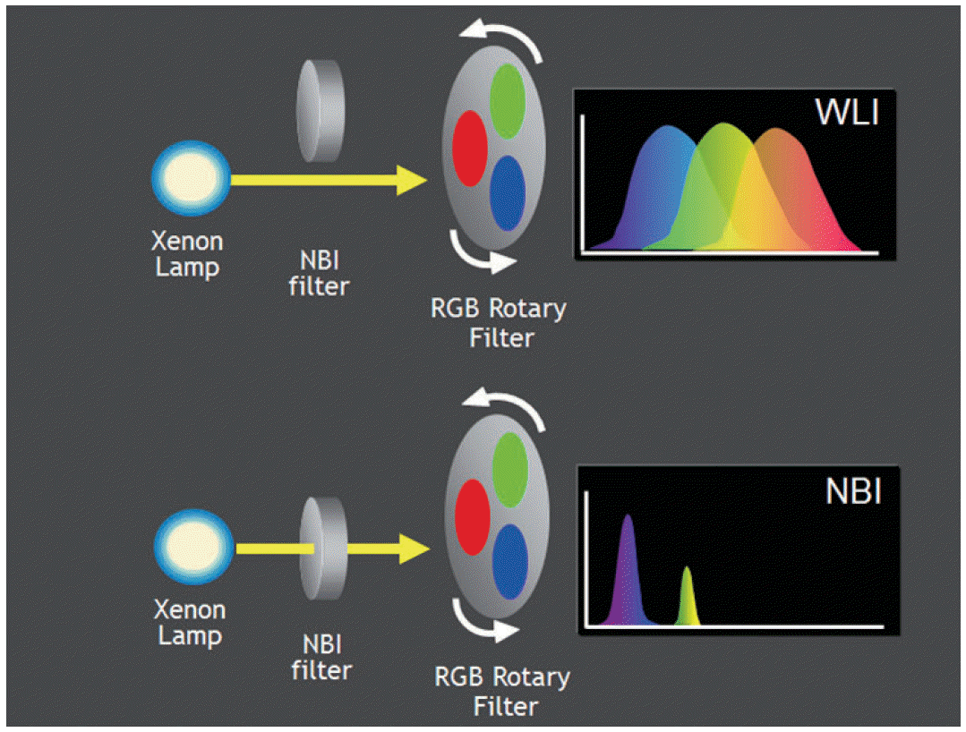
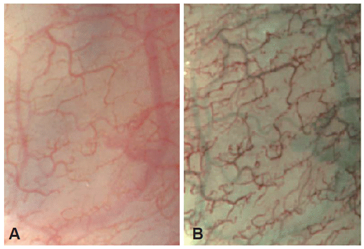
The NBI system configuration is shown in Fig. 1. In the light source unit, an NBI filter is placed between a xenon lamp and a red-green-blue rotary filter. The light moves in and out onto the optical axis, and the spectrum of the illumination can be transformed from broad-band blue, green, and red to narrow band blue and green as shown in the upper and lower graph of Fig. 1. Images of the membrane of the human tongue are shown in Fig. 2. The left and right images were taken under conventional white light and NBI, respectively. On the NBI image, the thin capillary network on the mucosal surface has a brownish appearance and thick blood vessels have a cyan appearance. Owing to NBIтs unique color allocation rules, an NBIтs final color image has a different color reproduction from the conventional one. As shown in Fig. 3, the narrow band blue image, which is taken at 415 nm in the center wavelength illumination, has the blue and green channel of an observation display. The narrow band green image, which is taken at 540 nm in the center wavelength, is allocated to the red channel of the display. This color allocation rule is the only one that has an enhancement effect on the capillary network.
Wave-particle duality is one of the characteristics of light. Light as a wave has a wavelength, which means the distance between peaks in each wave. Visible wavelengths range from 400 to 700 nm. A different wavelength is visually perceived as a different color. Generally, the level of saturation varies with wavelength. Blue light with a narrow bandwidth looks more vivid compared with a broad bandwidth because when light contains more wavelengths, saturation decreases. Light having a broad bandwidth within the range between 400 and 700 nm looks white.
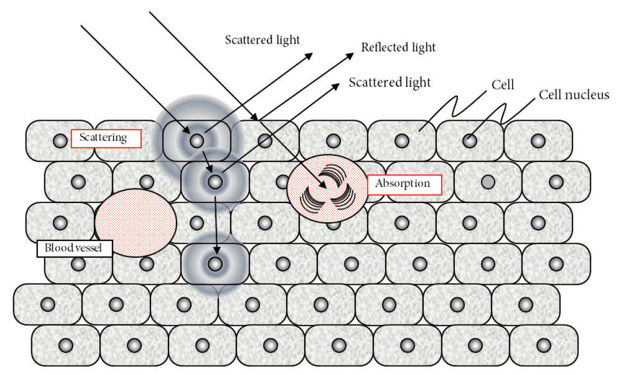
Interestingly, different wavelengths have different behaviors in biological tissue. In optical turbid medium, light scattering occurs. When light strikes small particles such as complex proteins, organelles, and cell structures, its light energy diffuses three-dimensionally. This is called light scattering. When there is a multitude of particles, multiple scattering occurs as scattered light scatters again by striking another particle. Light propagates diffusively because of this light scattering even with a flux of light. A schematic diagram of the interaction between light and living tissue is shown in Fig. 4.
When light enters biological tissue, some reflects off the surface and some diffuses within the body. Multiple scattering occurs among light and small particles such as cell nuclei, cell organelles, and nuclei in the tissue. As a result, light propagates diffusively through the tissue. The propagation of light is determined by its wavelength. While red light diffuses widely and deeply because of its long wavelength, blue light, having a short wavelength, diffuses with a smaller range. A part of the scattered light is absorbed by the blood. To be accurate, hemoglobin absorbs blue and green light. Hemoglobin is a type of chromophore. Therefore, the color of the gastrointestinal mucosa is mainly determined by hemoglobin. As mentioned above, NBI uses 415 and 540 nm in center wavelength narrow band illumination. These center wavelengths match with the two absorption peaks of the hemoglobin. Owing to strong scattering and absorption, blue narrow band illumination can reveal capillary networks well.
RESEARCH AND DEVELOPMENT HISTORY
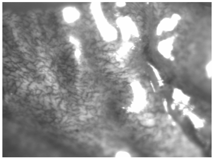
In May 1999, the idea of NBI was first conceived [1]. To confirm the feasibility of NBI, a study was conducted using a multi-spectrum camera capable of producing spectroscopic images and high-powered light source; I had volunteered as a test subject. The study revealed that the use of 415 nm narrowband light can improve the contrast of capillary images that have been difficult to observe with conventional white light. The first image of living tissue ever produced using NBI is shown in Fig. 5. Then, the development of a NBI endoscopy system started in co-operation with Dr. Sano of National Cancer Center Hospital East. On December 14, 1999, based on a study using the NBI prototype, we confirmed that it was a promising technology for endoscopic examination of hollow viscous mucosa, including the colon, stomach, and esophagus. We started to develop products in cooperation with not only Japanese endoscopists but also from around the world by improving the capacity of the prototype. EXERA II (OLYMPUS CLV-180 EVIS EXERA II XENON LIGHT SOURCE, OLYMPUS CV-180 EVIS EXERA II VIDEO SYSTEM CENTER) and EVIS LUCERA SPECTRUM (OLYMPUS CLV-260SL EVIS LUCERA SPECTRUM XENON LIGHT SOURCE, OLYMPUS CV-260SL EVIS LUCERA SPECTRUM VIDEO SYSTEM CENTER; Olympus, Tokyo, Japan) were introduced in 2005 and 2006, respectively, as the next generation system equipped with high-definition television and NBI.
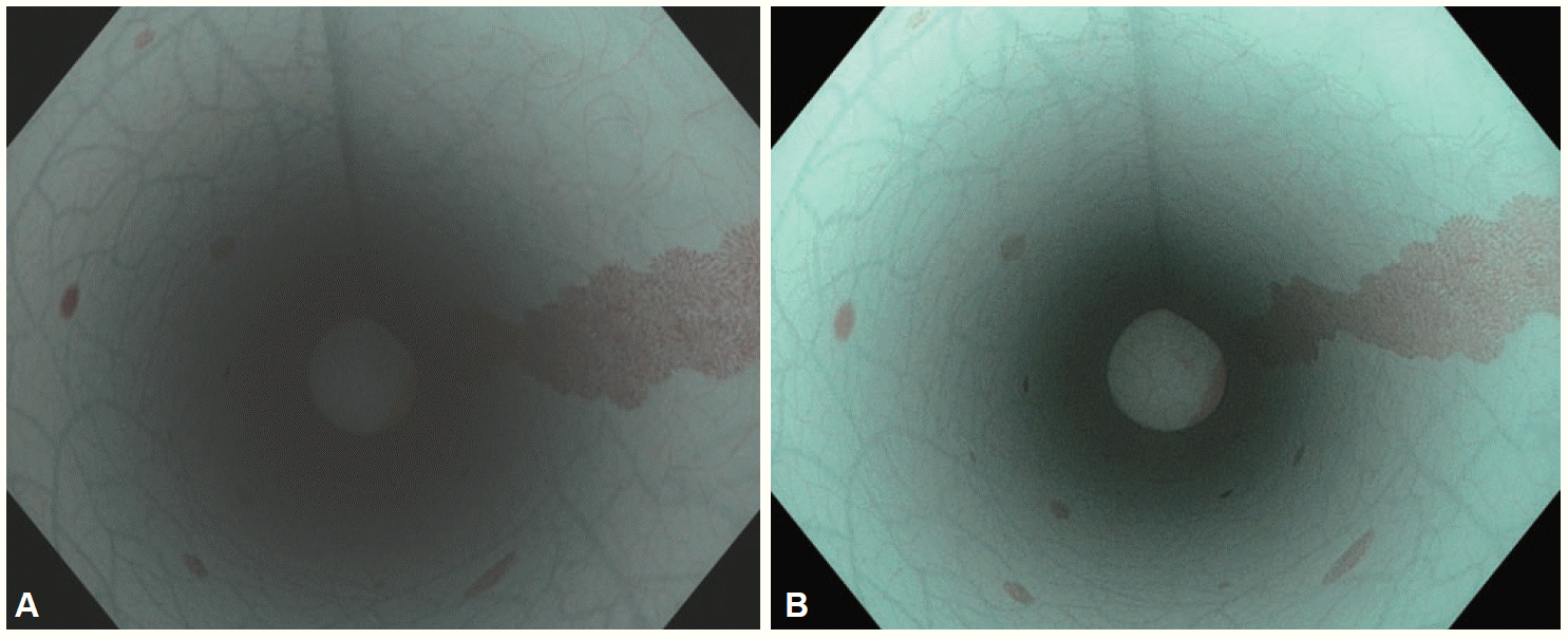
Since the first-generation NBI was introduced in the market, Olympus R&D has been committed to improving the performance of NBI. The first-generation NBI had a limitation in brightness: on gastric observation, the distal end of the endoscope needed to be carefully advanced to the mucosa to obtain sufficient brightness in the NBI mode. This insufficient brightness adversely affected the operability of the endoscope. To overcome this limitation, we made various modifications to increase the brightness of NBI. We developed a high-intensity discharge lamp, intensified the brightness of the lens in the light source, improved the sensitivity of the image sensor, and worked on image processing in the processor to reduce noise. As a result of these modifications at various parts of the system from the tip of the endoscope to the light source and signal processor, the second-generation NBI is able to deliver brightness more than one-and-a-half times as high as the first-generation NBI and to get twice the viewable distance in the lumen. The difference in the brightness of test models between the first-generation (Fig. 6A) and second-generation NBI (Fig. 6B) is shown in Fig. 6.
CONCLUSIONS
The NBI is still continually evolving since its development in 1999. We, Olympus R&D, are now improving its quality for improved ease of use. Understanding optical tissue characteristics and physician demands, market-available medical devices must be continually improved in a stepwise fashion. The NBI is progressing using this paradigm. Surely, the next innovation should be completely different from NBI. This does not seem to be an easy feat. However, it is valuable to challenge the status quo.