Skip Navigation
Skip to contents

Clin Endosc : Clinical Endoscopy

OPEN ACCESS

Articles

Page Path
HOME > Clin Endosc > Volume 48(6); 2015 > Article
Focused Review Series: Image Enhanced Endoscopy Advanced Imaging Technology in Biliary Tract Diseases:Narrow-Band Imaging of the Bile Duct
Hyun Jong Choi, Jong Ho Moon,, Yun Nah Lee
Clinical Endoscopy 2015;48(6):498-502.
DOI: https://doi.org/10.5946/ce.2015.48.6.498
Published online: November 30, 2015

Digestive Disease Center and Research Institute, Department of Internal Medicine, Soonchunhyang University School of Medicine, Bucheon, Korea

Correspondence: Jong Ho Moon Digestive Disease Center and Research Institute, Soonchunhyang University Bucheon Hospital, Soonchunhyang University School of Medicine, 170 Jomaru-ro, Wonmi-gu, Bucheon 14584, Korea Tel: +82-32-621-5094, Fax: +82-32-621-5080, E-mail: jhmoon@schmc.ac.kr
• Received: October 9, 2015   • Revised: November 4, 2015   • Accepted: November 4, 2015

Copyright © 2015 Korean Society of Gastrointestinal Endoscopy

This is an Open Access article distributed under the terms of the Creative Commons Attribution Non-Commercial License (http://creativecommons.org/licenses/by-nc/3.0/) which permits unrestricted non-commercial use, distribution, and reproduction in any medium, provided the original work is properly cited.

  • 8,280 Views
  • 113 Download
  • 8 Web of Science
  • 8 Crossref
  • 11 Scopus
prev next
  • Newly introduced direct peroral cholangioscopy and the development of video choledochoscopes have enabled more defined observation of bile duct mucosal lesions with clearer images. Narrow-band imaging (NBI) is a unique endoscopic imaging technology that provides enhanced endoscopic images of surface mucosal structures and its superficial microvessels. Advanced cholangioscopy and NBI are expected to be useful for precise evaluation and correct diagnosis of biliary tract diseases. However, the diagnostic value of advanced bile duct imaging with cholangioscopy requires further evaluation.
Image-enhanced endoscopic technologies, including narrow-band imaging (NBI) with magnification have been restrictively applied for evaluation of the bile duct. Thus, there are only limited data on the role of image-enhanced endoscopy in assessing bile duct mucosal lesions. However, the development of video choledochoscopes and the recent advances in cholangioscopy systems have permitted expanded performance of cholangioscopy and enhanced endoscopic evaluation of mucosal surface characteristics of biliary tract diseases. In this review, we present the recent advances in cholangioscopy systems and the current status of image-enhanced endoscopy for the evaluation of biliary tract diseases, particularly focusing on NBI cholangioscopy.
There are recent advancements in cholangioscopic systems for overcoming limitations of the conventional mother-baby endoscope system. One of the newly developed peroral cholangioscopy (POC) systems is the SpyGlass Direct Visualization System (Boston Scientific, Natick, MA, USA). This is a catheter-based, single-operator POC system that introduces four-way steering SpyScope (10 Fr outer diameter) with SpyGlass (a fiber optic device of 0.9 mm diameter with 6,000-pixel resolution power) through the working channel of therapeutic duodenoscope. The SpyGlass POC system has opened a new horizon in the cholangioscopic evaluation of biliary tract diseases. However, the image quality is not satisfactory for precise evaluation of bile duct lesions. In addition, image-enhanced endoscopy such as NBI is not available with this system.
Another newly introduced POC system is the direct POC using an ultraslim upper endoscope, which is directly inserted into the bile duct through the duodenal papilla [1-4]. This method uses the most widely available conventional endoscopy system. It provides good endoscopic visualization with the highest resolution among the current cholangioscopy systems. Direct POC also enables image-enhanced endoscopy such as NBI. However, insertion of a slim endoscope into the bile duct requires additional assisting accessories and some maneuvering skills because of the acute angle between the duodenum and the bile duct. Furthermore, direct POC is usually indicated in patients with dilated bile duct (>8 mm in diameter). Improvement of additional assisting accessories and development of multibending slim endoscope are expected to enable easier performance of direct POC [5-7].
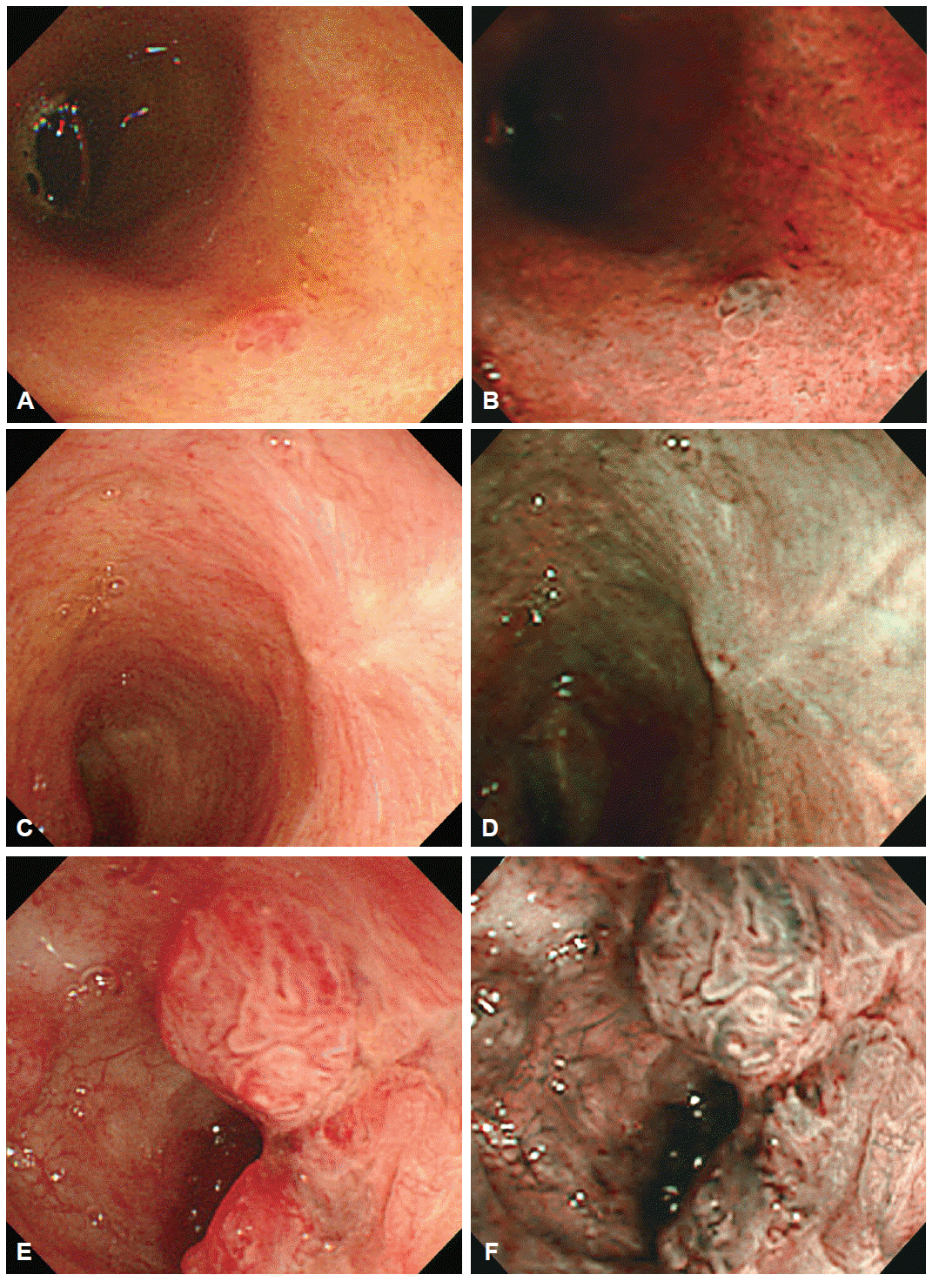
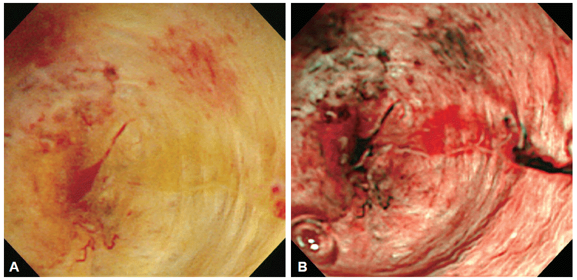
Cholangioscopic findings for biliary tract diseases have been reported in previous PTCS or POC studies. Normal bile duct mucosa is observed as a flat surface, with or without shallow pseudodiverticula (“dimple”) and fine networks of thin microvessels (Fig. 1). Benign inflammatory mucosal lesion may be observed as a slightly homogeneous papillogranular surface or scale-like appearance without primary mass suggesting hyperplasia, ulcer or scar change with convergence of folds, or bumpy surface with or without pseudodiverticula (Fig. 2). Cholangioscopic findings suggesting malignant lesion show thick irregular tortuous vessels (“tumor vessels”), irregular papillogranular or fine granular surface, fish-egg like appearance, and mucosal friability with easy oozing of blood (Fig. 3) [8-13]. Cholangioscopic evaluation may improve diagnostic yield to endoscopic retrograde cholangiopancreatography (ERCP)-based evaluation of bile duct lesions and be useful for pre-operative bile duct evaluation of tumor spreading. Osanai et al. [12] evaluated the diagnostic accuracy of POC (mother-baby endoscope system) in evaluating biliary tract lesions. The diagnostic accuracy, sensitivity, and specificity of POC in comparison with ERCP-guided forceps biopsy for indeterminate biliary diseases (n=38), were 92.1% vs. 85.7%, 96.4% vs. 81.5%, and 80.0% vs. 100%, respectively. The accuracy of ERCP, ERCP with POC, and ERCP with POC plus POC-guided biopsy, for the preoperative evaluation of tumor extension (n=49), were 73.5%, 83.5, and 92.9% respectively.
NBI is a unique technology, allowing image-enhanced endoscopy by using a single push button on the light source system. The NBI system is based on the modification of spectral features with an optical color separation filter that eliminates all illumination wavelengths except for two narrow wavelengths, to highlight surface structures and capillaries [14,15]. NBI provides enhanced visualization of surface mucosal structures and mucosal microvessels compared to conventional white light images. Therefore, NBI may be useful for precise observation and differentiation of bile duct mucosal lesions. Itoi et al. [16] evaluated the clinical usefulness of video cholangioscopy (mother-baby endoscope POC system) by using NBI in seven patients with bile duct cancer and five patients with benign biliary diseases. Regardless of whether the lesion is benign or malignant, the observation made with NBI was as good as or better than conventional white light observation to identify both the surface structure (p<0.01) and mucosal vessels (p<0.05). In particular, NBI allowed better observation of superficial tumor margin. NBI also detected four lesions that were not identified via conventional white light observation.
There are fundamental and technical limitations in the NBI enhanced cholangioscopy for evaluation of biliary tract diseases. First, it is impossible to evaluate submucosal or extraductal conditions with a cholangioscope. Therefore, NBI is only useful for assessing mucosal surface structures of biliary tract lesions. Submucosal spread or extraductal invasion of the tumor cannot be evaluated by cholangioscopy with NBI. Second, bile appears as dark red color and blood as dark color on NBI. Therefore, washing bile and blood during NBI evaluation is required to obtain clear endoscopic view [4,16]. Third, magnification function is not available in current cholangioscopy systems, unlike endoscopes used in the gastrointestinal tract. Magnifying endoscopy may provide more detailed images of bile duct mucosal structures and be helpful in cholangioscopic NBI evaluation [17]. In addition, cholangioscopic findings of neoplastic and non-neoplastic biliary tract diseases are not sufficiently established until now and studies that correlate NBI findings with histopathologic findings are lacking. Although cholangioscopic visual diagnosis with NBI has high sensitivity for bile duct mucosal lesions, a definite diagnosis should be confirmed by histopathologic findings [13,15,18].
ERCP-based cholangiographic evaluation with conventional cross-sectional images remains the primary and essential modality for diagnosing biliary tract diseases. Continuous improvement and development of direct cholangioscopic evaluation of the bile duct are ongoing to enhance diagnostic accuracy. The role of cholangioscopy with NBI has not been sufficiently evaluated and has limitations in assessing biliary tract diseases. Nevertheless, with continuous advancement in cholangioscopy and imaging technology, the value of cholangioscopy and enhanced image technology like NBI for the evaluation of biliary tract diseases is expected to grow in the near future.
Fig. 1.
Cholangioscopic view of a normal bile duct. (A) White light image. (B) Narrow-band imaging (NBI). Note how fine reticular superficial microvessels become more prominent with NBI.
ce-48-6-498f1.gif
Fig. 2.
Cholangioscopic view of benign bile duct lesions with white light images and narrow-band imaging, respectively. (A, B) Inflammatory polyp. (C, D) Scar change. (E, F) Inflammatory lesions.
ce-48-6-498f2.gif
Fig. 3.
Cholangioscopic view of malignant biliary structure. (A) White light image. (B) Narrow-band imaging (NBI). Note the thick “tumor vessels” enhanced by NBI.
ce-48-6-498f3.gif
  • 1. Larghi A, Waxman I. Endoscopic direct cholangioscopy by using an ultra-slim upper endoscope: a feasibility study. Gastrointest Endosc 2006;63:853–857.ArticlePubMed
  • 2. Choi HJ, Moon JH, Ko BM, et al. Overtube-balloon-assisted direct peroral cholangioscopy by using an ultra-slim upper endoscope (with videos). Gastrointest Endosc 2009;69:935–940.ArticlePubMed
  • 3. Moon JH, Ko BM, Choi HJ, et al. Intraductal balloon-guided direct peroral cholangioscopy with an ultraslim upper endoscope (with videos). Gastrointest Endosc 2009;70:297–302.ArticlePubMed
  • 4. Moon JH, Terheggen G, Choi HJ, Neuhaus H. Peroral cholangioscopy: diagnostic and therapeutic applications. Gastroenterology 2013;144:276–282.ArticlePubMed
  • 5. Itoi T, Sofuni A, Itokawa F, et al. Free-hand direct insertion ability into a simulated ex vivo model using a prototype multibending peroral direct cholangioscope (with videos). Gastrointest Endosc 2012;76:454–457.ArticlePubMed
  • 6. Itoi T, Nageshwar Reddy D, Sofuni A, et al. Clinical evaluation of a prototype multi-bending peroral direct cholangioscope. Dig Endosc 2014;26:100–107.ArticlePubMedPMC
  • 7. Lee YN, Moon JH, Choi HJ, et al. A newly modified access balloon catheter for direct peroral cholangioscopy by using an ultraslim upper endoscope (with videos). Gastrointest Endosc 2015 Aug 15 [Epub]. http://dx.doi.org/10.1016/j.gie.2015.08.021.Article
  • 8. Kim HJ, Kim MH, Lee SK, Yoo KS, Seo DW, Min YI. Tumor vessel: a valuable cholangioscopic clue of malignant biliary stricture. Gastrointest Endosc 2000;52:635–638.ArticlePubMed
  • 9. Seo DW, Lee SK, Yoo KS, et al. Cholangioscopic findings in bile duct tumors. Gastrointest Endosc 2000;52:630–634.ArticlePubMed
  • 10. Fukuda Y, Tsuyuguchi T, Sakai Y, Tsuchiya S, Saisyo H. Diagnostic utility of peroral cholangioscopy for various bile-duct lesions. Gastrointest Endosc 2005;62:374–382.ArticlePubMed
  • 11. Kawakami H, Kuwatani M, Etoh K, et al. Endoscopic retrograde cholangiography versus peroral cholangioscopy to evaluate intraepithelial tumor spread in biliary cancer. Endoscopy 2009;41:959–964.ArticlePubMed
  • 12. Osanai M, Itoi T, Igarashi Y, et al. Peroral video cholangioscopy to evaluate indeterminate bile duct lesions and preoperative mucosal cancerous extension: a prospective multicenter study. Endoscopy 2013;45:635–642.ArticlePubMedPDF
  • 13. Nishikawa T, Tsuyuguchi T, Sakai Y, et al. Preoperative assessment of longitudinal extension of cholangiocarcinoma with peroral video-cholangioscopy: a prospective study. Dig Endosc 2014;26:450–457.ArticlePubMed
  • 14. Itoi T, Neuhaus H, Chen YK. Diagnostic value of image-enhanced video cholangiopancreatoscopy. Gastrointest Endosc Clin N Am 2009;19:557–566.ArticlePubMed
  • 15. Ishida Y, Itoi T, Okabe Y. Can image-enhanced cholangioscopy distinguish benign from malignant lesions in the biliary duct? Best Pract Res Clin Gastroenterol 2015;29:611–625.ArticlePubMed
  • 16. Itoi T, Sofuni A, Itokawa F, et al. Peroral cholangioscopic diagnosis of biliary-tract diseases by using narrow-band imaging (with videos). Gastrointest Endosc 2007;66:730–736.ArticlePubMed
  • 17. Ishida Y, Okabe Y, Kaji R, et al. Evaluation of magnifying endoscopy using narrow band imaging using ex vivo bile duct (with video). Dig Endosc 2013;25:322–328.ArticlePubMed
  • 18. Azeem N, Gostout CJ, Knipschield M, Baron TH. Cholangioscopy with narrow-band imaging in patients with primary sclerosing cholangitis undergoing ERCP. Gastrointest Endosc 2014;79:773–779.ArticlePubMed

Figure & Data

REFERENCES

    Citations

    Citations to this article as recorded by  
    • Comparison of disposable digital single-operator cholangioscopy versus direct peroral cholangioscopy for the diagnosis of intraductal superficial lesions of the bile duct
      Il Sang Shin, Jong Ho Moon, Yun Nah Lee, Hee Kyung Kim, Jun Chul Chung, Tae Hoon Lee, Jae Kook Yang, Young Deok Cho, Sang-Heum Park
      Endoscopy.2024; 56(10): 749.     CrossRef
    • Detection and endoscopic classification of intraductal neoplasms of the bile duct by peroral cholangioscopy with narrow-band imaging (with videos)
      Il Sang Shin, Jong Ho Moon, Yun Nah Lee, Hee Kyung Kim, Jun Chul Chung, Tae Hoon Lee, Jae Kook Yang, Sang-Woo Cha, Young Deok Cho, Sang-Heum Park
      Gastrointestinal Endoscopy.2023; 97(5): 898.     CrossRef
    • Biliary Tree Diagnostics: Advances in Endoscopic Imaging and Tissue Sampling
      Matteo Ghisa, Angelo Bellumat, Manuela De Bona, Flavio Valiante, Marco Tollardo, Gaia Riguccio, Angelo Iacobellis, Edoardo Savarino, Andrea Buda
      Medicina.2022; 58(1): 135.     CrossRef
    • Association between bile area in the duodenal bulb and abdominal symptoms: Quantitative analysis using blue laser imaging
      Daiki Abe, Tsutomu Takeda, Daisuke Asaoka, Tomoyo Iwano, Ryota Uchida, Hisanori Utsunomiya, Shotaro Oki, Nobuyuki Suzuki, Atsushi Ikeda, Noboru Yatagai, Yoichi Akazawa, Kohei Matsumoto, Kumiko Ueda, Hiroya Ueyama, Mariko Hojo, Yuko Kojima, Shinji Nakamura
      Biomedical Reports.2022;[Epub]     CrossRef
    • NBI utility in oncologic surgery: An organ by organ review
      Francesca Boscolo Nata, Giancarlo Tirelli, Vincenzo Capriotti, Alberto Vito Marcuzzo, Erica Sacchet, Azzurra Nicole Šuran-Brunelli, Nicolò de Manzini
      Surgical Oncology.2021; 36: 65.     CrossRef
    • Biliary Strictures and Cholangiocarcinoma – Untangling a Diagnostic Conundrum
      Alexander Ney, Andres Garcia-Sampedro, George Goodchild, Pilar Acedo, Giuseppe Fusai, Stephen P. Pereira
      Frontiers in Oncology.2021;[Epub]     CrossRef
    • The endoscopist and malignant and non-malignant biliary obstruction
      S.P. Pereira, G. Goodchild, G.J.M. Webster
      Biochimica et Biophysica Acta (BBA) - Molecular Basis of Disease.2018; 1864(4): 1478.     CrossRef
    • Dilation of Strictures in Patients with Inflammatory Bowel Disease
      Nayantara Coelho-Prabhu, John A. Martin
      Gastrointestinal Endoscopy Clinics of North America.2016; 26(4): 739.     CrossRef

    • PubReader PubReader
    • ePub LinkePub Link
    • Cite
      CITE
      export Copy Download
      Close
      Download Citation
      Download a citation file in RIS format that can be imported by all major citation management software, including EndNote, ProCite, RefWorks, and Reference Manager.

      Format:
      • RIS — For EndNote, ProCite, RefWorks, and most other reference management software
      • BibTeX — For JabRef, BibDesk, and other BibTeX-specific software
      Include:
      • Citation for the content below
      Advanced Imaging Technology in Biliary Tract Diseases:Narrow-Band Imaging of the Bile Duct
      Clin Endosc. 2015;48(6):498-502.   Published online November 30, 2015
      Close
    • XML DownloadXML Download
    Figure
    • 0
    • 1
    • 2
    Advanced Imaging Technology in Biliary Tract Diseases:Narrow-Band Imaging of the Bile Duct
    Image Image Image
    Fig. 1. Cholangioscopic view of a normal bile duct. (A) White light image. (B) Narrow-band imaging (NBI). Note how fine reticular superficial microvessels become more prominent with NBI.
    Fig. 2. Cholangioscopic view of benign bile duct lesions with white light images and narrow-band imaging, respectively. (A, B) Inflammatory polyp. (C, D) Scar change. (E, F) Inflammatory lesions.
    Fig. 3. Cholangioscopic view of malignant biliary structure. (A) White light image. (B) Narrow-band imaging (NBI). Note the thick “tumor vessels” enhanced by NBI.
    Advanced Imaging Technology in Biliary Tract Diseases:Narrow-Band Imaging of the Bile Duct

    Clin Endosc : Clinical Endoscopy Twitter Facebook
    Close layer
    TOP