Skip Navigation
Skip to contents

Clin Endosc : Clinical Endoscopy

OPEN ACCESS

Articles

Page Path
HOME > Clin Endosc > Volume 52(6); 2019 > Article
Case Report Migration of a Percutaneous Endoscopic Gastrojejunostomy Tube into the Colon with Small Intestinal Telescoping
Shinya Taki1, Takao Maekita1,orcid, Mayumi Sakata2, Kazuhiro Fukatsu1, Yoshimasa Maeda1, Mikitaka Iguchi1, Hidefumi Ito2, Masayuki Kitano1
Clinical Endoscopy 2019;52(6):616-619.
DOI: https://doi.org/10.5946/ce.2019.016
Published online: October 16, 2019

1Department of Gastroenterology, School of Medicine, Wakayama Medical University, Wakayama, Japan

2Department of Neurology, School of Medicine, Wakayama Medical University, Wakayama, Japan

Correspondence: Takao Maekita Department of Gastroenterology, School of Medicine, Wakayama Medical University, 811-1 Kimiidera, Wakayama 641-0012, Japan Tel: +81-73-441-0627, Fax: +81-73-445-3616, E-mail: maekita@wakayama-med.ac.jp
• Received: December 20, 2018   • Revised: April 25, 2019   • Accepted: April 26, 2019

Copyright © 2019 Korean Society of Gastrointestinal Endoscopy

This is an Open Access article distributed under the terms of the Creative Commons Attribution Non-Commercial License (http://creativecommons.org/licenses/by-nc/3.0/) which permits unrestricted non-commercial use, distribution, and reproduction in any medium, provided the original work is properly cited.

  • 4,878 Views
  • 92 Download
  • 2 Web of Science
  • 4 Crossref
  • 1 Scopus
prev next
  • Continuous duodenal levodopa/carbidopa intestinal gel delivery by a gastrostomy infusion system improves control of Parkinson’s disease. The overall complication rates of percutaneous endoscopic gastrojejunostomy were reported to be 41% and 59% for immediate and delayed adverse events, respectively. A 72-year-old woman underwent percutaneous endoscopic gastrojejunostomy using the delivery system noted above. Abdominal pain and vomiting occurred 3 months later. Esophagogastroduodenoscopy showed a longitudinal ulcer extending from the lower gastric body to the ileum end, with small intestinal telescoping. Colonoscopy showed a large bezoar of food residue that was attached around the tip of the tube, reaching the ascending colon, which may have acted as an anchor. Thus, the gastric antrum and small intestine were shortened with telescoping. This complication was resolved by crushing the bezoar with forceps during colonoscopy and can be prevented by consuming a fiber-free diet and periodic exchanges of the tube using esophagogastroduodenoscopy.
In Parkinson’s disease (PD), existing medical therapy, including levodopa component preparation, may not be effective enough. In 1988, it was reported that consecutive administration of levodopa using a nasoduodenal tube to a treatment-resistant PD patient was effective against motor fluctuations [1]. Levodopa/carbidopa intestinal gel (LCIG, Duodopa®; AbbVie GK, Tokyo, Japan) administration to the small intestine by a gastrostomy infusion system was approved for the first time in Sweden in 2005. Delivery by percutaneous endoscopic gastrojejunostomy (PEG-J) provides more stable plasma levels and improves control of PD [2]. It has been increasingly performed in PD patients since it is therapeutically effective, relatively safe and easy to perform, simple to construct, and does not need periodic replacement. Since then, it has been approved in 48 countries including France, Germany, the UK, and the USA (as of June 2016). However, there is a non-negligible risk of adverse events (AEs) in association with the gastrostomy infusion system. Cheron et al. reported that the overall complication rates of PEG-J were 41% and 59% for immediate AEs (<24 h) and delayed AEs (0.1–74 months), respectively [3]. Epstein et al. reported that 42% of AEs occurred during the first 4 weeks [4]. Although most were minor complications such as site infection, irritation, and peristomal leakage after successful gastrostomy tube placement, severe complications were rarely reported.
Here we report a case of a rare, severe complication of PEG-J, in which the tube migrated into the colon, with small intestinal telescoping.
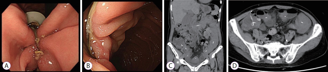
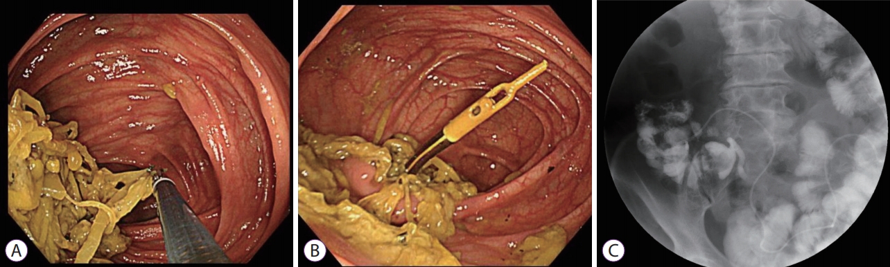
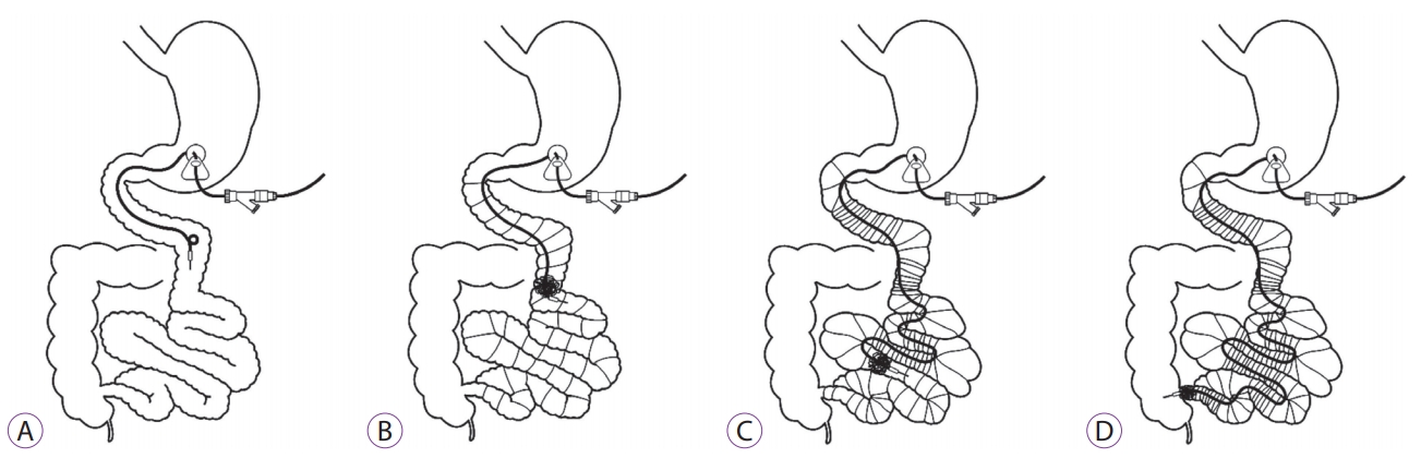
A 72-year-old woman with a 7-year history of advanced PD underwent PEG-J using the Duodopa system with the pull technique at the anterior wall of the lower gastric body (Fig. 1A). It was confirmed that the tip of the PEG-J tube was over the ligament of Treitz by contrast examination (Fig. 1B). Esophagogastroduodenoscopy (EGD) showed normal findings in the stomach and duodenum, and no technical difficulties or immediate post-procedural complications were encountered. Her PD symptoms improved, with remarkable reduction of the “off ” time, as well as improvement of motor symptoms (Unified Parkinson’s Disease Rating Scale motor scores were 23 before and 13 after LCIG). Three months after initial PEG-J tube placement, the patient developed abdominal pain and vomiting; however, there was no obvious abnormality in the administration flow rate of the LCIG and on the visible part of the tube EGD. and radiographic examination showed a longitudinal ulcer extending from the lower gastric body to the ileum end, with shortening of the gastric antrum that was being pulled down by the tube and small intestinal telescoping (Fig. 2A, B). Computed tomography showed small intestinal telescoping and the tube tip that reached the ileocecum (Fig. 2C, D). Colonoscopy showed a large bezoar (6 cm × 2 cm) that was attached around the tube tip, reaching the ascending colon (Fig. 3A, B). The bezoar, formed by persistent infusion of LCIG gel and food residues around the PEG-J tube tip, may have worked as an anchor. As a result, the gastric antrum and small intestine were shortened with telescoping (Fig. 4). It was impossible to remove the PEG-J tube from the body surface side; therefore, the PEG-J tube was cut in the body surface side and removed transanally by crushing the bezoar with forceps under colonoscopy. The patient resumed eating a few days after this procedure without any clinical AEs.
Patients with advanced PD and disabling motor fluctuations for whom optimized oral or transdermal PD medications are ineffective experienced a great improvement in their quality of life after the introduction of LCIG. Although PEG-J-related late complications, namely a bezoar and migration of the PEG-J tube, have previously been published [5,6], migration of the PEG-J tube in the colon with telescoping of the entire small intestine does not appear to have been reported previously.
Excessive traction on the stomach, duodenum, jejunum, and ileum by the PEG-J tube was considered the main reason for tube migration. Excessive traction on the stomach usually causes buried bumper syndrome; however, in this case migration of the PEG-J tube in the colon with telescoping of the entire small intestine occurred. Several factors may have contributed to this outcome. Firstly, the management of PEG-J tubes was not sufficiently diligent. It is not necessary to periodically exchange PEG-J tubes with LCIG infusion into the small intestine. In this case, the tubes were not changed, and there was no follow-up for up to 3 months after the initial procedure. In leaving the tube unchanged, a large bezoar formed by the accumulation of gel and food residues around the tube, from the tip to the loop, may have worked as an anchor. This resulted in long-term excessive traction by the PEG-J tube against the gastric and small intestinal walls, resulting in shortening of the small intestine with telescoping. Secondly, PD improvement may have contributed to migration of the PEG-J tube. Because PD patients usually show a decrease in gastrointestinal peristalsis [7], LCIG may ironically improve the motility of the small intestine, resulting in migration of the tube tip. Thirdly, a dietary fiber-rich meal may be a risk factor for bezoar formation. Constipation is one of the non-motor symptoms of PD, and the patient is typically encouraged to have dietary fiber-rich meals. Therefore, several cases of bezoars occurring after PEG-J construction have been reported. Marano reported duodenal occlusion with a food bezoar at the catheter tip, and perforation and fistula formation by mechanical stimulation of the small intestinal loop formation have been reported [8]. Lastly, the tube of the LCIG system is soft, and its pliability might have played a role in the migration of the tube tip into the colon. These considerations imply that in patients undergoing PEG-J, careful confirmation of the tube position and periodic replacements are important.
In conclusion, migration of a PEG-J tube for infusion of LCIG into the colon with telescoping of the small intestine can occur as a rare, late complication of PEG-J tube placement. This indicates that a fiber-free diet and periodic exchanges of the PEG-J tube by EGD could prevent this complication. In the future, it will be necessary to examine the appropriate exchange distance and improve the shape and materials of the tip of the tube to prevent the build-up of food residues. To date, this is the first report on such a complication of this procedure.
The authors would like to thank Drs. Takeichi Yoshida and Hiroshi Ishiguchi for their advice.
Fig. 1.
Esophagogastroduodenoscopy and contrast examination. (A) Endoscopic and (B) contrast examination images of the initial percutaneous endoscopic gastrojejunostomy.
ce-2019-016f1.jpg
Fig. 2.
Esophagogastroduodenoscopy and computed tomography (CT). (A) Endoscopic image of a longitudinal ulcer extending from the anterior wall of the lower gastric body and (B) duodenum to the ileum end, with shortening of the lesser curvature of the stomach and telescoping of the entire small intestine. (C) and (D) Coronal and horizontal CT images. The tube tip reaches the ileocecum.
ce-2019-016f2.jpg
Fig. 3.
Colonoscopy. (A) Colonoscopy image showing a large bezoar, about 6×2 cm2 in size, attached around the tip of the tube, reaching the ascending colon over the Bauhin’s valve. (B) After bezoar removal by forceps. (C) The contrast-enhanced image of the gastrointestinal tract shows telescoping of the small intestine.
ce-2019-016f3.jpg
Fig. 4.
Scheme for the presumed process of migration of the percutaneous percutaneous endoscopic gastrojejunostomy (PEG-J) tube into the colon with small intestinal telescoping. (A) The normal deployment of the PEG-J tube for the Duodopa® (AbbVie GK, Tokyo, Japan) system. (B) Attachment of food residues around the tube tip. (C) Shortening of the stomach and small intestinal telescoping by long-term excessive traction. (D) Migration of the tube tip into the colon.
ce-2019-016f4.jpg
  • 1. Kurlan R, Rothfield KP, Woodward WR, et al. Erratic gastric emptying of levodopa may cause “random” fluctuations of parkinsonian mobility. Neurology 1988;38:419–421.ArticlePubMed
  • 2. Nyholm D, Nilsson Remahl AI, Dizdar N, et al. Duodenal levodopa infusion monotherapy vs oral polypharmacy in advanced Parkinson disease. Neurology 2005;64:216–223.ArticlePubMed
  • 3. Cheron J, Deviere J, Supiot F, et al. The use of enteral access for continuous delivery of levodopa-carbidopa in patients with advanced Parkinson’s disease. United European Gastroenterol J 2017;5:60–68.ArticlePubMedPMC
  • 4. Epstein M, Johnson DA, Hawes R, et al. Long-term PEG-J tube safety in patients with advanced Parkinson’s disease. Clin Transl Gastroenterol 2016;7:e159.ArticlePubMedPMCPDF
  • 5. Klostermann F, Jugel C, Bömelburg M, Marzinzik F, Ebersbach G, Müller T. Severe gastrointestinal complications in patients with levodopa/carbidopa intestinal gel infusion. Mov Disord 2012;27:1704–1705.ArticlePubMed
  • 6. Schrader C, Böselt S, Wedemeyer J, Dressler D, Weismüller TJ. Asparagus and jejunal-through-PEG: an unhappy encounter in intrajejunal levodopa infusion therapy. Parkinsonism Relat Disord 2011;17:67–69.ArticlePubMed
  • 7. Abbott RD, Petrovitch H, White LR, et al. Frequency of bowel movements and the future risk of Parkinson’s disease. Neurology 2001;57:456–462.ArticlePubMed
  • 8. Marano M, Pizzicannella M, di Biase L, et al. Jejunal pulling syndrome: a peculiar LCIG complication. Parkinsonism Relat Disord 2018;52:113–114.ArticlePubMed

Figure & Data

REFERENCES

    Citations

    Citations to this article as recorded by  
    • Retrograde Migration of a Percutaneous Endoscopic Gastro-Jejunal Tube Into the Esophagus
      Binyamin R Abramowitz, Jude Noel, Sushil Ahlawat
      Cureus.2024;[Epub]     CrossRef
    • Weighted Tip Extensions Result in Fewer Gastrojejunostomy Tube Migrations and Increase Tube Lifespan
      Matthew A. Patetta, Kyung R. Kim, Clayton W. Commander, Peter R. Bream
      Journal of Vascular and Interventional Radiology.2023; 34(1): 124.     CrossRef
    • Understanding Parkinson's Disease: A Basic Overview
      Rachael L Anderson, Adelene Choo, Sue Sharrad, Ruth Withey
      Australasian Journal of Neuroscience.2023; 33(2): 9.     CrossRef
    • Parkinson hastalarında perkütan endoskopik gastrojejunostomi katateri takılan hastalarımızın takibi: Tek merkez deneyimi
      Nurten TÜRKEL KÜÇÜKMETİN, Tevfik SOLAKOĞLU
      Akademik Gastroenteroloji Dergisi.2022; 21(3): 157.     CrossRef

    • PubReader PubReader
    • ePub LinkePub Link
    • Cite
      CITE
      export Copy Download
      Close
      Download Citation
      Download a citation file in RIS format that can be imported by all major citation management software, including EndNote, ProCite, RefWorks, and Reference Manager.

      Format:
      • RIS — For EndNote, ProCite, RefWorks, and most other reference management software
      • BibTeX — For JabRef, BibDesk, and other BibTeX-specific software
      Include:
      • Citation for the content below
      Migration of a Percutaneous Endoscopic Gastrojejunostomy Tube into the Colon with Small Intestinal Telescoping
      Clin Endosc. 2019;52(6):616-619.   Published online October 16, 2019
      Close
    • XML DownloadXML Download
    Figure
    • 0
    • 1
    • 2
    • 3
    Migration of a Percutaneous Endoscopic Gastrojejunostomy Tube into the Colon with Small Intestinal Telescoping
    Image Image Image Image
    Fig. 1. Esophagogastroduodenoscopy and contrast examination. (A) Endoscopic and (B) contrast examination images of the initial percutaneous endoscopic gastrojejunostomy.
    Fig. 2. Esophagogastroduodenoscopy and computed tomography (CT). (A) Endoscopic image of a longitudinal ulcer extending from the anterior wall of the lower gastric body and (B) duodenum to the ileum end, with shortening of the lesser curvature of the stomach and telescoping of the entire small intestine. (C) and (D) Coronal and horizontal CT images. The tube tip reaches the ileocecum.
    Fig. 3. Colonoscopy. (A) Colonoscopy image showing a large bezoar, about 6×2 cm2 in size, attached around the tip of the tube, reaching the ascending colon over the Bauhin’s valve. (B) After bezoar removal by forceps. (C) The contrast-enhanced image of the gastrointestinal tract shows telescoping of the small intestine.
    Fig. 4. Scheme for the presumed process of migration of the percutaneous percutaneous endoscopic gastrojejunostomy (PEG-J) tube into the colon with small intestinal telescoping. (A) The normal deployment of the PEG-J tube for the Duodopa® (AbbVie GK, Tokyo, Japan) system. (B) Attachment of food residues around the tube tip. (C) Shortening of the stomach and small intestinal telescoping by long-term excessive traction. (D) Migration of the tube tip into the colon.
    Migration of a Percutaneous Endoscopic Gastrojejunostomy Tube into the Colon with Small Intestinal Telescoping

    Clin Endosc : Clinical Endoscopy Twitter Facebook
    Close layer
    TOP