Intraductal Ultrasonography Can Enhance the Success of Endoscopic Transpapillary Gallbladder Drainage in Patients with Acute Cholecystitis
Article information
See “A New Technique of Endoscopic Transpapillary Gallbladder Drainage Combined with Intraductal Ultrasonography for the Treatment of Acute Cholecystitis” by Ryota Sagami, Kenji Hayasaka, Tetsuro Ujihara, et al., on page [Related article:] 221-229.
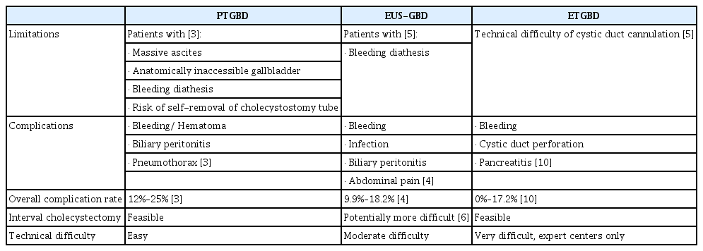
Acute cholecystitis is a common clinical condition that may present with a severity spectrum ranging from mild to severe life-threatening disease. Treatment is initiated with intravenous fluids, antibiotics and analgesia, followed by laparoscopic cholecystectomy performed either early or after an 8-week interval. Cholecystectomy represents definitive therapy but is not appropriate for patients who are unfit for surgery. Approximately 20% will develop more severe disease and fail to respond to medical therapy [1], and these individuals will require non-surgical gallbladder drainage (GBD). A detailed review of the current non-surgical options for acute cholecystitis management appears in a recent issue of this journal [2]. The choice of drainage technique will often depend on whether drainage is performed as a bridge to surgery (in patients expected to regain surgical fitness) or as definitive therapy in individuals with irreversible severe comorbidities. Other factors affecting the choice of drainage modality include patient factors, the presence of stones, local operator experience, and availability of equipment. Percutaneous transhepatic GBD (PTGBD) is well established, widely available, and technically easy to perform. However, the procedure has some limitations (Table 1) and an overall complication rate of 12%–25% [3]. In addition, long-term placement of an indwelling percutaneous cholecystostomy tube is associated with poor quality of life [3]. The advent of lumen-apposing metal stents (LAMS) has made transmural endoscopic ultrasound-guided GBD (EUS-GBD) a compelling alternative to PTGBD. EUS-GBD is associated with a comparable overall complication rate of 9.9% [4], but is relatively contraindicated in patients with coagulopathy [5]. EUS-GBD confers the unique advantage of enabling stone removal via the lumen of the LAMS. Saumoy et al. examined the widely held concern that cholecystoduodenal or cholecystogastric fistulae created via EUS-GBD would make interval cholecystectomy more difficult to perform [6]. They found no difference in the rate of successful laparoscopic cholecystectomy following EUS-GBD vs. PTGBD, although the numbers were small.

Characteristics of Percutaneous Transhepatic Gallbladder Drainage, Endoscopic Ultrasound-Guided Gallbladder Drainage, and Endoscopic Transpapillary Gallbladder Drainage
Endoscopic transpapillary GBD (ETGBD), first described by Kozarek in 1984 [7], is another alternative to PTGBD, especially in patients with ascites or coagulopathy. Its adverse effect profile (Table 1) is favorable, and it does not pose an anatomical challenge to interval cholecystectomy. The technique involves the placement of a stent or nasocystic drain across the major papilla. However, cannulation of the cystic duct during endoscopic retrograde cholangiopancreatography can be challenging due to factors such as the inability to locate the cystic duct origin at cholangiography, the presence of cystic duct stenoses or impacted stones within the gallbladder neck precluding catheter advancement, and tortuous valves of Heister [5]. Moreover, cholecystitis may worsen in the event that cystic duct cannulation is unsuccessful after contrast injection in the gallbladder. As such, ETGBD is often limited to expert tertiary centers.
ETGBD may be assisted by intraductal ultrasonography or cholangioscopy. Cholangioscopy enables direct visualization of the cystic duct orifice and wire cannulation to facilitate deployment of a stent draining the gallbladder across the major papilla. Biliary intraductal ultrasound (IDUS) was first described in 1992 by Yasuda et al. [8] for the characterization of indeterminate biliary strictures. Hayasaka et al. described its use for cystic duct localization and cannulation in 2017 [9]. In the study by Sagami et al. published in this issue of Clinical Endoscopy, the authors from the same institution proceeded to examine if IDUS improved ETGBD outcomes in a retrospective single-center study involving 100 patients with acute cholecystitis, employing propensity score matching of key parameters [10]. The technical success rate of ETGBD (defined as successful placement of the drainage catheter in the gallbladder) was significantly higher in the IDUS group compared to that in the non-IDUS group (92% vs. 76%, p=0.044). The addition of IDUS did not result in a significant difference in procedure length between the IDUS and non-IDUS groups (74.0 min vs. 66.7 min, p=0.310). The use of IDUS was associated with more complications (6%) than in the non-IDUS group (0%) (p<0.001), but the complications were mild and resolved with conservative treatment. Overall, ETGBD was unsuccessful in 20% of the patients. There were no differences in clinical outcomes between patients in the IDUS and non-IDUS groups.
Sagami et al. also describe in their paper a classification of anatomical variations of the cystic duct, subdivided by position with reference to the portal vein, by distance between the hepatic hilum and ampulla, and by the angle of cystic duct takeoff from its orifice [10]. The authors examined the relationship between cystic duct anatomical characteristics and technical success of gallbladder stent placement. They proposed that this information may help in educating trainee endoscopists and for future research into the optimal choice of accessories for cannulation based on the anatomical subtype of the cystic duct.
Sagami et al. are to be congratulated for a well-executed study which demonstrated that the addition of IDUS can increase the technical success rate of ETGBD [10]. They have also added to the body of knowledge by classifying the variations in cystic duct anatomy, which may guide further refinement in the technique. ETGBD has key utility for GBD in acutely ill individuals who are unfit for surgery; who also have bleeding diathesis, severe liver disease, or refractory ascites; and for whom other methods of GBD are potentially unsafe. In patients with concomitant choledocholithiasis, common bile duct stent placement may be performed via the same procedure. As with all transpapillary procedures, the risk of pancreatitis must be considered, although in this study, only mild pancreatitis was observed with a low incidence. ETGBD, however, remains a technically difficult procedure (by the admission of the authors), and therefore, its availability is likely to be confined to expert tertiary centers. The median procedure duration reported (IDUS 74.0 min, non-IDUS 66.7 min, p=0.310) conveys the difficulty inherent in many of these cases. We now see meta-analyses of comparisons between EUSGBD, ETGBD, and PTGBD for GBD in patients with high surgical risk, with superior outcomes in favor of EUS-GBD [11], which we expect will drive greater adoption of this procedure. There is, however, a clearly defined patient demographic, as described earlier, for whom ETGBD may be a suitable option.
ETGBD may represent the most appropriate option for acute GBD in certain ill individuals; this study by Sagami et al. showed that IDUS guidance increases the technical success of this potentially challenging procedure [10]. Endoscopists who perform ETGBD may wish to look into the utility of IDUS guidance in their own practice.
Notes
Conflicts of Interest: The authors have no financial conflicts of interest.
Author Contributions
Conceptualization: Clement Chun Ho Wu, Christopher Jen Lock Khor
Data curation: CCHW, CJLK
Formal analysis: CCHW, CJLK
Project administration: CCHW, CJLK
Supervision: CJLK
Writing-review&editing: CCHW, CJLK
