INTRODUCTION
Since the introduction of the first capsule endoscope in 2000, capsule endoscopy (CE) has become an essential noninvasive modality for the investigation and diagnosis of small-bowel (SB) diseases [1,2]. The capsule endoscope could move through the entire gastrointestinal (GI) tract and facilitate the detection of SB mucosal abnormalities, which conventional endoscopes could not reach. Furthermore, the ease of use, patient comfort, and safety have led to the extensive use of CE [3]. Over time, CE has become the first-line investigation tool for obscure GI bleeding, and has been an important method for the evaluation of Crohnās disease, evaluation of SB tumors, and surveillance of polyposis syndromes [4,5]. However, CE also presents several challenges such as the time-consuming and tedious reading process, lack of active locomotion, inability to obtain biopsies, and inability to perform therapeutic interventions such as drug delivery. To overcome these drawbacks, novel technologies are being developed by several research groups, especially the application of artificial intelligence (AI) in the field of CE as part of recent evolutions in AI. Here, we present AI approaches for the detection of SB abnormalities using CE, and introduce recent studies about innovative technologies in CE.
ARTIFICIAL INTELLIGENCE IN CAPSULE ENDOSCOPY
Role of artificial intelligence in capsule endoscopy
Together with the numerous medical data, the evolution of computer technology has led to recent advances in AI using deep learning in the medical field [6]. Computer-aided diagnosis (CAD) systems using esophagogastroduodenoscopy (EGD) and colonoscopy images have become a vigorous research field, and these systems have demonstrated promising performance in the field of gastroenteroloy [7-9]. Typically, a CE video includes an average number of 50,000ā60,000 frames in a single examination, requiring an average of 30ā120 min of reading time by physicians, depending on the experience level of the reader [5,10]. Because physicians passively read numerous images with intense focus and attention, CE reading is a time-consuming and tedious process. Furthermore, SB abnormalities may present in only one or two frames of the video and appear with a wide diversity of color, shape, and size. This highlights the inherent risk of oversights during manual reading by physicians. With a substantial number of CE images, the use of AI in CE is an attractive solution for reducing the reading time and simplifying the identification of specific landmarks and suspicious abnormalities. There is growing evidence for the clinical implications of AI in the field of CE [11].
Application of artificial intelligence in capsule endoscopy
Since late 2000, AI has been developed to detect SB abnormalities based on CE images. Early studies concentrated on technical issues and usually used support vector machine (SVM) [12-16] or multilayer perceptron network [17-19] as AI classifiers. Studies from the biocomputational field have shown good performance in detecting polyps/tumors [12,13], ulcers [14,15], celiac disease [16,20], hookworms [21], angioectasia [22], and bleeding [19,23]. However, these studies used data from a limited number of patients with insufficient clinical information in terms of inclusion and exclusion criteria. Therefore, more robust evidence is needed to apply the proposed CAD system from the biocomputational field to the clinical situation. Along with the evolution of deep learning algorithm, convolutional neural network (CNN), which extracts specific features by convolutional and pooling layers and performs back-propagation to make the best-feature map, has become the main deep learning algorithm for image analysis [24]. The CNN system has shown outstanding performance in the detection or characterization of esophageal [7], gastric [8], and colorectal abnormalities [9], and has been actively investigated for the utilization of CE in clinical practice (Table 1).
For the detection of ulcers or erosion in CE, Aoki et al. designed a CNN-based program for detecting mucosal erosions and ulceration in CE using 5,360 CE images, which were all manually annotated with rectangular bounding boxes [25]. This CNN model processed 10,440 SB images including 440 images of erosions and ulcerations as the validation dataset in 233 sec, and showed promising performance with area under the receiver-operating characteristic curve (AUROC) of 0.958, sensitivity of 88.2%, specificity of 90.9%, and accuracy of 90.8%. Furthermore, they demonstrated the clinical usefulness of the established CNN system for relieving the reviewerās workload without missing SB mucosal breaks [26]. This study compared the detection rate of mucosal breaks and the reading time between endoscopist-only readings and endoscopist readings after first being screened by established CNN using 20 CE videos. When endoscopists analyzed CE images detected by CNN, the mean reading time was significantly reduced (expert 3.1 min, trainee 5.2 min vs. expert 12.2 min, trainee 20.7 min); however, the detection rate was not decreased (expert 87%, trainee 55% vs. expert 84%, trainee 47%), thus showing the potential of the application of the CNN system as the first screening tool in clinical practice. Klang et al. reported the performance of a CNN model to detect Crohnās disease ulcers using 17,640 CE images from 49 patients [27]. Unlike Aoki et al., they did not use bounding boxes or other markings to specify the lesion, and tested the developed CNN model with both 5-fold cross validation and individual patient-level experiment, which trained datasets from 48 different patients and tested the dataset of one individual patient [25]. This CNN model showed good results with AUROC of 0.99 and accuracy ranging from 95.4% to 96.7% for 5-fold cross validation, and AUROC of 0.94ā0.99 for individual patient-level experiments.
For the detection of SB angioectasia, two studies were reported in 2018 and 2019. Leenhardt et al. proposed a CNN model for the detection of angioectasia using 300 typical angioectasia images and 300 normal images [28]. This model reached a sensitivity of 100%, a specificity of 96%, and a 39-min-long reading process for a full-length SB video. Tsuboi et al. trained a CNN system using 2,237 CE images of angioectasia, and assessed its diagnostic accuracy with 10,488 SB images including 488 images of angioectasia [29]. The AUROC for detecting angioectasia was 0.998, and the sensitivity and specificity were 98.8% and 98.4%, respectively. In 2019, Aoki et al. reported a CNN system for detecting blood content, and compared its performance with that of the suspected blood indicator (SBI), which automatically tags images with suspicious hemorrhages in the reading system [30]. The dataset consisted of 27,847 total CE images, including a training dataset of 6,503 images depicting blood content from 29 patients and a validation dataset of 10,208 images with 208 images depicting blood content. This CNN system outperformed the conventional SBI in terms of sensitivity (96.6% vs. 76.9%), specificity (99.9% vs. 99.8%), and accuracy (99.9% vs. 99.3%). The AUROC of the CNN system was 0.9998, and the CNN system took 250 sec to read 10,208 test images.
To detect protruding lesions and classify them into polyps, nodules, epithelial tumors, submucosal tumors, and venous structures, Saito et al. developed a CNN model using 30,584 CE images from 292 patients [31]. When this CNN model analyzed 17,507 test images (including 7,507 images of protruding lesions from 73 patients), the AUROC was 0.91 and the sensitivity and specificity were 90.7% and 79.8%, respectively. In the analysis of the classification of protruding lesions, the sensitivity for the detection of polyps, nodules, epithelial tumors, submucosal tumors, and venous structures were 86.5%, 92.0%, 95.8%, 77.0%, and 94.4%, respectively.
Although the above-mentioned studies showed good performance of CNN for detecting diverse SB lesions, they only focused on detecting one category of abnormalities. In 2014, Iakovidis et al. developed an automatic lesion detection software using SVM, and the average performance with AUROC for the detection of various abnormalities such as angioectasias, ulcers, polyps, and hemorrhage was 89.2% [32]. However, they used only 137 CE images including 77 pathologies, which were insufficient to evaluate the diagnostic accuracy. Recently, Ding et al. reported a CNN model that could differentiate various abnormal lesions such as inflammation, ulcer, polyps, lymphangiectasia, bleeding, vascular disease, protruding lesion, lymphatic follicular hyperplasia, diverticulum, and parasite from normal mucosa using 113 million CE images from 6,970 patients at 77 medical centers [33]. The CNN model showed a significantly higher level of sensitivity for the identification of abnormalities than conventional analysis by endoscopists in per-patient analysis (99.9% vs. 74.6%) and per-lesion analysis (99.9% vs. 76.9%). In addition, the CNN model significantly reduced the reading time compared with conventional reading by endoscopists (5.9 min vs. 96.6 min), thus showing the outstanding effectiveness of CNN models. Finally, in a systematic review and meta-analysis, Soffer et al. analyzed 10 studies that provided sufficient data for a quantitative meta-analysis of the CNN technique [34]. The pooled sensitivity and specificity for ulcer detection were 0.95 and 0.94, respectively, and the pooled sensitivity and specificity for bleeding or the bleeding source were 0.98 and 0.99, respectively. However, there was high heterogeneity between studies and most studies had a high risk of bias.
Challenges and future direction for the application of artificial intelligence in the field of capsule endoscopy
Although many research groups have obtained remarkable results on the use of AI in the field of CE, AI has not yet been applied in real-world patient management beyond clinical studies. Several obstacles need to be overcome for the clinical implementation of AI. First, most published studies to date were performed retrospectively and used data from a single center or a small number of centers, which leads to inherent selection and spectrum bias and restricts the generalization of the established CNN system. The AI system for medical applications, especially CNN, is highly dependent on training data and a high quality of data for model development is essential. In addition, because the mechanism of the AI system is difficult to explain (black box, lack of interpretability), the validation of the AI system is an important step in evaluating AI performance. Investigations without prospective or external validation have the risk of overfitting (meaning that the learning model is customized too closely to the distinct training dataset), thus failing to predict future observations. Therefore, for meticulous evaluation and verification of the clinical relevance of the CNN system, further multicenter, prospective studies and external validation with irrelevant data for model development are mandatory. Second, in most studies, CNN systems were trained and validated using still CE images rather than videos, and clear and accurate images rather than insufficiently prepared images with significant bubble, debris, and bile. Furthermore, light limitation, low resolution (320Ć320 pixels), and various orientations of SB lesions due to the free mobilization of the capsule endoscope in real-world practice could worsen the quality of CE images. The performance of the published CNN system could not be guaranteed in actual clinical settings. Third, most studies developed a CNN system using data from a specific kind of CE. Because each CE system has different image processing characteristics, it is questionable whether the established CNN system can be adopted for other CE systems. Therefore, acceptance of various kinds of CE systems and use of CE data from a large variety of clinical situations are crucial for the clinical application of upcoming CNN systems.
There are several unsolved issues for the clinical application of AI in CE, such as AI application in other medical fields. Before the incorporation of AI in community use, cost-effectiveness and the satisfaction of both patients and physicians should be demonstrated. We believe that the AI system will be used as a supplement, but not a replacement, in the medical filed. Therefore, a system for educating physicians on AI implementation and for helping them understand the technology must be established. Further, legal and ethical problems concerning the responsibility of AI application and significant reimbursement concerns must be addressed.
OTHER TECHNICAL ADVANCEMENTS IN CAPSULE ENDOSCOPY
Potential for novel capsule endoscopy in clinical practice
Beyond SB examination, colon capsule endoscopy (CCE) has the advantage of being noninvasive and painless. The second-generation CCE-2 capsule has double-headed lenses, longer recording capacity, and variable frame capture rates. In a meta-analysis of studies with 1,292 patients, Spada et al. compared the results of CCE-2 and colonoscopy [35]. The sensitivity and specificity of CCE-2 for the detection of polyps larger than 6 mm was 86.0% and 88.1%, respectively, and all invasive cancers that were observed by colonoscopy were also identified by CCE-2, showing the potential role of CCE-2 as an alternative screening method [35]. In controlled studies, CCE-2 also showed acceptable performance for the assessment of the colonic mucosa in patients with Crohnās disease and ulcerative colitis [36,37].
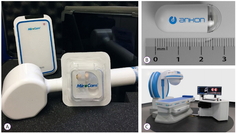
The major challenge of conventional CE is that physicians could not control the movement, orientation, and speed of the capsule. These limitations interfere with the navigation and meticulous observation of the area of interest, and reduce the diagnostic and therapeutic efficacy of CE. Because capsule locomotion and navigation could improve mucosal visualization, and further enable biopsy of the target lesion and treatment delivery in the long term, various research groups have developed CE, which is controlled by physicians with two approaches [38]. The first approach is a self-propelled mechanism including crawling or swimming as an internal locomotion mechanism. The second approach is a mechanism externally propelled by magnetic force as an external locomotion mechanism [39]. However, restricted power capacity and limited propulsion force are the main obstacles to the clinical implementation of internal locomotion mechanisms beyond experimental studies. Conversely, external propulsion using magnetic power is more practical and has emerged in several types of capsule endoscopes for navigating the device to the desired area (Fig. 1). Recently, magnetically guided CE was evaluated as a screening tool for gastric cancer in 3,182 asymptomatic patients, and seven patients (0.22%) were diagnosed with gastric cancer, accounting for 0.74% (7/948) of patients aged ā„50 years, showing the potential role of magnetic CE [40]. In a multicenter blinded study involving 350 patients with upper abdominal complaints, Liao et al. compared the detection of focal lesions between robotically assisted magnetically guided CE and conventional EGD [41]. The diagnostic accuracy was comparable between the two examinations, and >95% patients preferred magnetically guided CE because of its noninvasiveness. The prospective study by Ching et al. demonstrated that magnetically assisted CE (MACE) had better diagnostic yield than EGD in patients investigated for iron deficiency anemia [42]. Beg et al. evaluated the performance of MACE for the detection of Barrettās esophagus and esophageal varix using a handheld magnet for the capture of the capsule, and MACE correctly diagnosed 15 of 16 cases of Barrettās esophagus and 11 of 15 cases of esophageal varix [43]. These results show the feasibility of magnetically guided CE for the diagnosis of GI disease, and further clinical trials with respect to superior performance, cost-effectiveness, and safety are warranted for clinical implementation.
Recent studies on capsule prototypes
With respect to the locomotion of the capsule, Fontana et al. developed single-camera spherical capsule endoscope for colorectal screening [44]. The spherical shape of this novel capsule endoscope was designed to reduce friction during its locomotion in the colon. The interaction between the integrated permanent magnet in the capsule and the external electromagnet leads to its actuation. Fu et al. proposed a magnetically actuated micro-robotic capsule with hybrid motion such as screw jet motion, paddling motion, and fin motion [45]. This capsule endoscope is moved by an electromagnetic actuation system, which generates a rotational magnetic field and an alternate magnetic field. Guo et al. reported a spiral robotic capsule with a modular structure guided by an external magnetic field [46]. This robotic capsule comprises a guided robot and an auxiliary robot, both of which have two helical diversion grooves with different spiral directions between them. The two capsules move relative to each other under the same external magnetic field. After observing the guide robot, the treatment robot is swallowed and docked with the guide robot, which reduces the time needed to navigate the target lesion.
Several approaches have recently been proposed to enhance the imaging capability of present-day CE. Jang et al. developed a capsule endoscope with four Video Graphics Array cameras [47]. Each camera has a 120Ā° field of view, which enables capturing full 360Ā° high-resolution images of 640Ć480 pixels at four frames per second. Moving apart from conventional white-light imaging, Demosthenous et al. proposed CE for the detection of fluorescence released by very low levels of indocyanine green fluorophores [48]. Because physicians would only need to examine whether the observed fluorescence level exceeded a predefined threshold, early-stage SB cancer could be cost-effectively screened by eliminating the present CE reviewing process.
Several research groups have attempted to develop CE with biopsy capabilities. Micro-jaw forceps and two multiscale magnetic-based robotic devices including centimeter-scaled untethered magnetically actuated soft capsule endoscope (MASCE) and a submillimeter-scale self-folding micro-gripper have been previously reported [49,50]. Son et al. recently developed B-MASCE, which enables fine-needle aspiration biopsy [51]. B-MASCE was developed to enable axial jabbing motion of the needle and rolling locomotion in the stomach for biopsy. A magnetic field is employed for the control and torque of the magnet in CE, and four soft legs guide the penetration of the needle into the target lesion.
In addition to diagnostic capsules, several therapeutic capsule prototypes have been proposed by various research groups. Stewart et al. introduced SonoCAIT for an ultrasound (US)-mediated targeted drug delivery [52]. The proposed capsule consists of a US transducer, drug delivery channel, vision module, and multichannel external tether, and use US to release drugs and/or to enhance drug uptake via sonoporation for drug delivery to the target lesion. When drug-filled microbubbles arrive at the target lesion, the drug is released by US. Leung et al. developed a capsule for hemostasis using an inflated balloon [53]. This capsule was composed of a gas generation chamber, an acid injector, and a circuit box with flexible joints. The balloon was inflated by acid injection into a gas generation chamber filled with base powder, resulting in hemostasis by tamponade at the bleeding site.
CONCLUSIONS
With its rapid evolution, CE has become an important method for the investigation of obscure GI bleeding, Crohnās disease, SB tumors, and polyposis syndrome. However, the time-consuming and tedious reading process and lack of active locomotion are major challenges for the widespread use of CE in clinical practice. Here, we reviewed recent CNN-based approaches for CE that have been applied to detect various SB abnormalities including erosion/ulcers, angioectasias, blood content, and protruding lesions. Although published studies showed the promising diagnostic accuracy of the CNN system, several challenges need to be overcome for its clinical application in real-world practice. Further multicenter, prospective studies and external validation will provide robust evidence for the performance of the CNN system in CE. Moreover, in the colon, magnetically guided CE showed the potential for clinical use as an alternative method to conventional endoscopy. Further technical advancements in CE in terms of active locomotion, image enhancement, and therapeutic approaches are being actively investigated and could be integrated into patient management in the near future.