Endoscopic ultrasound-guided fine-needle aspiration (EUS-FNA) is the procedure of choice for tissue diagnoses of the gastrointestinal tract. The most common complications are bleeding, abdominal pain, and pancreatitis, with an overall rate of 0.98% [1]. Needle fracture is a rare complication [2-5].
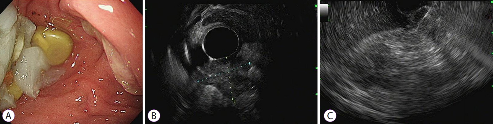
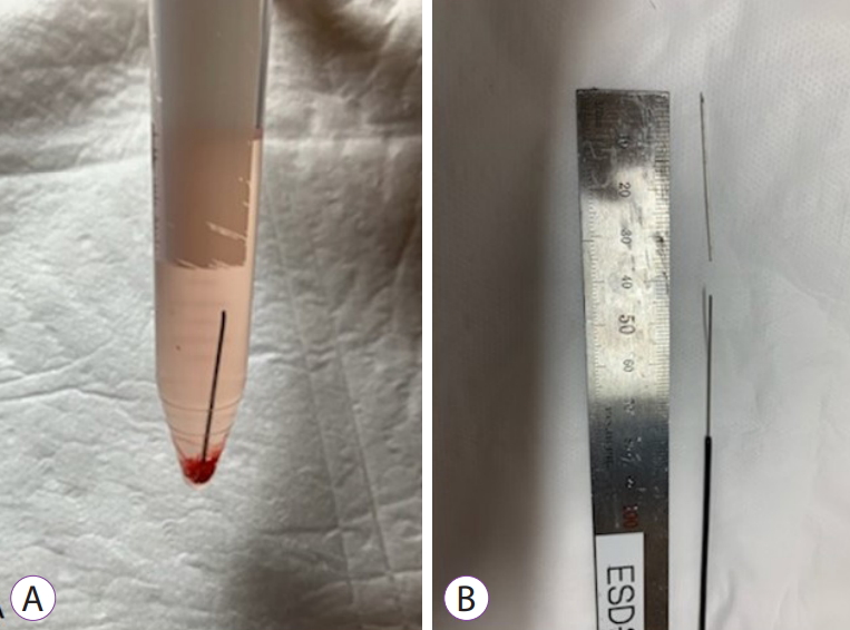
A 77-year-old man was transferred to our hospital because of bowel obstruction. Abdominal computed tomography showed a markedly distended stomach due to a duodenal mass, causing gastric outlet obstruction. Histopathology of an endoscopic biopsy specimen obtained at outside hospital revealed chronic ulceration. Upper endoscopy failed to provide clear visualization of the obstructive lesion due to the duodenal deformity (Fig. 1A). After changing to a side-view scope, a round mass with a smooth surface and soft consistency was seen obstructing the duodenal lumen, causing passage failure of the scope. Under conscious sedation, the radial and linear EUS (GF-UCT260; Olympus Ltd., Tokyo, Japan) demonstrated a poorly demarcated 28├Ś27 mm sized hypoechoic mass in the duodenum. A 22-gauge needle (Acquire; Boston Scientific, Spencer, IN, USA) was used for tissue acquisition (Fig. 1B, C). The tip of the scope was torqued, and the elevator was maximally erected for targeting. The first two punctures were to some extent successful, with small amounts of tissue sampling. Once punctured, to-and-fro movements were performed more than 10 times, including the fanning technique. On the third puncture, the scope was suddenly pushed back into the stomach, and the needle tip was not visualized on the screen, indicating it was wedged in the duodenal mass, and the needle body extending from the pylorus to the antrum was visualized in the stomach. The needle handle was locked, and several attempts to withdraw the needle into the sheath failed. With a forceful pulling of the needle backward with firm grip, the needle was slipped into the sheath, without any complication. The aspirated sample was being pushed and slid into the saline tube using the stylet, and a fragment of the fractured needle was dropped in the tube (Fig. 2A, B). Finally, the patient was diagnosed with duodenal cancer based on the pathologic confirmation of adenocarcinoma, and subsequently, a metal stent was placed into the stenotic lesion because of the patientŌĆÖs refusal to undergo surgery.
Table 1 shows a summary of previous reports associated with needle fractures during EUS-FNA [2-5]. All previous cases of fractures occurred during the second or third passage of the needle into the target lesion, but in this case, it occurred after complete retrieval of the needle from the linear scope. The length of the fractured fragment was approximately 4 cm, similar to all previous cases except one [2]. Needle fractures have occurred in needle calibers ranging from 22 to 19 gauge. The accumulated physical force might have attributed to needle breakage. Plausible explanations of this phenomenon are as follows. First, a maximal strain is exerted on the specific point of the needle when the elevator is erected to the greatest degree possible for performing EUS-FNA. Second, the repeated to-and-fro movement and/or the fanning technique can influence the curved point of the needle physically vulnerable for breakage. Finally, the forceful maneuver to retrieve the needle into the sheath might aggravate the maximal resistance force on the bent point, as in our case. It is presumed that in our case, unusual breakage of the needle might be mainly caused by combined factors, including extreme bending of the needle and vigorous retrieval maneuver of the needle.
This is the first report in the literature on a needle fracture during EUS-FNA in Korea. We informed the manufacturer about the accidental needle fracture for further analyses. With the recently increasing necessity for EUS-FNA, clinicians must be aware of the rare but potential risk of needle fractures when excessive needle bending or multiple punctures are required with numerous to-and-fro motions for the tissue aspiration, in particular, of the duodenal second portion or uncinate process of the pancreas. Furthermore, in cases of duodenal invasion, endoscopic biopsy should be performed initially, if available, to minimize EUS-FNA-associated complications.