Skip Navigation
Skip to contents

Clin Endosc : Clinical Endoscopy

OPEN ACCESS

Articles

Page Path
HOME > Clin Endosc > Volume 55(3); 2022 > Article
Brief Report Metastasis of breast cancer presenting as enlarged folds in the stomach
So Eun Jeun1orcid, Gwang Ha Kim1,2orcid, Moon Won Lee1,2orcid, Sojeong Lee3orcid
Clinical Endoscopy 2022;55(3):463-464.
DOI: https://doi.org/10.5946/ce.2020.239
Published online: November 6, 2020

1Department of Internal Medicine, Pusan National University College of Medicine, Busan, Korea

2Biomedical Research Institute, Pusan National University Hospital, Busan, Korea

3Department of Pathology, Pusan National University College of Medicine, Busan, Korea

Correspondence: Gwang Ha Kim Department of Internal Medicine, Pusan National University College of Medicine and Biomedical Research Institute, Pusan National University Hospital, 179 Gudeok-ro, Seo-gu, Busan 49241, Korea E-mail: doc0224@pusan.ac.kr
• Received: August 28, 2020   • Revised: September 11, 2020   • Accepted: September 12, 2020

Copyright © 2022 Korean Society of Gastrointestinal Endoscopy

This is an Open Access article distributed under the terms of the Creative Commons Attribution Non-Commercial License (http://creativecommons.org/licenses/by-nc/4.0/) which permits unrestricted non-commercial use, distribution, and reproduction in any medium, provided the original work is properly cited.

  • 5,104 Views
  • 189 Download
prev next
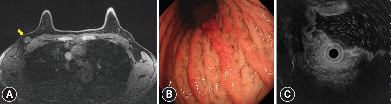
A 45-year-old female patient presented for the evaluation of intermittent abdominal pain and weight loss (15 kg over 4 months). The patient recently underwent a needle biopsy for a 2 cm-sized mass in the right breast (Fig. 1A) and was diagnosed with invasive lobular carcinoma. On endoscopy, diffusely nodular, enlarged folds with hyperemia were observed on the greater curvature of the gastric body (Fig. 1B). On air inflation, the stomach maintained relatively good distensibility. Endoscopic ultrasonography revealed the thickening of the submucosal layer (Fig. 1C). Endoscopic biopsy using a bite-on-bite technique revealed only chronic gastritis; therefore, a strip biopsy was performed. Discohesive tumor cells had infiltrated the submucosa of the stomach (Fig. 2A), and the tumor cells were immunopositive for gross cystic disease fluid protein-15, human milk fat globule protein membrane-2, estrogen receptor, and progesterone receptor (Fig. 2B). These results were consistent with the histopathological findings of the right breast cancer. Therefore, the gastric lesion was diagnosed as a metastasis of breast cancer to the stomach. The patient underwent a radical mastectomy for right breast cancer, but gastrectomy was impossible owing to the presence of metastatic nodules in the small bowel and peritoneum. At the time of writing this report, the patient is receiving systemic chemotherapy (paclitaxel 175 mg/m2 every 3 weeks).
Breast cancer is reported to be the second most common metastatic cancer of the gastrointestinal tract after lung cancer.1 The incidence rate of breast cancer metastasis to the stomach is <1%. Gastric metastasis from breast cancer shows nonspecific endoscopic features such as an elevated mucosal lesion, erosion, or ulcer, and it can rarely appear as enlarged folds on endoscopy, as in the present case.2 In case of such folds, a strip biopsy or endoscopy ultrasound-guided fine needle biopsy is sometimes needed to obtain adequate tissue sample. Because breast cancer metastases to the stomach are morphologically similar to poorly cohesive gastric carcinomas, especially in invasive lobular carcinoma, an immunohistochemical examination is warranted for a definite diagnosis.3 For the treatment of breast cancer metastasis to the stomach, systemic agents such as cytotoxic chemotherapeutic agents or hormonal agents are used. Surgical resection of the stomach has a limited role in palliative treatments such as relieving obstructive symptoms.1
Fig. 1.
(A) Breast magnetic resonance imaging shows a 2 cm-sized mass in the right breast (arrow). (B) On endoscopy, diffusely nodular, enlarged folds with hyperemia are seen on the greater curvature of the gastric body. On air inflation, the stomach maintains relatively good distensibility. (C) Endoscopic ultrasonography reveals the thickening of the submucosal layer.
ce-2020-239f1.jpg
Fig. 2.
(A) Strip biopsy shows that discohesive tumor cells infiltrate the submucosa of the stomach (hematoxylin & eosin, ×40; boxed area: hematoxylin & eosin, ×400). (B) The tumor cells are immunopositive for gross cystic disease fluid protein-15 (immunohistochemical stain, ×200).
ce-2020-239f2.jpg
  • 1. McLemore EC, Pockaj BA, Reynolds C, et al. Breast cancer: presentation and intervention in women with gastrointestinal metastasis and carcinomatosis. Ann Surg Oncol 2005;12:886–894.ArticlePubMedPDF
  • 2. Yim K, Ro SM, Lee J. Breast cancer metastasizing to the stomach mimicking primary gastric cancer: a case report. World J Gastroenterol 2017;23:2251–2257.ArticlePubMedPMC
  • 3. Honma N, Horii R, Iwase T, et al. Clinical importance of estrogen receptor-beta evaluation in breast cancer patients treated with adjuvant tamoxifen therapy. J Clin Oncol 2008;26:3727–3734.ArticlePubMed

Figure & Data

REFERENCES

    Citations

    Citations to this article as recorded by  

      • PubReader PubReader
      • ePub LinkePub Link
      • Cite
        CITE
        export Copy Download
        Close
        Download Citation
        Download a citation file in RIS format that can be imported by all major citation management software, including EndNote, ProCite, RefWorks, and Reference Manager.

        Format:
        • RIS — For EndNote, ProCite, RefWorks, and most other reference management software
        • BibTeX — For JabRef, BibDesk, and other BibTeX-specific software
        Include:
        • Citation for the content below
        Metastasis of breast cancer presenting as enlarged folds in the stomach
        Clin Endosc. 2022;55(3):463-464.   Published online November 6, 2020
        Close
      • XML DownloadXML Download
      Figure
      • 0
      • 1
      Metastasis of breast cancer presenting as enlarged folds in the stomach
      Image Image
      Fig. 1. (A) Breast magnetic resonance imaging shows a 2 cm-sized mass in the right breast (arrow). (B) On endoscopy, diffusely nodular, enlarged folds with hyperemia are seen on the greater curvature of the gastric body. On air inflation, the stomach maintains relatively good distensibility. (C) Endoscopic ultrasonography reveals the thickening of the submucosal layer.
      Fig. 2. (A) Strip biopsy shows that discohesive tumor cells infiltrate the submucosa of the stomach (hematoxylin & eosin, ×40; boxed area: hematoxylin & eosin, ×400). (B) The tumor cells are immunopositive for gross cystic disease fluid protein-15 (immunohistochemical stain, ×200).
      Metastasis of breast cancer presenting as enlarged folds in the stomach

      Clin Endosc : Clinical Endoscopy Twitter Facebook
      Close layer
      TOP