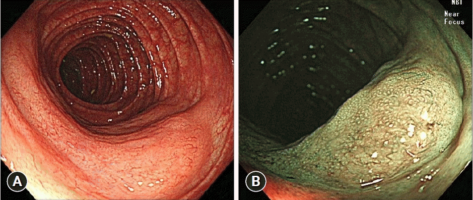
A focally flat-elevated lesion in distal transverse colon resembling a subepithelial tumor
Article information
Quiz
A 72-year-old male with intermittent left upper quadrant pain was admitted to our hospital with an abnormal lesion in the distal transverse colon detected by abdominopelvic computed tomography (APCT) at a local clinic. He had undergone a surveillance colonoscopy one year previously. At that time, his colonoscopy finding was completely normal. Physical examination of the abdomen revealed mild tenderness on the left upper quadrant of the abdomen. The complete blood count and blood chemistry results were normal. However, the carcinoembryonic antigen was 4.8 ng/mL, which was slightly elevated compared to the reference range (0–3.5 ng/mL).
APCT revealed focal thickening of the distal transverse colon, with enlarged pericolic lymph nodes (Fig. 1). Colonoscopy and narrow-band imaging (NBI) revealed a focally elevated lesion in the distal transverse colon resembling a subepithelial tumor (Fig. 2). Biopsies were performed, and pathological findings with hematoxylin and eosin and immunohistochemical (IHC) staining for CD20, CD3 and Ki-67 are as follows (Fig. 3). What was the most likely diagnosis?

Abdominopelvic computed tomography with axial view (A) and coronal view (B) revealed wall thickening of the distal transverse colon (white arrows) and infiltration along the mesentery around the distal transverse colon.

Colonoscopy. A flat, elevated lesion was observed in the distal transverse colon on colonoscopy (A), and an abnormal vasculature was observed on narrow-band imaging (B).

Histological findings. (A) Atypical lymphoid cell proliferation was seen in the deep mucosa and submucosa, and many lymphoepithelial lesions were observed (hematoxylin and eosin stain, ×100). (B, C) Lymphoid cells was positive for CD20 and negative for CD3 (immunohistochemical stain, x100). (D) Ki-67 labeling index was 7% (Ki-67 stain, x200).
Answer
The final diagnosis was a low-grade B-cell lymphoma of the colon. At first, transverse colon cancer, expressed as interval cancer, was suspected based on APCT findings (Fig. 1) and elevated carcinoembryonic antigen levels. Colonoscopy (Fig. 2) revealed a flat, elevated lesion with an abnormal pit pattern in the distal transverse colon, resembling a subepithelial tumor. NBI revealed the abnormal vascularity of the lesions. After obtaining tissue from the lesion, hematoxylin and eosin staining was performed, which showed proliferation of atypical lymphoid cells up to the submucosal layer and many lymphoepithelial lesions. On IHC staining, CD20 was positive, CD3 was negative, and Ki-67 labeling index was 7% (Fig. 3).
Non-Hodgkin’s lymphoma of the gastrointestinal tract accounts for 4% to 20% of all non-Hodgkin’s lymphoma cases and is the most common extranodal site; however, most cases are gastric lymphoma, and primary colonic lymphoma is uncommon.1,2 Colonic lymphoma is often asymptomatic and diagnosed during screening colonoscopy; however, in some cases, it is diagnosed by examination accompanied by complaints of abdominal pain, diarrhea, weight loss, and hematochezia.3 Colonic lymphoma may be observed in various endoscopic findings. Lesions may be single or multiple and sometimes observed as flat, elevated, or polypoid lesions.3,4 Ulceration can also be identified in colonic lymphoma, but is rare when compared with gastric mucosa-associated lymphoid tissue lymphoma. The surface can be seen as smooth or granular.4 Using NBI, the clinician can identify the branch-like capillary pattern of the lesion.3,5 At last, as in the present case, it can also be observed as an elevated lesion with the appearance of a subepithelial tumor. Therefore, caution should be exercised when observing lesions during colonoscopy.
In IHC staining, CD20 is a pan B-cell marker, and CD3 is a pan T-cell marker, helping to distinguish lymphocyte types according to their positive and negative finding.6 Ki-67 labeling index, which reflects the proliferation of tumor cells, is generally considered low grade if less than 20% and high grade if it exceeds 30%.6 However, the proliferation rate can vary, and Ki-67 alone is not used for grading.2,3,6
Notes
Conflicts of Interest
Sang-Bum Kang is currently serving as a KSGE Publication Committee member; however, he had not involved in the peer reviewer selection, evaluation, or decision process of this article. Shin-Hee Lee has no potential conflicts of interest.
Funding
None.
Acknowledgments
The patient provided informed consent and agreed to publication of the images.
Author Contributions
Conceptualization: SBK; Data curation: SHL, SBK; Supervision: SBK; Writing–original draft: SHL, SBK; Writing–review & editing: SBK.
