Clin Endosc > Volume 56(4); 2023 > Article
Uozumi, Abe, Makiguchi, Nonaka, Suzuki, Yoshinaga, and Saito: Complications of endoscopic resection in the upper gastrointestinal tract

Abstract

Endoscopic resection (ER) is widely utilized as a minimally invasive treatment for upper gastrointestinal tumors; however, complications could occur during and after the procedure. Post-ER mucosal defect leads to delayed perforation and bleeding; therefore, endoscopic closure methods (endoscopic hand-suturing, the endoloop and endoclip closure method, and over-the-scope clip method) and tissue shielding methods (polyglycolic acid sheets and fibrin glue) are developed to prevent these complications. During duodenal ER, complete closure of the mucosal defect significantly reduces delayed bleeding and should be performed. An extensive mucosal defect that comprises three-quarters of the circumference in the esophagus, gastric antrum, or cardia is a significant risk factor for post-ER stricture. Steroid therapy is considered the first-line option for the prevention of esophageal stricture, but its efficacy for gastric stricture remains unclear. Methods for the prevention and management of ER-related complications in the esophagus, stomach, and duodenum differ according to the organ; therefore, endoscopists should be familiar with ways of preventing and managing organ-specific complications.

INTRODUCTION

Endoscopic resection (ER), including endoscopic mucosal resection (EMR) and endoscopic submucosal dissection (ESD), is a well-established therapy for gastrointestinal neoplasms with a negligible risk of lymph node metastasis.1-7 EMR is a technically easy and time-saving procedure for small lesions; however, the procedure cannot resect lesions with sizes larger than that of the endoscopy snare. In contrast, ESD, which is technically challenging and time-consuming, allows for en bloc resection regardless of the tumor size.8-15 Although ER is widely known as a minimally invasive treatment, complications could occur during and after the procedure. Despite mostly being managed conservatively, some complications are still difficult to treat and cause severe conditions. This review focuses on the prevention and management of complications of upper gastrointestinal ER and summarizes the literature regarding this topic.

ESOPHAGEAL ER

Indications for the treatment of superficial esophageal cancer is described in the ESD/EMR guidelines published by the Japan Gastroenterological Endoscopy Society (JGES).16 According to a meta-analysis of 3,796 patients in 21 retrospective studies, ER for superficial esophageal squamous cell cancer showed favorable long-term outcomes with a five-year overall survival and disease-specific survival of 87.3% and 97.7%, respectively.4 Major complications related to esophageal ER are perforation, bleeding, and stricture. A meta-analysis of eight studies, involving 1,081 patients, revealed that ESD had a lower local recurrence rate and higher perforation rate than EMR; however, there was no significant difference in postoperative stricture and bleeding rates between the two techniques.17

Intraoperative perforations

Intraoperative perforation rates of EMR and ESD were reported to be 0% to 1.6% and 2.6% to 6.9%, respectively.9,18-21 The risk of intraoperative perforation is much higher in cases with large tumor sizes or for procedures in less experienced facilities.22,23 Esophageal ESD is technically challenging because of the following reasons: (1) the narrow lumen of the esophagus decreases the efficacy of gravity counter-traction;8 (2) a resected specimen retracts distally, thereby making it difficult to maintain good traction and orientation;8 (3) the thin wall of the esophagus increases the risk of perforation;8 and (4) localized muscle layer defects lead to unexpected extensive perforations, although these are extremely rare.24 Traction-assisted ESD, which helps maintain a good endoscopic view, is useful in overcoming the challenges mentioned above.25 According to CONNECT-E, a multicenter randomized controlled trial (RCT), no perforation occurred in traction-assisted ESD using dental floss, whereas 4.3% of the cases had perforations in conventional ESD.26 The ESD/EMR guidelines for esophageal cancer published by the JGES weakly recommend traction-assisted ESD.16
Clip closure technique and conservative therapy (e.g., fasting and antibiotic therapy) are the first lines for intraoperative perforation.27 Clip placement should begin at the defect’s end distal to the endoscope to provide an optimal endoscopic view.28 Recently, the usefulness of the application of a polyglycolic acid (PGA) sheet has also been reported.29,30

Delayed perforations

Delayed perforations after esophageal ESD can occur, owing to tissue necrosis and degeneration due to excessive energy during the procedure.31 A literature that describes the management of delayed perforations is limited to case reports,31,32 in which the temporary use of self-expanding metallic stents (SEMSs) is reported as a nonsurgical treatment.31 There is no consensus on whether surgical intervention or an endoscopic approach should be performed to manage delayed perforations after esophageal ESD; therefore, a therapeutic decision should be made based on the patient’s condition.

Delayed bleeding

Delayed bleeding after esophageal ESD is a relatively rare complication with an incidence rate of 0% to 1.3%.21,22,33 The risk factors for delayed bleeding are not well described in previous reports. Delayed bleeding after esophageal ESD is usually managed with endoscopic hemostasis and does not need surgical intervention.33

Stricture

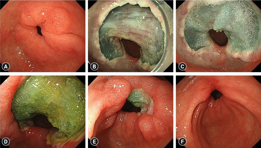
According to Shi et al.,34 the rate of post-ER stricture in patients who underwent esophageal ER for superficial esophageal carcinoma was 11.6% (42/362), and the median time from ER to stricture was 28 days (21–90 days). Moreover, the incidence of stricture increases as the circumferential range of the mucosal defect increases (≤1/2 circumferential mucosal defect, 0.7%; 1/2–3/4 circumferential mucosal defect, 27.6%; and ≥3/4 circumferential mucosal defect, 94.1%). A widespread mucosal defect involving three-quarters of the circumference of the esophagus is a strong risk factor for the development of post-ER stricture.34-36 Several preventive methods and management strategies have been developed for post-ER stricture (Table 1).37-44

1) Stricture prevention methods

Ezoe et al.45 reported that prophylactic endoscopic balloon dilation (EBD) decreases the incidence of post-ESD stricture from 92% to 59% in patients with a mucosal defect covering more than three-quarters of the circumference, and there were no complications after 166 prophylactic EBD procedures were performed for 29 patients. Although prophylactic EBD might be effective in decreasing the incidence of stricture, it could not prevent stricture formation in almost half of the cases. Moreover, several EBD sessions are needed to prevent stricture, thereby leading to a substantial cost and burden for the patients.
SEMS placement has been explored as an option to prevent and treat benign esophageal strictures. Wen et al.37 conducted an RCT on patients with mucosal defects covering more than three-quarters of the circumference, and the incidence of stricture in a stent group was significantly lower than that of a non-stent group (18.2% vs. 72.7%, p<0.05). Stent placement is a simple procedure and might be effective in preventing stenosis; however, there is a risk of stent migration and perforation.
Steroids exert anti-inflammatory effects and reduce collagen synthesis and fibroblast proliferation. Nonaka et al.46 investigated the healing process of esophageal mucosal defects with and without steroid injection using porcine models. The characteristic process underlying the formation of the stenotic luminal ridge is the proliferation of spindle-shaped myofibroblasts at the ulcer bed. Local steroid injection modifies this healing process and contributes to preventing esophageal stricture. Hanaoka et al.38 conducted a prospective study to evaluate the efficacy of single-session local triamcinolone injection (LTI); they revealed that a study group had a significantly lower stricture rate (10% vs. 66%, p<0.001) and lower number of EBD sessions (median value 0, range 0–2 vs. median value 2, range 0–15, p<0.001) than a historical control group. Yamaguchi et al.39 compared clinical outcomes between oral prednisolone administration (OPA) and preemptive EBD groups in a retrospective study. Post-ESD esophageal stricture was significantly less frequent in the OPA group than in the preemptive EBD group (5.3% vs. 31.8%, p<0.05). Recently, Tanaka et al.40 reported the results of an open-label, multicenter RCT (JCOG1217) investigating the superiority of OPA over LTI for preventing post-ESD stricture. In this study, primary endpoint was stricture-free survival (SFS), and it was 88.5% (95% confidence interval [CI], 81.6%–92.9%) in an LTI group and 94.8% (89.4%–97.5%) in an OPA group. OPA was not superior to LTI for SFS (hazard ratio, 0.672; 90% CI, 0.361–1.250], one-sided p=0.144); This study concluded that LTI is a standard treatment for the prevention of esophageal stricture after ESD for non-circumferential esophageal cancer.
The efficacy of LTI for preventing post-ESD stricture is limited in the case of whole circumferential resection.47 Chu et al.41 reported that LTI plus OPA reduces the incidence of stricture in patients with mucosal defects involving whole or near-whole circumference (>90%) compared with no treatment (18.2% vs. 83.3%, p<0.001), with no refractory stricture occurring in the treatment group. Although the combination therapy showed preferable results for preventing stricture after near-whole circumferential resection, it should be noted that one patient experienced delayed perforation in the treatment group.
Preventive methods other than steroid interventions for postoperative esophageal stricture include tissue shielding methods with a PGA sheet42,48,49 and application of autologous cell sheet transplantation.43,44 A pilot study on the efficacy of PGA sheet for mucosal defects of more than three-quarters of the circumference demonstrated that the incidence of stricture was 37.5% (3/8).49 Sakaguchi et al.42 conducted a single-center retrospective analysis of 500 consecutive cases of esophageal ESD, excluding cervical esophageal cancer and complete circumferential resection. Postoperative stricture rates for PGA sheet with steroid injection, PGA-sheet-alone, and control groups were 18.9%, 41.4%, and 51.7%, respectively (p=0.015).42 Although the PGA-sheet-alone is insufficient in preventing post-ER stricture, a combination of steroid injection and PGA sheet might be effective for non-circumferential resection.
Ohki et al.43,44 developed a technique for endoscopic transplantation of cultured autologous oral mucosal epithelial cell sheets to a post-ER mucosal defect. The incidence of stricture was 10.0% (1/10) in cases with mucosal defect involving more than half of the circumference, and the autologous cell sheets completely epithelialized the mucosal defect after a median of three weeks.43,44 The problems of this novel method include costs of the cell sheet, long procedure time for transplantation, and limited amount of oral mucosa that can be harvested.
Currently, although several other innovative methods have been developed, steroid therapies are still the first-line options for preventing esophageal strictures.

2) Management of strictures

EBD is the standard approach for the treatment of postoperative stricture.27 The success rate of EBD for post-ER stricture is 89.7% (26/29), and median time to treatment success is 4.4 months.50 A balloon dilates the stiff tissue of the postoperative scar with mechanical force; therefore, EBD carries an inevitable risk of perforation.51,52 Takahashi et al.51 reported perforation rates of EBD for post-ESD stricture to be 9.2% (7/76) per patient and 1.1% (7/648) per procedure. Stricture dilation with a balloon >15 mm is a significant risk factor for perforation.53 Therefore, the size of the balloon dilater should be started from 12 mm or less to reduce the risk of perforation during EBD.
Although EBD is an effective approach for postoperative stricture, there are several cases of strictures refractory to repetitive EBD. For these cases, additional treatment should be considered, and radical incision and cutting might be effective methods for refractory strictures.54

GASTRIC ER

The treatment indications for early gastric cancer (EGC) are described in the ESD/EMR guidelines published by the JGES.55 A Japanese multicenter prospective cohort study using a web registry system, including 10,926 lesions from 9,715 patients (J-WEB/EGC), investigated the short- and long-term outcomes of ER for EGC.7,15 En-bloc and R0 resection rates were 99.2% and 91.6%, respectively.15 Moreover, the incidence of adverse events was acceptable, with postoperative bleeding and intraoperative perforation rates of 4.4% and 2.3%, respectively. The five-year overall survival and disease-specific survival rates were 89.0% (95% CI, 88.3%–89.6%) and 99.5% (95% CI, 99.3%–99.6%), respectively.7 ER for EGC showed favorable short- and long-term outcomes and was widely utilized as a standard approach. In gastric ER, the major complications included perforation, bleeding, and stricture.

Intraoperative perforations

The J-WEB/EGC cohort revealed that intraoperative perforations during gastric ESD occurred in 2.3% of lesions (218/10,821); however, most of them could be treated with conservative management, and only seven cases required emergency surgery.15 Endoscopic clip closure is the first-line therapeutic choice for intraoperative perforations.56,57 Other methods (such as endoloop and endoclip closure method,58 over-the-scope clip [OTSC] method,59 and PGA sheet and fibrin glue60) are used for perforations that are difficult to close completely with the simple clip closure method. The American Gastroenterological Association’s expert review recommends using standard through-the-scope clips (TTSCs) or OTSCs for perforations ≤2 cm and endoscopic suturing or a combination of TTSCs and endoloops for perforations >2 cm.28 The most important factor regarding the indication for emergency surgery is the condition of the patient, including vital signs and symptoms.27,28
Risk factors for intraoperative perforations during gastric ESD include the involvement of the upper third of the stomach, severe fibrosis, invasion depth, and tumor size ≥20 mm.61,62 ESD for gastric tumors in the upper third of the stomach is challenging, owing to frequent intraoperative bleeding and thinner wall thickness than that of other parts of the stomach.62 CONNECT-G, a multicenter RCT, demonstrated a lower incidence of perforations in traction-assisted ESD than in conventional ESD (0.3% vs. 2.2%, p=0.01). The traction-assisted technique can reduce the risk of perforations by improving the visibility of the submucosal dissection plane, which helps in the identification of muscle direction during submucosal dissection.63

Delayed perforations

Delayed perforations after gastric ESD are rare, but can cause peritonitis, which requires emergency surgery. A single-center retrospective study of 4,943 cases showed that delayed perforations after gastric ESD occurred only in seven (0.1%) cases, and the median time until delayed perforations was 11 hours (range, 6–172 hours).64 Gastric tube cases, lesions located in the upper stomach, and excessive electrocautery are associated with delayed perforations.64,65 Symptoms of peritoneal irritation and rebound tenderness are critical physical signs, indicating a necessity for emergency surgery.65 Although emergency surgery can be required with a high probability,65,66 several cases of delayed perforations were successfully managed conservatively.67,68 Suzuki et al.64 reported that early detection of the onset of delayed perforations within 24 hours after the procedure might be helpful in avoiding emergency surgery. Although no report has described effective prevention methods for delayed perforations, owing to their rarity, complete closure of the mucosal defect theoretically contributes to reducing the incidence of delayed perforations.

Delayed bleeding

Delayed bleeding is a major complication related to gastric ESD. Although administering proton pump inhibitors (PPIs) and prophylactic coagulation after ESD are reportedly effective for preventing delayed bleeding,69-71 the incidence of delayed bleeding remains as high as 2.6% to 5.8%.15,72,73 A meta-analysis indicated that significant risk factors for delayed bleeding after gastric ESD were the use of antithrombotic agents, chronic kidney disease, resected specimen size >30 mm, and use of histamine-2 receptor antagonists instead of PPIs.74 Hatta et al.75 reported the BEST-J score, which is a novel scoring system for predicting delayed bleeding using risk factors, such as comorbidities, tumor factors, and use of antithrombotic agents. This prediction model stratifies the risk of delayed bleeding into four levels, according to the total score of risk factors: low-risk (0–1 point; 2.8%), intermediate-risk (2 points; 6.1%), high-risk (3–4 points; 11.4%), and very high-risk (≥5 points; 29.7%) (Table 2).75 Notably, the management of antithrombotic agents in the BEST-J score is based on the 2014 JGES guideline,76 which does not include the management of direct oral anticoagulants.
Second-look endoscopy (SLE) has been empirically performed to prevent delayed bleeding; however, three RCTs revealed that routine SLE did not reduce delayed bleeding in patients with an average risk of bleeding.77-79 Furthermore, in a single-center retrospective study, there was no statistical difference in the incidence of delayed bleeding between SLE and non-SLE groups in patients taking antithrombotic agents (21.7% vs. 21.9%, respectively).80 There is no prospective study investigating the efficacy of SLE in a high-risk group, and the indication of SLE in high-risk patients remains unclear.
Several researchers have developed innovative approaches, such as tissue shielding methods (PGA sheets and fibrin glue) (Fig. 1),81,82 endoscopic closure of artificial ulcers (the endoloop and endoclip closure method, OTSCs, and hand-suturing method),83-86 and the use of hemostatic powder,87,88 for the prevention of delayed bleeding. These methods are particularly used in high-risk patients.
Kataoka et al.81 conducted an RCT that investigated the efficacy of PGA sheets and fibrin glue in high-risk patients. There was no significant difference in delayed bleeding rates between PGA and control groups (4.5% vs. 5.7%, p>0.99). Although this study enrolled high-risk patients, delayed bleeding occurred in only 5.7% of the control group; this incidence rate is equivalent to that of low-risk patients.81 This result implies that the inclusion criteria were not optimal. Therefore, further well-designed and advanced large-scale studies are required to provide evidence for PGA sheets in the prevention of delayed bleeding.
Endoscopic closure of mucosal defects (including the endoloop and endoclip closure method,83 OTSCs,84 and hand-suturing method85,86) has a clinical potential to reduce the risk of delayed bleeding (Table 3). However, it is difficult to achieve complete closure of a gastric mucosal defect and avoid dehiscence because of the thickness of the gastric wall. Endoloop and endoclip closure method is an easy-to-perform and low-cost procedure, but it is not satisfactory in keeping the mucosal defect closed.83 Ego et al.83 reported that the endoloop and endoclip closure method did not prevent delayed bleeding in patients with antithrombotic therapy compared with control individuals (11.5% vs. 11.9%, p=0.89), and sustained closure rate in patients who underwent SLE was 47.8% (33/69). Endoscopic hand-suturing (EHS) demonstrated an incredibly high rate of complete closure (97%–100%) and sustained closure of mucosal defects (84%–100%).85,86 Akimoto et al.85 reported that there was no delayed bleeding in patients taking antithrombotic agents without perioperative cessation (0%, 0/20). EHS is a promising method for achieving complete and sustained closure, thereby allowing for the prevention of delayed bleeding. However, EHS is a technically challenging and time-consuming procedure. Further improvement is required to utilize EHS in clinical practice. Polysaccharide hemostatic powder (PHP) is a novel topical hemostatic method used for non-variceal upper gastrointestinal bleeding,89 and its efficacy for preventing post-ER bleeding has been investigated.87,88 Jung et al.87 conducted an RCT to investigate the efficacy of PHP in preventing post-ESD bleeding among patients with a high risk of bleeding (resected specimen size >40 mm and regular use of antithrombotic agents). This study showed that there was no significant difference in post-ESD bleeding rates between PHP and control groups (5.5% vs. 7.1%, p=0.74). However, a subgroup analysis of patients who discontinued antithrombotic agents during ESD showed that post-ESD bleeding rate tended to be lower in the PHP group than in the control group (0% vs. 6.3%, p=0.06).
Considering the increasing population of patients taking antithrombotic agents, the development of an effective preventive method for delayed bleeding is desirable. However, there is no RCT demonstrating the efficacy of preventive approaches. This topic is still under investigation, and further innovative methods are required.

Stricture

It is reported that stricture occurs in 0.8% to 2.5% of cases following gastric ESD.90-93 An extensive mucosal defect that comprises three-quarters of the circumference in the antrum or cardia is a significant risk factor for stenosis.91 An EBD is empirically performed to alleviate stricture and stasis symptoms; however, it carries a certain risk of perforation (7.8%–14.3%).91,94 Moreover, cases of post-gastric ESD stricture require repeated EBD until the stricture is eliminated, and there are cases of strictures refractory to repeated EBD. Contrary to esophageal stricture, there is limited evidence for preventive methods, such as local steroid injection and prophylactic EBD for stricture following gastric ESD.94,95 Further investigations should be conducted to determine the optimal approach for preventing strictures.

DUODENAL ER

According to a large-scale national database in Japan, the incidence of duodenal cancer was 23.7 per 1,000,000 person-years.96 Almost half of duodenal cancers were detected at the localized stage (56.4%, 1,694/3,005), and 48.0% (813/1,694) of localized duodenal cancers were endoscopically resected.96 Although duodenal cancer is rare, the number of ER procedures for duodenal tumors has been increasing with the development of endoscopic modalities.97 ER of duodenal neoplasms is still challenging due to the anatomic characteristics of the duodenum, such as the tortuous duodenal lumen, thin wall, and exposure to bile and pancreatic juices. Several previous studies have shown preferable clinical outcomes of underwater EMR (UEMR) and cold snare polypectomy, and these novel ER techniques are among therapeutic options for duodenal neoplasms.98-102 Major complications associated with duodenal ER are perforation and bleeding, which substantially differ among the procedures (Table 4).98-105

Perforations

A multicenter retrospective observational study, including 3,107 cases (187, 579, 1,324, and 1,017 cases of cold polypectomy, UEMR, conventional EMR, and ESD, respectively) revealed that intraoperative perforation and delayed perforation rates were significantly higher in an ESD group than in other groups.103 Fukuhara et al.106 analyzed the clinical course of 32 patients with intraoperative perforations during duodenal ESD. Either simple closure using clips or the string clip suturing technique was attempted to close a perforation, and complete closure was achieved in 13 of 32 patients (40.6%). They found that if the whole mucosal defect, including the perforation, was closed completely, the clinical course was significantly better than that of incomplete closure and was equivalent to that of cases without perforations. Moreover, incomplete closure cases with an endoscopic nasobiliary and pancreatic duct drainage tube (ENBPD tube) could be managed without additional intervention. However, one of eight cases with neither complete closure nor an ENBPD tube required percutaneous drainage for an abscess. An ENBPD tube might prevent mucosal defect exposure to pancreatic and bile juices and help to avoid a worse clinical course in a case without complete closure of the mucosal defect.

Delayed perforations

Delayed perforation is a more severe complication than intraoperative perforation and requires emergency surgery. A meta-analysis revealed that the incidence of delayed perforations in a closure group tended to be lower than that in an unclosed group, although it was not statistically different (1.6% vs. 3.8%, p=0.13; risk ratio, 0.39; 95% CI, 0.12–1.32).107 Complete closure of the mucosal defect might help to reduce the risk of delayed perforations; however, it is sometimes difficult to achieve complete closure for a lesion in the medial or anterior wall and in lesion size >40 mm.108 In order to overcome these difficulties, various endoscopic closure techniques (the clip with string method, endoloop and endoclip closure method, OTSCs, and PGA sheets) were developed.109-113 Moreover, external drainage of bile and pancreatic juices using an ENBPD tube might be a therapeutic option for preventing delayed perforation in cases of difficult complete mucosal defect closure. Fukuhara et al.114 reported that no patients developed delayed perforation (0/21) with immediate insertion of ENPBD tubes after duodenal ESD, whereas 4.1% (2/49) of the patients who did not undergo ENBPD tube insertion developed delayed perforations. It should be noted that ENBPD has a risk of post-endoscopic retrograde cholangiopancreatography pancreatitis and mucosal defect perforation when inserting a side-viewing endoscope. Therefore, the indications for ENBPD should be limited to cases in which endoscopic closure is impossible or difficult.

Delayed bleeding

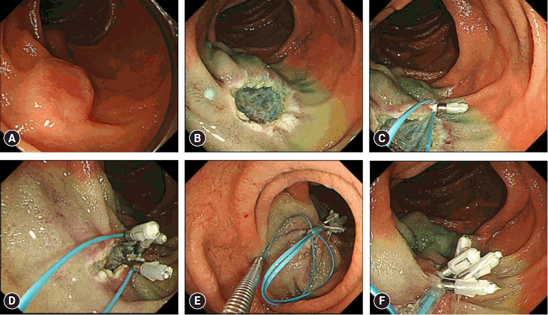
The incidence of delayed bleeding in cold polypectomy, UEMR, conventional EMR, and ESD are 0% to 0.5%, 0% to 2.1%, 0% to 2.6%, and 4.7% to 5.2%, respectively.98-105 A meta-analysis revealed that the incidence of delayed bleeding was significantly lower in a closure group than in an unclosed group (2.0% vs. 17.3%, p<0.01; risk ratio, 0.14; 95% CI, 0.06–0.33).107 Simple endoscopic clip closure is the standard method for closing mucosal defect after ER of duodenal neoplasms. However, it is technically challenging to achieve complete closure of extensive mucosal defects, particularly after ESD. Thus, various endoscopic closure techniques (the clip with string method, endoloop and endoclip closure method, OTSCs, and PGA sheets) are reported to prevent delayed bleeding after ER of duodenal neoplasms (Fig. 2).109-113 These studies are single-arm descriptive studies or case reports with small samples. A clinician can determine a preferred method based on procedural difficulty and device availability and cost. A database analysis using propensity-score matching demonstrated that vonoprazan significantly reduced the incidence of delayed bleeding compared with PPIs following duodenal ESD.115 However, it should be noted that this study did not consider the effect of endoscopic preventive procedures after duodenal ESD.
In summary, endoscopic closure of post-ER mucosal defect is highly recommended for preventing delayed bleeding. The indications for ER of duodenal neoplasms should be determined based on the technical difficulty of defect closure, as well as resection.

CONCLUSIONS

We reviewed complications related to ER of upper gastrointestinal neoplasms. Despite the low incidence of complications, it is critical for clinicians to properly manage adverse events during and after ER because serious complications can result in emergency surgery. Although basic management has been standardized for many of the complications, appropriate preventive methods have not yet been established, particularly for strictures after extensive esophageal and gastric ESD and for delayed bleeding after gastric ESD. Further innovative and large-scale prospective studies are warranted to solve the issues and provide better patient cases in real clinical practice.

NOTES

Conflicts of Interest
The authors have no potential conflicts of interest.
Funding
None.
Author Contributions
Conceptualization: TU; Data curation: TU; Formal analysis: TU; Supervision: SA, MEM, SN, HS, SY, YS; Writing–original draft: TU; Writing–review & editing: all authors.

Fig. 1.
Tissue shielding method with polyglycolic acid sheet and fibrin glue. (A) A 15-mm type 0–IIc lesion was located in the lesser curvature of the pyloric ring. (B) Endoscopic submucosal dissection (ESD) was performed, and en bloc resection was achieved. (C) Polyglycolic acid sheet and fibrin glue were attached to the post-ESD ulcer. (D) Postoperative day (POD) 5. (E) POD 15. (F) POD 60. There were no adverse events after the ESD.
ce-2023-024f1.jpg
Fig. 2.
Endoloop and endoclip closure method. (A) A 12-mm type 0–IIa lesion was located in the second part of the duodenum. (B) Endoscopic mucosal resection was performed, and en bloc resection was achieved. (C) The endoloop was anchored to the distal side of the mucosal defect. (D) The endloop was placed with the endoclips along the edge of the mucosal defect. (E) The mucosal defect was closed by tightening the fixed endoloop. (F) Complete closure was successfully achieved.
ce-2023-024f2.jpg
Table 1.
Reports about esophageal stricture preventive methods
Study Study design Preventive method Control group Luminal circumference No. of cases Stricture rate No. of EBD
Hanaoka et al. (2012)38 Single-arm, prospective study Triamcinolone injection Historical control without any prophylactic therapy ≥3/4 Circumferential mucosal defect 30/29 10% vs. 66% (p<0.001) Median 0 vs. 2 (p<0.001)
Yamaguchi et al. (2011)39 Single-center retrospective study Oral steroid administration Prophylactic EBD ≥3/4 Circumferential mucosal defect 19/22 5.3% vs. 31.8% (p=0.03) Mean 1.7 vs. 15.6 (p<0.001)
Tanaka et al. (2022)40 Multicenter, phase 3 RCT (JCOG1217) Oral steroid administration Triamcinolone injection ≥1/2 Circumferential cancer 140/141 Stricture-free survival 94.8% vs. 88.5% (p=0.14) Total 26 (10 patients) vs. 38 (14 patients) (p=0.38)
Chu et al. (2019)41 Single-center retrospective study Triamcinolone injection with oral steroid administration Without any prophylactic therapy ≥2/3 Circumferential mucosal defect 34/36 14.7% vs. 52.8% (p=0.001) Mean 0.2 vs. 3.3 (p<0.001)
Sakaguchi et al. (2020)42 Single-center retrospective study a) PGA sheet with steroid injection Without any prophylactic therapy ≥1/2 Circumferential cancer 37/29/29 18.9% vs. 41.4% vs. 51.7% (p=0.015) Mean 2.2 vs. 2.6 vs. 5.0 (p=0.066)
b) PGA sheet only
Ohki et al. (2012)43, (2015)44 Open-label, single-arm Autologous cell sheet NA ≥1/2 Circumferential mucosal defect 9 11.1% 21
Wen et al. (2014)37 RCT SEMS Without any prophylactic therapy ≥3/4 Circumferential mucosal defect 11/11 18.2% vs. 72.7% (p=0.03) Mean 0.45 vs. 3.9 (p=0.036)

EBD, endoscopic balloon dilation; RCT, randomized control trial; PGA, polyglycolic acid; NA, not applicable; SEMS, self-expanding metallic stent.

Table 2.
Delayed bleeding in gastric endoscopic resection
Bleeding risk Score
Prediction model BEST-J score75 2.8% (low-risk: 0–1 points) -1 Point: interruption of each kind of antithrombotic agents
6.1% (intermediate-risk: 2 points) 1 Point: cilostazol, multiple tumors, tumor size ≥30 mm, lower tumor location
11.4% (high-risk: 3–4 points) 2 Points: aspirin and P2Y12 receptor antagonist
29.7% (very high-risk: ≥5 points) 3 Points: chronic kidney disease with hemodialysis
4 Points: warfarin and DOAC

DOAC, direct oral anticoagulants.

Table 3.
Reports on endoscopic mucosal closure methods to prevent delayed bleeding in gastric endoscopic resection
Study design No. of cases Antithrombotic therapy Procedure time (min) Resected specimen size (mm) Complete closure Sustained closure Delayed bleeding
Endoscopic hand suturing
 Goto et al. (2020)86 Prospective, single arm 30 patients, 30 lesions 50.0 (15/30) 49.5±16.2 36.0±7.1 97 (29/30) 83.3 (25/30) 10.0 (3/30)
 Akimoto et al. (2022)85 Prospective, single arm 20 patients, 22 lesions 100 (20/20) 36 (24–60) 30 (12–51) 100 (22/22) 100 (22/22) 0
Endoloop and endoclip
 Ego et al. (2021)83 Retrospective 110 patients, 131 lesions 100 (110/110) 15 (4–60) 34.8±11.2 86.3 (113/131) 47.8 (33/69) 11.5 (15/131)
OTSC
 Maekawa et al. (2015)84 Retrospective, case series 12 patients, 12 lesions 0 15.1 39.3 91.7 (11/12) 91.7 (11/12) 0

Values are presented as % (number/total number), mean±standard deviation, median (range), or mean only.

OTSC, over-the-scope clip.

Table 4.
Endoscopic resection of duodenal tumors
Cold polypectomy101-103 UEMR98-100,103 Conventional EMR103,104,105 ESD103,104,105
Intraoperative perforation (%) 0 0–0.5 0–0.8 9.3–13.8
Delayed perforation (%) 0 0–0.2 0–0.2 1.7–2.3
Delayed bleeding (%) 0–0.5 0–2.1 0–2.6 4.7–5.2
En bloc resection (%) 79.1–91.2 78.6–89.8 86.8–96 94.8–98.3
R0 resection (%) 40.5–58.8 56.0–66.9 61.2–87 78.7–85.1

UEMR, underwater endoscopic mucosal resection; EMR, endoscopic mucosal resection; ESD, endoscopic submucosal dissection.

REFERENCES

1. Yamashina T, Ishihara R, Nagai K, et al. Long-term outcome and metastatic risk after endoscopic resection of superficial esophageal squamous cell carcinoma. Am J Gastroenterol 2013;108:544–551.
crossref pmid pdf
2. Tanabe S, Ishido K, Matsumoto T, et al. Long-term outcomes of endoscopic submucosal dissection for early gastric cancer: a multicenter collaborative study. Gastric Cancer 2017;20(Suppl 1):45–52.
crossref pmid pdf
3. Berger A, Rahmi G, Perrod G, et al. Long-term follow-up after endoscopic resection for superficial esophageal squamous cell carcinoma: a multicenter Western study. Endoscopy 2019;51:298–306.
crossref pmid
4. Yeh JH, Huang RY, Lee CT, et al. Long-term outcomes of endoscopic submucosal dissection and comparison to surgery for superficial esophageal squamous cancer: a systematic review and meta-analysis. Therap Adv Gastroenterol 2020;13:1756284820964316.
crossref pmid pmc pdf
5. Abdelfatah MM, Barakat M, Ahmad D, et al. Long-term outcomes of endoscopic submucosal dissection versus surgery in early gastric cancer: a systematic review and meta-analysis. Eur J Gastroenterol Hepatol 2019;31:418–424.
crossref pmid
6. Nonaka S, Oda I, Tada K, et al. Clinical outcome of endoscopic resection for nonampullary duodenal tumors. Endoscopy 2015;47:129–135.
crossref pmid
7. Suzuki H, Ono H, Hirasawa T, et al. Long-term survival after endoscopic resection for gastric cancer: real-world evidence from a multicenter prospective cohort. Clin Gastroenterol Hepatol 2023;21:307–318.
crossref pmid
8. Oyama T, Tomori A, Hotta K, et al. Endoscopic submucosal dissection of early esophageal cancer. Clin Gastroenterol Hepatol 2005;3(7 Suppl 1):S67–S70.
crossref pmid
9. Ishihara R, Iishi H, Uedo N, et al. Comparison of EMR and endoscopic submucosal dissection for en bloc resection of early esophageal cancers in Japan. Gastrointest Endosc 2008;68:1066–1072.
crossref pmid
10. Oka S, Tanaka S, Kaneko I, et al. Advantage of endoscopic submucosal dissection compared with EMR for early gastric cancer. Gastrointest Endosc 2006;64:877–883.
crossref pmid
11. Park YM, Cho E, Kang HY, et al. The effectiveness and safety of endoscopic submucosal dissection compared with endoscopic mucosal resection for early gastric cancer: a systematic review and metaanalysis. Surg Endosc 2011;25:2666–2677.
crossref pmid pdf
12. Nakamoto S, Sakai Y, Kasanuki J, et al. Indications for the use of endoscopic mucosal resection for early gastric cancer in Japan: a comparative study with endoscopic submucosal dissection. Endoscopy 2009;41:746–750.
crossref pmid
13. Lian J, Chen S, Zhang Y, et al. A meta-analysis of endoscopic submucosal dissection and EMR for early gastric cancer. Gastrointest Endosc 2012;76:763–770.
crossref pmid
14. Cao Y, Liao C, Tan A, et al. Meta-analysis of endoscopic submucosal dissection versus endoscopic mucosal resection for tumors of the gastrointestinal tract. Endoscopy 2009;41:751–757.
crossref pmid
15. Suzuki H, Takizawa K, Hirasawa T, et al. Short-term outcomes of multicenter prospective cohort study of gastric endoscopic resection: ‘real-world evidence’ in Japan. Dig Endosc 2019;31:30–39.
crossref pmid pdf
16. Ishihara R, Arima M, Iizuka T, et al. Endoscopic submucosal dissection/endoscopic mucosal resection guidelines for esophageal cancer. Dig Endosc 2020;32:452–493.
crossref pmid pdf
17. Guo HM, Zhang XQ, Chen M, et al. Endoscopic submucosal dissection vs endoscopic mucosal resection for superficial esophageal cancer. World J Gastroenterol 2014;20:5540–5547.
crossref pmid pmc
18. Furue Y, Katada C, Tanabe S, et al. Effectiveness and safety of endoscopic aspiration mucosectomy and endoscopic submucosal dissection in patients with superficial esophageal squamous-cell carcinoma. Surg Endosc 2019;33:1433–1440.
crossref pmid pdf
19. Fujishiro M, Yahagi N, Kakushima N, et al. Endoscopic submucosal dissection of esophageal squamous cell neoplasms. Clin Gastroenterol Hepatol 2006;4:688–694.
crossref pmid
20. Takahashi H, Arimura Y, Masao H, et al. Endoscopic submucosal dissection is superior to conventional endoscopic resection as a curative treatment for early squamous cell carcinoma of the esophagus (with video). Gastrointest Endosc 2010;72:255–264.
crossref pmid
21. Teoh AY, Chiu PW, Yu Ngo DK, et al. Outcomes of endoscopic submucosal dissection versus endoscopic mucosal resection in management of superficial squamous esophageal neoplasms outside Japan. J Clin Gastroenterol 2010;44:e190–e194.
crossref pmid
22. Tsujii Y, Nishida T, Nishiyama O, et al. Clinical outcomes of endoscopic submucosal dissection for superficial esophageal neoplasms: a multicenter retrospective cohort study. Endoscopy 2015;47:775–783.
crossref pmid
23. Odagiri H, Yasunaga H, Matsui H, et al. Hospital volume and adverse events following esophageal endoscopic submucosal dissection in Japan. Endoscopy 2017;49:321–326.
crossref pmid
24. Koyama Y, Abe S, Itoi T. Extensive perforation of the esophagus caused by a localized muscle layer defect during endoscopic submucosal dissection. Dig Endosc 2022;34:e40–e41.
crossref pmid pdf
25. Abe S, Wu SY, Ego M, et al. Efficacy of current traction techniques for endoscopic submucosal dissection. Gut Liver 2020;14:673–684.
crossref pmid pmc
26. Yoshida M, Takizawa K, Nonaka S, et al. Conventional versus traction-assisted endoscopic submucosal dissection for large esophageal cancers: a multicenter, randomized controlled trial (with video). Gastrointest Endosc 2020;91:55–65.
crossref pmid
27. Yamamoto Y, Kikuchi D, Nagami Y, et al. Management of adverse events related to endoscopic resection of upper gastrointestinal neoplasms: review of the literature and recommendations from experts. Dig Endosc 2019;31 Suppl 1:4–20.
crossref pmid pdf
28. Lee JH, Kedia P, Stavropoulos SN, et al. AGA clinical practice update on endoscopic management of perforations in gastrointestinal tract: expert review. Clin Gastroenterol Hepatol 2021;19:2252–2261.
crossref pmid
29. Takahashi R, Yoshio T, Horiuchi Y, et al. Endoscopic tissue shielding for esophageal perforation caused by endoscopic resection. Clin J Gastroenterol 2017;10:214–219.
crossref pmid pdf
30. Seehawong U, Morita Y, Nakano Y, et al. Successful treatment of an esophageal perforation that occurred during endoscopic submucosal dissection for esophageal cancer using polyglycolic acid sheets and fibrin glue. Clin J Gastroenterol 2019;12:29–33.
crossref pmid pdf
31. Omae M, Konradsson M, Baldaque-Silva F. Delayed perforation after endoscopic submucosal dissection treated successfully by temporary stent placement. Clin J Gastroenterol 2018;11:118–122.
crossref pmid pdf
32. Matsuda Y, Kataoka N, Yamaguchi T, et al. Delayed esophageal perforation occurring with endoscopic submucosal dissection: a report of two cases. World J Gastrointest Surg 2015;7:123–127.
crossref pmid pmc
33. Urabe Y, Hiyama T, Tanaka S, et al. Advantages of endoscopic submucosal dissection versus endoscopic oblique aspiration mucosectomy for superficial esophageal tumors. J Gastroenterol Hepatol 2011;26:275–280.
crossref pmid
34. Shi Q, Ju H, Yao LQ, et al. Risk factors for postoperative stricture after endoscopic submucosal dissection for superficial esophageal carcinoma. Endoscopy 2014;46:640–644.
crossref pmid
35. Katada C, Muto M, Manabe T, et al. Esophageal stenosis after endoscopic mucosal resection of superficial esophageal lesions. Gastrointest Endosc 2003;57:165–169.
crossref pmid
36. Ono S, Fujishiro M, Niimi K, et al. Predictors of postoperative stricture after esophageal endoscopic submucosal dissection for superficial squamous cell neoplasms. Endoscopy 2009;41:661–665.
crossref pmid
37. Wen J, Lu Z, Yang Y, et al. Preventing stricture formation by covered esophageal stent placement after endoscopic submucosal dissection for early esophageal cancer. Dig Dis Sci 2014;59:658–663.
crossref pmid pdf
38. Hanaoka N, Ishihara R, Takeuchi Y, et al. Intralesional steroid injection to prevent stricture after endoscopic submucosal dissection for esophageal cancer: a controlled prospective study. Endoscopy 2012;44:1007–1011.
crossref pmid
39. Yamaguchi N, Isomoto H, Nakayama T, et al. Usefulness of oral prednisolone in the treatment of esophageal stricture after endoscopic submucosal dissection for superficial esophageal squamous cell carcinoma. Gastrointest Endosc 2011;73:1115–1121.
crossref pmid
40. Tanaka M, Ono H, Takizawa K. A randomized controlled phase III trial comparing oral prednisolone administration and local triamcinolone injection therapy for the prevention of esophageal stricture after endoscopic submucosal dissection, JCOG 1217 (Abstract). United European Gastroenterol J 2022;10(suppl 8):9–184.
pmc
41. Chu Y, Chen T, Li H, et al. Long-term efficacy and safety of intralesional steroid injection plus oral steroid administration in preventing stricture after endoscopic submucosal dissection for esophageal epithelial neoplasms. Surg Endosc 2019;33:1244–1251.
crossref pmid pdf
42. Sakaguchi Y, Tsuji Y, Shinozaki T, et al. Steroid injection and polyglycolic acid shielding to prevent stricture after esophageal endoscopic submucosal dissection: a retrospective comparative analysis (with video). Gastrointest Endosc 2020;92:1176–1186.
crossref pmid
43. Ohki T, Yamato M, Ota M, et al. Prevention of esophageal stricture after endoscopic submucosal dissection using tissue-engineered cell sheets. Gastroenterology 2012;143:582–588.
crossref pmid
44. Ohki T, Yamato M, Ota M, et al. Application of regenerative medical technology using tissue-engineered cell sheets for endoscopic submucosal dissection of esophageal neoplasms. Dig Endosc 2015;27:182–188.
crossref pmid
45. Ezoe Y, Muto M, Horimatsu T, et al. Efficacy of preventive endoscopic balloon dilation for esophageal stricture after endoscopic resection. J Clin Gastroenterol 2011;45:222–227.
crossref pmid
46. Nonaka K, Miyazawa M, Ban S, et al. Different healing process of esophageal large mucosal defects by endoscopic mucosal dissection between with and without steroid injection in an animal model. BMC Gastroenterol 2013;13:72.
crossref pmid pmc pdf
47. Kadota T, Yano T, Kato T, et al. Prophylactic steroid administration for strictures after endoscopic resection of large superficial esophageal squamous cell carcinoma. Endosc Int Open 2016;4:E1267–E1274.
crossref pmid pmc
48. Sakaguchi Y, Tsuji Y, Fujishiro M, et al. Triamcinolone injection and shielding with polyglycolic acid sheets and fibrin glue for postoperative stricture prevention after esophageal endoscopic resection: a pilot study. Am J Gastroenterol 2016;111:581–583.
crossref pmid pdf
49. Sakaguchi Y, Tsuji Y, Ono S, et al. Polyglycolic acid sheets with fibrin glue can prevent esophageal stricture after endoscopic submucosal dissection. Endoscopy 2015;47:336–340.
crossref pmid
50. Yoda Y, Yano T, Kaneko K, et al. Endoscopic balloon dilatation for benign fibrotic strictures after curative nonsurgical treatment for esophageal cancer. Surg Endosc 2012;26:2877–2883.
crossref pmid pdf
51. Takahashi H, Arimura Y, Okahara S, et al. Risk of perforation during dilation for esophageal strictures after endoscopic resection in patients with early squamous cell carcinoma. Endoscopy 2011;43:184–189.
crossref pmid
52. Kishida Y, Kakushima N, Kawata N, et al. Complications of endoscopic dilation for esophageal stenosis after endoscopic submucosal dissection of superficial esophageal cancer. Surg Endosc 2015;29:2953–2959.
crossref pmid pdf
53. Tsujii Y, Hayashi Y, Kawai N, et al. Risk of perforation in balloon dilation associated with steroid injection for preventing esophageal stricture after endoscopic submucosal dissection. Endosc Int Open 2017;5:E573–E579.
crossref pmid pmc
54. Muto M, Ezoe Y, Yano T, et al. Usefulness of endoscopic radial incision and cutting method for refractory esophagogastric anastomotic stricture (with video). Gastrointest Endosc 2012;75:965–972.
crossref pmid
55. Ono H, Yao K, Fujishiro M, et al. Guidelines for endoscopic submucosal dissection and endoscopic mucosal resection for early gastric cancer (second edition). Dig Endosc 2021;33:4–20.
crossref pmid pdf
56. Ikehara H, Gotoda T, Ono H, et al. Gastric perforation during endoscopic resection for gastric carcinoma and the risk of peritoneal dissemination. Br J Surg 2007;94:992–995.
crossref pmid pdf
57. Minami S, Gotoda T, Ono H, et al. Complete endoscopic closure of gastric perforation induced by endoscopic resection of early gastric cancer using endoclips can prevent surgery (with video). Gastrointest Endosc 2006;63:596–601.
crossref pmid
58. Abe S, Oda I, Mori G, et al. Complete endoscopic closure of a large gastric defect with endoloop and endoclips after complex endoscopic submucosal dissection. Endoscopy 2015;47 Suppl 1 UCTN:E374–E375.
crossref
59. Kobara H, Mori H, Nishiyama N, et al. Over-the-scope clip system: a review of 1517 cases over 9 years. J Gastroenterol Hepatol 2019;34:22–30.
crossref pmid pdf
60. Nagami Y, Shiba M, Arakawa T. Use of PGA sheets in the endoscopic closure of a perforation after endoscopic submucosal dissection for gastric-tube cancer. Am J Gastroenterol 2016;111:768.
crossref pdf
61. Jeong JY, Oh YH, Yu YH, et al. Does submucosal fibrosis affect the results of endoscopic submucosal dissection of early gastric tumors? Gastrointest Endosc 2012;76:59–66.
crossref
62. Ohta T, Ishihara R, Uedo N, et al. Factors predicting perforation during endoscopic submucosal dissection for gastric cancer. Gastrointest Endosc 2012;75:1159–1165.
crossref pmid
63. Yoshida M, Takizawa K, Suzuki S, et al. Conventional versus traction-assisted endoscopic submucosal dissection for gastric neoplasms: a multicenter, randomized controlled trial (with video). Gastrointest Endosc 2018;87:1231–1240.
crossref pmid
64. Suzuki H, Oda I, Sekiguchi M, et al. Management and associated factors of delayed perforation after gastric endoscopic submucosal dissection. World J Gastroenterol 2015;21:12635–12643.
crossref pmid pmc
65. Hanaoka N, Uedo N, Ishihara R, et al. Clinical features and outcomes of delayed perforation after endoscopic submucosal dissection for early gastric cancer. Endoscopy 2010;42:1112–1115.
crossref pmid
66. Kang SH, Lee K, Lee HW, et al. Delayed perforation occurring after endoscopic submucosal dissection for early gastric cancer. Clin Endosc 2015;48:251–255.
crossref pmid pmc pdf
67. Ono H, Takizawa K, Kakushima N, et al. Application of polyglycolic acid sheets for delayed perforation after endoscopic submucosal dissection of early gastric cancer. Endoscopy 2015;47 Suppl 1 UCTN:E18–E19.
crossref pmid
68. Ikezawa K, Michida T, Iwahashi K, et al. Delayed perforation occurring after endoscopic submucosal dissection for early gastric cancer. Gastric Cancer 2012;15:111–114.
crossref pmid pdf
69. Takizawa K, Oda I, Gotoda T, et al. Routine coagulation of visible vessels may prevent delayed bleeding after endoscopic submucosal dissection: an analysis of risk factors. Endoscopy 2008;40:179–183.
crossref pmid
70. Uedo N, Takeuchi Y, Yamada T, et al. Effect of a proton pump inhibitor or an H2-receptor antagonist on prevention of bleeding from ulcer after endoscopic submucosal dissection of early gastric cancer: a prospective randomized controlled trial. Am J Gastroenterol 2007;102:1610–1616.
crossref pmid
71. Yang Z, Wu Q, Liu Z, et al. Proton pump inhibitors versus histamine-2-receptor antagonists for the management of iatrogenic gastric ulcer after endoscopic mucosal resection or endoscopic submucosal dissection: a meta-analysis of randomized trials. Digestion 2011;84:315–320.
crossref pdf
72. Zullo A, Manta R, De Francesco V, et al. Endoscopic submucosal dissection of gastric neoplastic lesions in Western countries: systematic review and meta-analysis. Eur J Gastroenterol Hepatol 2021;33:e1–e6.
crossref pmid
73. Ngamruengphong S, Ferri L, Aihara H, et al. Efficacy of endoscopic submucosal dissection for superficial gastric neoplasia in a large cohort in North America. Clin Gastroenterol Hepatol 2021;19:1611–1619.
pmid
74. Libânio D, Costa MN, Pimentel-Nunes P, et al. Risk factors for bleeding after gastric endoscopic submucosal dissection: a systematic review and meta-analysis. Gastrointest Endosc 2016;84:572–586.
crossref pmid
75. Hatta W, Tsuji Y, Yoshio T, et al. Prediction model of bleeding after endoscopic submucosal dissection for early gastric cancer: BEST-J score. Gut 2021;70:476–484.
crossref pmid pmc
76. Fujimoto K, Fujishiro M, Kato M, et al. Guidelines for gastroenterological endoscopy in patients undergoing antithrombotic treatment. Dig Endosc 2014;26:1–14.
crossref pmid pdf
77. Mochizuki S, Uedo N, Oda I, et al. Scheduled second-look endoscopy is not recommended after endoscopic submucosal dissection for gastric neoplasms (the SAFE trial): a multicentre prospective randomised controlled non-inferiority trial. Gut 2015;64:397–405.
crossref pmid
78. Ryu HY, Kim JW, Kim HS, et al. Second-look endoscopy is not associated with better clinical outcomes after gastric endoscopic submucosal dissection: a prospective, randomized, clinical trial analyzed on an as-treated basis. Gastrointest Endosc 2013;78:285–294.
crossref pmid
79. Kim JS, Chung MW, Chung CY, et al. The need for second-look endoscopy to prevent delayed bleeding after endoscopic submucosal dissection for gastric neoplasms: a prospective randomized trial. Gut Liver 2014;8:480–486.
crossref pmid pmc
80. Uozumi T, Sumiyoshi T, Tomita Y, et al. Does second-look endoscopy reduce the bleeding after gastric endoscopic submucosal dissection for patients receiving antithrombotic therapy? BMC Cancer 2021;21:946.
crossref pmid pmc pdf
81. Kataoka Y, Tsuji Y, Hirasawa K, et al. Endoscopic tissue shielding to prevent bleeding after endoscopic submucosal dissection: a prospective multicenter randomized controlled trial. Endoscopy 2019;51:619–627.
crossref pmid
82. Kawata N, Ono H, Takizawa K, et al. Efficacy of polyglycolic acid sheets and fibrin glue for prevention of bleeding after gastric endoscopic submucosal dissection in patients under continued antithrombotic agents. Gastric Cancer 2018;21:696–702.
crossref pmid pdf
83. Ego M, Abe S, Nonaka S, et al. Endoscopic closure utilizing endoloop and endoclips after gastric endoscopic submucosal dissection for patients on antithrombotic therapy. Dig Dis Sci 2021;66:2336–2344.
crossref pmid pdf
84. Maekawa S, Nomura R, Murase T, et al. Complete closure of artificial gastric ulcer after endoscopic submucosal dissection by combined use of a single over-the-scope clip and through-the-scope clips (with videos). Surg Endosc 2015;29:500–504.
crossref pmid pmc pdf
85. Akimoto T, Goto O, Sasaki M, et al. Endoscopic hand suturing for mucosal defect closure after gastric endoscopic submucosal dissection may reduce the risk of postoperative bleeding in patients receiving antithrombotic therapy. Dig Endosc 2022;34:123–132.
crossref pmid pdf
86. Goto O, Oyama T, Ono H, et al. Endoscopic hand-suturing is feasible, safe, and may reduce bleeding risk after gastric endoscopic submucosal dissection: a multicenter pilot study (with video). Gastrointest Endosc 2020;91:1195–1202.
crossref pmid
87. Jung DH, Moon HS, Park CH, et al. Polysaccharide hemostatic powder to prevent bleeding after endoscopic submucosal dissection in high risk patients: a randomized controlled trial. Endoscopy 2021;53:994–1002.
crossref pmid
88. Hahn KY, Park JC, Lee YK, et al. Efficacy of hemostatic powder in preventing bleeding after gastric endoscopic submucosal dissection in high-risk patients. J Gastroenterol Hepatol 2018;33:656–663.
crossref pmid pdf
89. Prei JC, Barmeyer C, Bürgel N, et al. EndoClot polysaccharide hemostatic system in nonvariceal gastrointestinal bleeding: results of a prospective multicenter observational pilot study. J Clin Gastroenterol 2016;50:e95–e100.
pmid
90. Tsunada S, Ogata S, Mannen K, et al. Case series of endoscopic balloon dilation to treat a stricture caused by circumferential resection of the gastric antrum by endoscopic submucosal dissection. Gastrointest Endosc 2008;67:979–983.
crossref pmid
91. Coda S, Oda I, Gotoda T, et al. Risk factors for cardiac and pyloric stenosis after endoscopic submucosal dissection, and efficacy of endoscopic balloon dilation treatment. Endoscopy 2009;41:421–426.
crossref pmid
92. Iizuka H, Kakizaki S, Sohara N, et al. Stricture after endoscopic submucosal dissection for early gastric cancers and adenomas. Dig Endosc 2010;22:282–288.
crossref pmid
93. Kakushima N, Tanaka M, Sawai H, et al. Gastric obstruction after endoscopic submucosal dissection. United European Gastroenterol J 2013;1:184–190.
crossref pmid pmc pdf
94. Sumiyoshi T, Kondo H, Minagawa T, et al. Risk factors and management for gastric stenosis after endoscopic submucosal dissection for gastric epithelial neoplasm. Gastric Cancer 2017;20:690–698.
crossref pmid pdf
95. Kishida Y, Kakushima N, Takizawa K, et al. Effects of steroid use for stenosis prevention after wide endoscopic submucosal dissection for gastric neoplasm. Surg Endosc 2018;32:751–759.
crossref pmid pdf
96. Yoshida M, Yabuuchi Y, Kakushima N, et al. The incidence of non-ampullary duodenal cancer in Japan: the first analysis of a national cancer registry. J Gastroenterol Hepatol 2021;36:1216–1221.
crossref pmid pdf
97. Goda K, Kikuchi D, Yamamoto Y, et al. Endoscopic diagnosis of superficial non-ampullary duodenal epithelial tumors in Japan: multicenter case series. Dig Endosc 2014;26 Suppl 2:23–29.
crossref pmid
98. Yamasaki Y, Uedo N, Akamatsu T, et al. Nonrecurrence rate of underwater EMR for ≤20-mm nonampullary duodenal adenomas: a multicenter prospective study (D-UEMR Study). Clin Gastroenterol Hepatol 2022;20:1010–1018.
crossref pmid
99. Kiguchi Y, Kato M, Nakayama A, et al. Feasibility study comparing underwater endoscopic mucosal resection and conventional endoscopic mucosal resection for superficial non-ampullary duodenal epithelial tumor < 20 mm. Dig Endosc 2020;32:753–760.
crossref pmid pdf
100. Yamasaki Y, Uedo N, Takeuchi Y, et al. Underwater endoscopic mucosal resection for superficial nonampullary duodenal adenomas. Endoscopy 2018;50:154–158.
crossref pmid
101. Maruoka D, Arai M, Ishigami H, et al. Cold polypectomy for nonampullary duodenal adenoma. Endoscopy 2015;47 Suppl 1 UCTN:E477–E478.
crossref pmid
102. Hamada K, Takeuchi Y, Ishikawa H, et al. Safety of cold snare polypectomy for duodenal adenomas in familial adenomatous polyposis: a prospective exploratory study. Endoscopy 2018;50:511–517.
crossref pmid
103. Kato M, Takeuchi Y, Hoteya S, et al. Outcomes of endoscopic resection for superficial duodenal tumors: 10 years’ experience in 18 Japanese high volume centers. Endoscopy 2022;54:663–670.
crossref pmid
104. Kuroki K, Sanomura Y, Oka S, et al. Clinical outcomes of endoscopic resection for superficial non-ampullary duodenal tumors. Endosc Int Open 2020;8:E354–E359.
crossref pmid pmc
105. Yahagi N, Kato M, Ochiai Y, et al. Outcomes of endoscopic resection for superficial duodenal epithelial neoplasia. Gastrointest Endosc 2018;88:676–682.
crossref pmid
106. Fukuhara S, Kato M, Iwasaki E, et al. Management of perforation related to endoscopic submucosal dissection for superficial duodenal epithelial tumors. Gastrointest Endosc 2020;91:1129–1137.
crossref pmid
107. Tsutsumi K, Kato M, Kakushima N, et al. Efficacy of endoscopic preventive procedures to reduce delayed adverse events after endoscopic resection of superficial nonampullary duodenal epithelial tumors: a meta-analysis of observational comparative trials. Gastrointest Endosc 2021;93:367–374.
crossref pmid
108. Mizutani M, Kato M, Sasaki M, et al. Predictors of technical difficulty for complete closure of mucosal defects after duodenal endoscopic resection. Gastrointest Endosc 2021;94:786–794.
crossref pmid
109. Yahagi N, Nishizawa T, Akimoto T, et al. New endoscopic suturing method: string clip suturing method. Gastrointest Endosc 2016;84:1064–1065.
crossref pmid
110. Ye LP, Mao XL, Zheng HH, et al. Safety of endoscopic resection for duodenal subepithelial lesions with wound closure using clips and an endoloop: an analysis of 68 cases. Surg Endosc 2017;31:1070–1077.
crossref pmid pdf
111. Mori H, Shintaro F, Kobara H, et al. Successful closing of duodenal ulcer after endoscopic submucosal dissection with over-the-scope clip to prevent delayed perforation. Dig Endosc 2013;25:459–461.
crossref pmid pdf
112. Doyama H, Tominaga K, Yoshida N, et al. Endoscopic tissue shielding with polyglycolic acid sheets, fibrin glue and clips to prevent delayed perforation after duodenal endoscopic resection. Dig Endosc 2014;26 Suppl 2:41–45.
crossref pmid
113. Takimoto K, Imai Y, Matsuyama K. Endoscopic tissue shielding method with polyglycolic acid sheets and fibrin glue to prevent delayed perforation after duodenal endoscopic submucosal dissection. Dig Endosc 2014;26 Suppl 2:46–49.
crossref pmid
114. Fukuhara S, Kato M, Iwasaki E, et al. External drainage of bile and pancreatic juice after endoscopic submucosal dissection for duodenal neoplasm: feasibility study (with video). Dig Endosc 2021;33:977–984.
crossref pmid pdf
115. Abe H, Hatta W, Ogata Y, et al. Prevention of delayed bleeding with vonoprazan in upper gastrointestinal endoscopic treatment. J Gastroenterol 2021;56:640–650.
crossref pmid pdf
TOOLS
PDF Links  PDF Links
PubReader  PubReader
ePub Link  ePub Link
XML Download  XML Download
Full text via DOI  Full text via DOI
Download Citation  Download Citation
  Print
Share:      
METRICS
2
Web of Science
3
Crossref
3
Scopus
2,910
View
158
Download
Related articles
Endoscopic resection penetrating the muscularis propria for gastric gastrointestinal stromal tumors: advances and challenges  
Awareness of green endoscopy is low among healthcare professionals performing gastrointestinal endoscopy  
Whitish gastric mucosa on upper gastrointestinal endoscopy  2024 March;57(2)
Current status and clinical outcome of endoscopic hemostatic powder in gastrointestinal bleeding: a retrospective multicenter study  
Nasopharyngeal examination during transoral upper gastrointestinal endoscopy  2024 January;57(1)
Editorial Office
Korean Society of Gastrointestinal Endoscopy
#817, 156 Yanghwa-ro (LG Palace, Donggyo-dong), Mapo-gu, Seoul, 04050, Korea
TEL: +82-2-335-1552   FAX: +82-2-335-2690    E-mail: CE@gie.or.kr
Copyright © Korean Society of Gastrointestinal Endoscopy.                 Developed in M2PI
Close layer