Comparison of Clinical Outcomes between Plastic Stent and Novel Lumen-apposing Metal Stent for Endoscopic Ultrasound-Guided Drainage of Peripancreatic Fluid Collections
Article information

Abstract
Background/Aims
Endoscopic ultrasound (EUS)-guided transmural drainage for peripancreatic fluid collections (PFCs) has gained wide acceptance as a nonsurgical intervention. Although a lumen-apposing metal stent (LAMS) was recently introduced, there are few data comparing the clinical outcomes between LAMS and plastic stent (PS) drainage.
Methods
Endoscopy databases of all patients who had undergone EUS-guided drainage for PFCs were searched and the clinical outcomes of EUS-guided drainage according to stent-type used were compared.
Results
A total of 27 patients (median age, 56 years) with PFCs underwent EUS-guided transmural drainage between January 2011 and December 2017. Of these, 17 underwent PS placement and 10 underwent LAMS placement. There was no significant difference in the technical success rate between the 2 groups (94.1% vs. 100%, p=1.0). Procedure time was shorter in the LAMS group compared to that in the PS group (10.6±2.5 min vs. 21.4±9.5 min, p=0.002). Among subjects with clinical success, recurrence of PFC after stent removal occurred in 5 of 12 patients with PS and 4 of 10 with LAMS, without statistical difference (41.7% vs. 40.0%, p=1.0).
Conclusions
Although our study showed similar clinical outcomes for LAMS and PS, further prospective trials are required to validate the superiority of LAMS.
INTRODUCTION
Peripancreatic fluid collections (PFCs) have been defined as circumscribed collections around the pancreas. These are known to develop as a result of peripancreatic inflammation and rupture of pancreatic side ducts following acute or chronic pancreatitis, trauma, and surgery [1]. Although most fluid collections without symptoms do not require specific management due to spontaneous resolution, surgical or non-surgical intervention may be required if the size of the cyst increases or accompanying symptoms are present [1-3]. Among several management options, including surgical, percutaneous, and endoscopic approaches, endoscopic ultrasound (EUS)-guided transmural drainage has been steadily increasing in popularity, and has become a standard procedure for the treatment of PFCs as a result of increasing expertise in the therapeutic use of EUS [2].
Traditionally, EUS-guided drainage of PFCs has been performed with EUS-guided cystic wall puncture followed by guidewire insertion, tract dilation, and placement of a plastic stent (PS) under fluoroscopic guidance. Although PS with a double-pigtail design diminishes the risk of migration, there are limitations associated with PS use. These include frequent occlusions due to the small stent diameter, a condition that requires multiple stent placement and multiple revisions to achieve resolution [1,2,4-6]. Recently, a fully-covered self-expandable metal stent (FCSEMS) was used to facilitate PFC drainage owing to a larger lumen diameter. However, FCSEMS is dedicated to draining the biliary or pancreatic duct and does not have antimigratory features, which requires placement of an additional PS.
A lumen-apposing metal stent (LAMS) with a biflanged wide-lumen design was introduced specifically for PFC drainage, and the preliminary data appear to be promising. As the name suggests, a LAMS approximates the wall of the fluid collection to the wall of the stomach or duodenum within a short distance; thus, it may reduce stent migration and serve as a conduit for direct endoscopic necrosectomy. Previous studies have shown lower rates of stent occlusion, stent migration, and perforation, and variable rates of bleeding with LAMS compared to that with PS [1,2,7]. Although the LAMS is a dedicated device for PFC drainage and has technical advantages over PS, there are few data comparing clinical outcomes associated with PS and LAMS. The purpose of our study was to compare the clinical outcomes in patients with PFCs who were treated with EUS-guided transmural drainage using PS or LAMS through a retrospective analysis.
PATIENTS AND METHODS
Patients
As a retrospective analysis of a single-center experience, data collection of patients with PFCs who underwent EUS-guided drainage was performed using the electronic medical record system of our hospital between 2011 and 2017. The study was conducted in accordance with the Declaration of Helsinki and was approved by the Institutional Review Board of Kyungpook National University Chilgok Hospital (IRB File No. 2018-02-013).
Using a prospectively maintained database, all patients undergoing EUS-guided drainage of pancreatic pseudocyst (PP) or walled-off necrosis (WON) were considered eligible for inclusion. Cross-sectional imaging via computed tomography (CT) or magnetic resonance imaging was performed prior to EUS-guided drainage. Both PP and WON were defined according to the revised 2012 Atlanta classification [8]. Inclusion criteria were age >18 years, presence of symptoms with large size of PFCs necessitating management, absence of coagulopathy (defined as international normalized ratio <1.5 and platelet counts >50,000/mm3), and greater than 6 months of follow-up.
By reviewing medical records from all eligible patients, data regarding the baseline demographics, etiology, size, and type of PFC, type of stent used, procedural indications, technical details, presence of recurrence, and adverse events (AEs) were collected.
All endoscopic procedures were performed under conscious sedation using meperidine, propofol, and/or midazolam after obtaining written informed consent for the procedure. EUS procedures were performed by 2 experienced endoscopists (MKJ and CMC), who had performed over 200 procedures of EUS-guided tissue sampling, with a linear echoendoscope (GF-UCT240; Olympus Medical Systems, Tokyo, Japan) and fluoroscopy. After avoiding intervening blood vessels with color Doppler and ensuring a short distance between the wall of the collection and bowel wall, puncture for PFCs was accomplished using a 19 G needle (Boston Scientific Co., Natick, MA, USA), followed by advancement of a long 0.035-inch guidewire into the fluid collection and coiling under fluoroscopic guidance. After removing the needle, the tract was then dilated with a needle knife catheter, balloon dilator, or cystotome at the discretion of the endoscopist. The participating endoscopist determined the type, size, and number of stents. Before the introduction of LAMS (SPAXUS; Taewoong Medical, Seoul, Korea) in October 2016 (Fig. 1), double-pigtail PSs (7 or 10 Fr; Cook Medical, Winston-Salem, NC, USA) were used for transmural drainage.
Lumen-apposing fully covered metal stent (SPAXUS; Taewoong Medical, Seoul, Korea).
During follow-up after the drainage procedure, all patients were monitored for clinical outcomes. CT was obtained from all patients within 3 months after the drainage procedure to evaluate the resolution of the PFCs. Stents were removed endoscopically at the discretion of the treating endoscopist after resolution of the PFCs. Thereafter, follow-up CT was performed at an interval of 3 or 6 months to evaluate recurrence.
Definitions and outcome measurements
Procedure time was measured from the time when the needle was advanced into the PFC to the time when the stent was appropriately placed between the walls of the PFCs and the lumen. Technical success was defined as the ability to access and drain a PFC by placement of the stent. Clinical success was defined by resolution of symptoms in combination with a decrease in the PFC size (complete resolution or reduction in size >75% or <2 cm) on follow-up imaging. Recurrence was defined as reappearance of fluid collections on follow-up CT after clinical success, irrespective of symptoms.
Bleeding was defined as necessitating transfusion or requiring hemostatic procedures and type and severity was defined according to the American Society for Gastrointestinal Endoscopy lexicon [9].
The primary outcome measure was the comparison between the 2 stents with respect to the rates of technical success, clinical success, and recurrence after the resolution of PFCs. Secondary measures were the comparison of the procedure time and AEs. Additionally, risk factors for recurrence were analyzed using univariate and logistic regression analyses.
Statistical analysis
Categorical data were expressed as frequencies and proportions and compared using the chi-square test or Fisher’s exact test as indicated. Continuous variables were summarized as means with standard deviation or median with ranges, and the t-test, or the Wilcoxon rank-sum test was performed for comparison. Univariate and multivariate analyses were conducted to evaluate potential predictors of PFC recurrence by using logistic regression, while controlling for possible related factors. All statistical analyses were performed using SPSS Statistics version 22.0 (IBM, Armonk, NY, USA). Two-tailed p-values of less than 0.05 were considered statistically significant.
RESULTS
Baseline characteristics of the enrolled patients
A total of 27 patients with PFCs were identified and underwent EUS-guided transmural drainage between 2011 and 2017. Of these, 17 underwent EUS-guided drainage using PS (PS group) and 10 using LAMS (LAMS group). The median age was 56 (range, 34–86) years, and 22 (81.5%) were male. The etiologies of PFCs were acute or chronic pancreatitis (59.3%) and AEs after surgery (25.9%) or EUS-guided fine-needle aspiration (14.8%). Based on the Atlanta classification, 25 patients had PPs (92.6%) and 2 patients had WON (7.4%). PFCs were located in the head (18.5%), body (22.2%), body with tail (7.4%), and tail (51.9%). The mean size of PFCs was 80.8 mm in the long axis (range, 43–199 mm). Baseline characteristics of the 2 groups are presented in Table 1. There were no statistically significant differences in characteristics between the 2 groups except the tools used for tract dilation and duration of follow-up.

Baseline Characteristics between Plastic Stent and LAMS
Clinical outcomes in patients with PFCs
Overall clinical course and clinical outcomes are shown in Fig. 2 and Table 2, respectively. Overall technical success was achieved in 26 patients (96.3%). Technical failure occurred in 1 patient in the PS group due to the thick wall of the PP. There was no statistical difference in the rate of technical success between the PS and LAMS groups (94.1% vs. 100%, p=1.0). In the PS group, 1 and 3 patients were lost to follow-up after stent insertion and stent removal, respectively. Clinical success was obtained in all patients with technical success in both groups. Although the duration of stent stay in the PS group was longer than in the LAMS group, there was no statistical difference (232.7±412.9 vs. 39.9±13.1, p=0.156). Stent removal failed in 1 patient in the LAMS group due to tight stent embedding. The procedure time was shorter in the LAMS group in contrast to that in the PS group (10.6±2.5 min vs. 21.4±9.5 min, p=0.002).
Clinical outcomes for enrolled patients with peripancreatic fluid collections (PFCs). EUS, endoscopic ultrasound; LAMS, lumen-apposing metal stent.
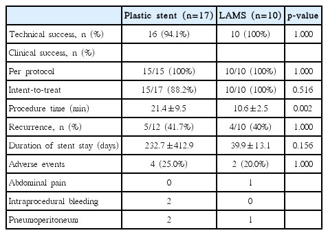
Clinical Outcomes between Plastic Stent and Lumen-Apposing Metal Stent
Overall, procedure-related AEs occurred in 6 of 27 patients (22.2%): 4 in the PS group and 2 in the LAMS group (23.5% vs. 20.0%, p=1.000). Further details regarding the nature of the AEs are described in Table 2. AEs included pneumoperitoneum in 3 patients, intraprocedural bleeding in 2, and abdominal pain in 1. Patients with pneumoperitoneum had no symptoms and spontaneous resolution without specific management. Intraprocedural hemorrhage was observed in 2 patients in the PS group during tract dilation using a needle knife catheter and was self-limited after stent placement. There was no procedure-related mortality.
Recurrence and related risk factors
Recurrence of PFCs after clinical success occurred in 5 patients in the PS group (5/12, 41.7%) and 4 in the LAMS group (4/10, 40%), with no statistically significant difference (p=1.000). EUS-guided transmural drainage with PS was performed in each patient of both groups. In 1 patient in the PS group, surgery was performed due to the recurrence of PFC with splenic infarction. Spontaneous regression of recurred PFC during follow-up was accomplished in 3 patients in the PS group and 1 in the LAMS group. In a univariate analysis of recurrence, only the tail location of the PFC was a statistically significant factor (Table 3). However, no significant risk factors were identified in multivariate analysis.
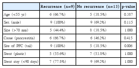
Univariate Analysis of Recurrence after Drainage
DISCUSSION
EUS-guided drainage for PFCs has been increasingly used as a minimally invasive procedure and is performed as an effective alternative to surgical treatment in many centers. Previous studies have shown similar success rates with meaningful advantages in comparison to percutaneous or surgical drainage [5,10,11]. EUS-guided drainage has a technical success rate of more than 90% and a clinical success rate of 75% to 90%, depending on the type of stent used [12-14].
LAMS is a new dedicated fully-covered SEMS for drainage of PFCs and overcomes the demerits of PS and FCSEMS. In addition to the wide biflanged shape of LAMS, a wide-diameter lumen provides a conduit for direct endoscopic necrosectomy as well as improved drainage, which obviates the need for repeated endoscopies. In our study, LAMS was placed in 2 patients with WON and both underwent clinically successful endoscopic necrosectomy. Currently, various types of LAMS have been introduced including a wide flared-end NAGITM stent (Taewoong Medical), BCF stent (MITech Co., Ltd., Seoul, Korea), and anchoring stent (AXIOS; Xlumena Inc., Mountain View, CA, USA). Several studies have shown that LAMSs have a very high rate of technical (89%–100%) and clinical (93%–100%) success [1,15-19]. LAMS provides stent stability, minimizes the risk of migration due to an anchoring effect, and enables a larger lumen for passage for easy direct access into the cavity for necrosectomy. Our study used SPAXUSTM as LAMS (Fig. 1), which has wide anchoring flanges that can be folded back after placement to prevent migration and maintain tight apposition.
Although metal stents have been used to compensate for the demerits of PSs, few studies have compared the clinical outcomes between the 2 stents; results indicating the better stent are still controversial. There is no definitive evidence so far in published literature that favors the LAMS when compared with the PS in resolving PFCs. The choice of stent is based on the discretion of the endoscopist rather than evidence-based findings. In a recent comparative study between PS and LAMS, which enrolled 103 patients with PFCs including 84 who were drained using PSs and 19 using LAMSs, clinical success was similar between the 2 groups (96% vs. 94%, p=0.78) [1]. EUS-guided drainage for PFCs had a high technical success rate with either PS or LAMS in our study. This result consistently matches reports from other studies [4,15,20,21]. Previous published studies have reported that the technical success rate of PP drainage using PSs ranges from 84% to 98%, whereas the rate of success when LAMSs are used is 78% to 100% [1,2]. Although many previous studies, including our study, showed no difference in clinical outcomes, different types of metal stents used and different times of evaluation may affect the rate of clinical success. Therefore, further controlled prospective comparative studies are required to validate the superiority of LAMS over PS.
While some reports have shown that more AEs occurred in PS placements (odds ratio, 2.9; 95% confidence interval, 1.4–6.3) compared to that in metal stent placements [2,22], other studies have shown no difference in AEs between the 2 stents (16% vs. 23%) in a systematic review [20]. Although our study showed no differences in the rates of technical and clinical success between the 2 stents, the procedure time was much shorter when using LAMS compared with PS (10.6±2.5 min vs. 21.4±9.5 min, p=0.002). The shorter procedure time in the LAMS group might be attributed to the simpler process requiring 1 stent and 1 guidewire, and little chance of AEs as well as additional benefits such as decreased patient discomfort and less radiation exposure may be expected [23]. However, the rate of procedure-related AEs was similar between the 2 groups in our study.
In previous comparative studies, bleeding in the metal stent was a major concern as procedure-related AEs. It is speculated that the reason for a high rate of bleeding in LAMS is perhaps the rapid decompression of the cystic cavity afforded by the large caliber of the LAMS, causing friction or irritation of the vasculature within the cavity [24]. Other studies have proposed that the wide lumen of the LAMS may allow for more entry of gastric acid into the cyst cavity, in which low pH fluid may irritate the exposed intracavitary vessels and promote bleeding [1]. However, no episode of bleeding occurred in the LAMS group in our study. Thus, further prospective studies may be required to elucidate which subsets of patients and patient characteristics may have increased risk of bleeding.
Although the result of our study did not reveal the superiority of LAMS in clinical outcomes and recurrence of PFC management, the reduced procedure time is a strength of our study, which was attributed from easy procedure technique.
There are some limitations of our study. First, the retrospective review of a small number of patients and heterogeneity of baseline patient characteristics between the 2 groups are the main limitations. Only 2 cases with WON were included in the LAMS group. Therefore, we were not able to equally select the stent for the procedure and had no choice to adjust the duration of follow-up or stent duration, which may have affected the results. Second, before the introduction of LAMS in October 2016, all procedures were performed with double-pigtail PSs; thereafter, LAMS was used for all patients with PFCs. Thus, there is a possibility that external factors such as the development of devices may have acted as a bias. Furthermore, the duration of follow-up was different between 2 groups and exerted bias in baseline characteristics. Third, the number of patients with clinical success and patients included in the recurrence analysis were somewhat different. Four patients in the PS group did not follow up after stent insertion in 1 or after stent removal in 3. Therefore, we could not judge the recurrence. Finally, the mean duration from the time of stent insertion to the time of the first follow-up CT between the PS and LAMS groups (66.8 days vs. 29.4 days, p=0.032) was significantly different. These differences may have acted as a bias in evaluating recurrence.
In conclusion, our study showed that PS and LAMS have similar clinical outcomes for drainage of PFCs. However, LAMS was associated with a shorter procedure time. At present, it is suggested that LAMS is an ideal stent and is highly recommended for treating PFCs in terms of antimigration and direct access to WON. Further prospective randomized studies including a larger number of cases are needed to validate the optimal management strategy for patients with PFCs.
Notes
Conflicts of Interest: The authors have no financial conflicts of interest.
Author Contributions
Conceptualization: Ho Cheol Shin, Chang Min Cho
Data curation: HCS, CMC
Formal analysis: HCS, CMC
Investigation; CMC, Min Kyu Jung
Supervision: CMC, MKJ, Seong Jae Yeo
Writing-original draft: HCS, CMC
Writing-review&editing: CMC, MKJ, SJY