Evaluation of Non-invasive Fibrosis Markers in Predicting Esophageal Variceal Bleeding
Article information

Abstract
Background/Aims
Esophageal variceal bleeding (EVB) is an important cause of mortality and morbidity in liver cirrhosis. In this study, we aimed to predict the possibility of EVB in patients with cirrhosis using a non-invasive score.
Methods
A total of 359 patients with cirrhosis were divided into two groups based on the presence or absence of EVB. ChildTurcotte-Pugh (CTP) score, a model for end-stage liver disease, aspartate aminotransferase to alanine aminotransferase ratio, aspartate aminotransferase to platelet ratio index (APRI), fibrosis-4-index (FIB-4), aspartate aminotransferase to alanine aminotransferase ratio/platelet ratio index (AARPRI), and S-index were measured for all participants. Receiver operating characteristic curves were obtained for all parameters, and the optimal cut-off value was determined in predicting EVB.
Results
In patients with EVB, the number of platelets (PLT) were low (p<0.001) and APRI, AARPRI, FIB-4, and S-index were significantly higher than those in patients without EBV. APRI, AARPRI, FIB-4, PLT, and S-index were statistically significant predictors of EVB (p<0.05).
Conclusions
FIB-4 and AARPRI, which are non-invasive markers of fibrosis, can be used to predict EVB. In addition, the 66.5 109/L cut-off value for PLT is important for EVB.
INTRODUCTION
Liver cirrhosis (LC) occurs because of widespread hepatocyte necrosis, development of regenerative nodules, fibrous tissue hyperplasia, and increased intrahepatic vascular pressure due to long-term and recurrent damage of one or more factors. Portal hypertension (PH), which develops as a result of increased intrahepatic vascular resistance in cirrhosis, results from the combination of damage in hepatic sinusoids and disruption of the balance between vasodilator and vasoconstrictor agents [1].
Normal portal pressure gradient values range from 1 to 5 mmHg, and values greater than 5 mmHg indicate the presence of PH [2]. A wide range of spontaneous portosystemic shunts and consequently esophageal varices (EV) may also occur as complications of long-standing PH in patients with LC [3]. The prevalence of gastroesophageal varices in LC is approximately 50% and is associated with the severity of liver disease. Esophageal variceal bleeding (EVB) occurs at a rate of 5% annually and is associated with a high mortality rate (15–25% in 6 weeks) [4,5].
Evaluation of the hepatic venous pressure gradient (HVPG) is the gold standard for diagnosing and measuring the degree of PH [6,7]. The most common method used to detect EV is endoscopic examination; however, it may not be cost-effective to perform EV screening in clinical practice because it is an invasive procedure and less than 50% of patients with cirrhosis have EV [4].
Evaluating the severity of PH and EV is essential for the management and prognosis of the disease in patients with cirrhosis. There are some adversities with these methods. For example, HVPG measurement can only be performed in specialized centers and endoscopic examination to detect EV is not a risk-free procedure. However, since EV development results from liver fibrosis and is due to increased intrahepatic resistance, non-invasive fibrosis markers (NFM) can be useful in detecting EV. Regarding this, aspartate aminotransferase to platelet ratio index (APRI) and fibrosis-4-index (FIB-4) are recommended and validated by the World Health Organization guidelines for evaluating hepatic fibrosis [8]. Owing to their easy, non-invasive, simple, and practical methods for detecting hepatic fibrosis, the model for end-stage liver disease (MELD), aspartate aminotransferase to alanine aminotransferase ratio (AAR), APRI, FIB-4, fibrosis index, and King score have been evaluated for the early detection of EV [9-14].
In this study, we aimed to predict the possibility of EVB using non-invasive fibrosis indicators to reduce the complications associated with the number of endoscopic scans, increase the cost-effectiveness, and determine the best time for invasive procedures.
PATIENTS AND METHODS
Study population
This study was conducted between 2015 and 2020 by scanning the files of patients followed-up for LC, online hospital data, and endoscopy records in the gastroenterology clinic of the institution. A total of 413 patients over the age of 18 years who had cirrhosis and detected EV in upper gastrointestinal endoscopy were included in the preliminary examination. Data on age, sex, cirrhosis etiology, MELD and Child-Turcotte-Pugh (CTP) classification scores, platelet (PLT) and biochemical results, endoscopically defined EV degrees, EVB history of the previous year were recorded. A total of 54 patients with portosystemic shunt, previous gastrointestinal surgery, liver metastasis, hepatocellular carcinoma, portal, hepatic and splenic vein thrombosis, myeloproliferative disease, pre-splenectomy, and those with a history of transjugular intrahepatic portosystemic shunt were excluded from the study.
Endoscopic examination
Endoscopies were performed by a gastroenterologist using Fujinon EG-580RD (Fujifilm Europe, Düsseldorf, Germany) brand gastroscopy devices. Varices grading was defined as grade I, II, and III [15].
Calculation of non-invasive fibrosis markers
The following formulas were used to calculate the investigated non-invasive markers:
1) AAR = aspartate aminotransferase (AST)/alanine aminotransferase (ALT) ratio
2) FIB-4 = (year of age × AST)/(PLT × the square root of ALT)
3) APRI = (AST/upper limit of normal) × 100/PLT (109/L)
5) S-index = 1000 × gamma glutamyl transferase/(PLT × albumin2)
6) aspartate aminotransferase to alanine aminotransferase ratio/platelet ratio index (AARPRI) = AAR/(PLT count (109/L)/150)
Statistical analysis
All data were analyzed using SPSS Statistics version 21 (IBM Co., Armonk, NY, USA). The consistency of continuous variables to normal distribution was evaluated using visual (histogram and probability plots) and analytical methods (Kolmogorov-Smirnov/Shapiro-Wilk tests).
Categorical variables are reported as numbers, percentages, normally distributed data as arithmetic means and standard deviations, and skewed-distributed data as median (minimum-maximum) values. An independent sample t-test was used for the comparative analysis between two independent groups of normally distributed data, and the Mann-Whitney U test was used in the non-compliant data. In the comparative analysis for categorical variables between independent groups, the Pearson’s or Fisher’s test was chosen from the chi-square (χ2) test. Receiver operating characteristic curve (ROC) analysis was performed to calculate the esophageal bleeding predictive value of NFM (APRI, AARPRI, FIB-4, AAR, and S-index), CTP score, and MELD. The area under the ROC curve (AUC) results were considered as follows: 0.9–1, excellent; 0.8–0.9, good; 0.7–0.8, fair; 0.6–0.7, poor; and 0.5–0.6, failed [16]. The results following ROC analysis, AUC and cut-off values, sensitivity, and specificity of these cut-off values, likelihood ratio (LR), odds ratio (OR), positive predictive value (PPV), and negative predictive value (NPV) were also presented. A p-value <0.05 was considered statistically significant.
Ethical approval
The study protocol was carried out with the approval of the Ethics Committee of the Ordu Training and Research Hospital, Ordu (Turkey) (No: 16/2018).
RESULTS
Demographic characteristics of the study population
In the analysis based on the presence of bleeding, no significant difference was found between the groups in terms of age and sex (p>0.05). The most common etiologic factor of cirrhosis in both groups was hepatitis B virus (Table 1).

Comparison Clinicodemographic and Laboratory Parameters in Study Population
In the group with EVB, grade I esophageal bleeding was not detected, while grade III esophageal varices were found in 84.1% (n=143) of cases.
Prediction of EVB with non-invasive markers
There was no significant difference between the groups in terms of albumin, AST, ALT, CTP score, MELD, and AAR (p>0.05). The number of PLT was significantly lower in the group with bleeding (p<0.001). APRI, AARPRI, FIB-4, and S-index were significantly higher in the group with bleeding (p=0.002, <0.001, <0.001, and 0.025, respectively).
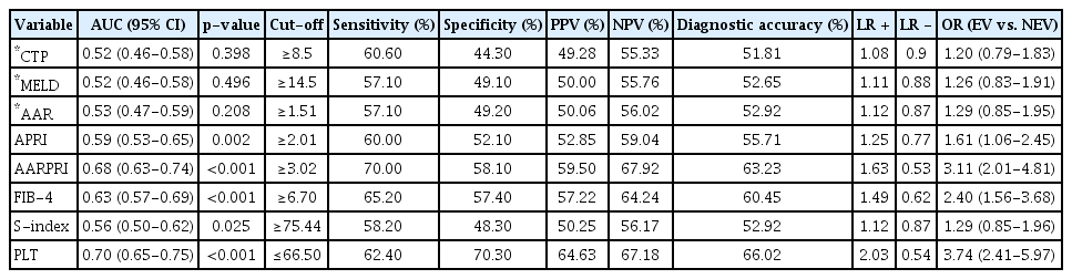
The predictive power of the CTP score, MELD, AAR, APRI, AARPRI, FIB-4, PLT, and S-index were evaluated by ROC analysis. AUC values and significance obtained from ROC analysis are presented in Table 2. MELD, CTP score, and AAR were not significant predictors of variceal bleeding (p>0.05). The AUC values obtained for FIB-4 (AUC=0.63), APRI (AUC=0.59), and S-index (AUC=0.56) were significant but weak (Fig. 1). The highest significant AUC value was obtained for PLT (AUC=0.7) (Fig. 2) and AARPRI (AUC=0.68). Sensitivity, specificity, + LR, - LR, PPV, NPV, and OR of the cut-off values obtained for the parameters found significant by ROC analysis were calculated. It was observed that the values of PLT ≤66.5 109/L were better than other parameters (sensitivity, 62%; specificity 70%; +LR, 2.03; PPV, 64.3%; NPV, 67.18; OR, 3.74).
Performance of Non-Invasive Markers for Prediction of Esophageal Variceal Bleeding

Receiver operating characteristic curves for the APRI, AAR, FIB4, AARPRI, and S-index. Larger results of APRI, AAR, FIB- 4, AARPRI, and S-index indicate more diagnostic positive tests for esophageal variceal bleeding. AAR, aspartate aminotransferase to alanine aminotransferase ratio; APRI, aspartate aminotransferase-to-platelet-ratio index; AARPRI, aspartate aminotransferase to alanine aminotransferase ratio/platelet ratio index; FIB-4, fibrosis-4 index.

Receiver operating characteristic curves for the platelet. A small number of platelets indicate a more diagnostic positive test for esophageal varices bleeding.
DISCUSSION
EVB is a fatal complication of cirrhotic PH. However, there is no apparent clinical finding for EV in most patients with cirrhosis, even during the decompensation period.
Today, clinicians are concerned with identifying some non-invasive biochemical markers with high sensitivity and specificity that are cheaper and easier to obtain to reduce the number of upper gastrointestinal endoscopies for screening and treating EV in liver patients. These non-invasive biomarkers are applied using routine laboratory tests that do not require extra cost and special devices or additional biochemical tests [17]. NFM can also predict the development of EV, as PH is a result of increased liver vascular resistance secondary to liver fibrosis [18]. Few studies have investigated non-invasive approaches to estimate the risk and development of EV and EVB, and this issue remains controversial [19-21].
While Iwata et al., reported that AAR was related to the severity of esophageal varices [22], there are some publications in the literature indicating that the AAR index has limited predictive value for severe EV [23]. In our study, the AAR index did not show a significant difference between the groups.
The APRI and FIB-4 index are two classic non-invasive scores with good diagnostic efficacy for cirrhosis [24, 25]. Zhang et al., determined that the AUC value of the APRI (0.729) was higher than that of the other three indices (AAR, FIB-4, and S-index) in determining the presence and severity of EV, and the APRI score greater than 1.4 can be used as a reference indicator for the early intervention of severe EV [23]. In addition, the APRI score was shown to be an independent predictor of recurrent EVB in a case-control study [26]. In a study that included only alcoholic cirrhotic patients, the average APRI values were significantly higher in the EVB group [27]. In our study, the APRI index was significantly higher in the EVB group than in the non-EVB group (p=0.002). However, the APRI score was found to be poorly associated with the prediction of EVB (OR, 1.61).
In the study by Zhang et al., the FIB-4 index was an independent predictor of EV, and the AUROC value was determined to be 0.64 [23]. In a large meta-analysis, the AUC value of FIB-4 was reported as 0.77 in predicting the presence of EV [28]. A study conducted by Kraja et al. [29] defined the FIB-4 score as a strong and significant predictor of EV presence. In the same study, the FIB-4 score was defined as an important predictor for EV at a 3.23 cut-off value (AUC, 0.66) and a weak predictor for EVB at a 5.02 cut-off value (AUC, 0.51). They interpreted FIB-4 as the most effective NFM of the liver that can be used as the first screening tool for cirrhotic patients [29]. In a study of patients with alcoholic cirrhosis, the mean FIB-4 score was found to be significantly higher in EVB group than in the non-EVB group (8.0 and 3.9, respectively), thereby predicting EVB with a diagnostic accuracy of 63.86% [27]. Similarly, in the current study, we also found that the FIB-4 score was significantly higher in the EBV group than in the non-EVB group (p<0.001). In addition, FIB-4 had a diagnostic accuracy rate of 60.45% at a cut-off value of 6.70. The predictive power of FIB-4 of EVB was found to be higher than the APRI score (OR, 2.40 and 1.61, respectively).
According to the Baveno VI consensus criteria, it was stated that endoscopic examination was not required in patients with transient elastography value less than 20 kPa and platelet value more than 150×109/L and annual PLT level monitoring was recommended [30]. Sarangapani et al., stated that a platelet count lower than 150×109/L was an independent predictor of the presence of EV [31]. Madonia et al., associated thrombocytopenia with recurrent variceal bleeding and stated that it was below the 80×109/L level as the lower limit [32]. In the current study, the number of platelets, determined as the strongest predictor of EVB (AUC, 0,70; OR, 3,74), had a 62% sensitivity and 70% specificity at a cut-off value of 66.5×10⁹/L.
In a study where the S-Index and FIB-4 score were evaluated by ROC analysis for the predictive power of EVB, the S-index was found to be stronger than the FIB-4 score (AUC, 0.695 and 0.673, respectively). However, it has been stated that EV is not an independent predictor (all p>0.01) [23]. In our study, the S-index was found to be significantly higher in patients with EVB than in patients without EVB (p=0.025). In addition, the power of the S-index to predict EVB was very weak (AUC, 0.56; OR, 1.29).
In a limited number of studies in which the AARPRI score was examined, a significant relationship was found between the liver fibrosis rate and the AARPRI score [33,34]. However, to the best of our knowledge, no study has evaluated the relationship between EV and AARPRI scores. In our study, AARPRI was found to have moderate power to predict the likelihood of EVB (AUC, 0.68; OR, 3,11).
In addition, to predict the likelihood of EVB, AARPRI was found to have a 63.33% diagnostic accuracy rate with a 1.63 likelihood at a 3.02 cut-off value (70% sensitivity and 58% specificity).
There were some limitations in this study. First, due to the retrospective nature of the study, we could not investigate the predictive values of these markers for EVB in patients without a previous history of EVB. Second, since the treatment records of patients were not fully available, it was not possible to distinguish patients who received prophylactic treatment for EVB.
In conclusion, the current study showed that FIB-4, AARPRI, and PLT as non-invasive liver fibrosis markers will contribute to our clinical knowledge in predicting EVB. However, larger studies are needed to implement a routine use of these markers in clinical practice. We hope that this study will guide future studies on the use of non-invasive methods as a screening method to predict EVB.
Notes
Conflicts of Interest: The authors have no potential conflicts of interest.
Funding
None.
Author Contributions
Conceptualization: Sami Cifci, Nergiz Ekmen
Data curation: SC, NE
Formal analysis: SC, NE
Investigation: SC, NE
Methodology: SC, NE
Project administration: SC, NE
Ono Supervision: SC, NE
Validation: SC, NE
Writing-original draft: SC, NE
Writing-review&editing: SC, NE