CME for
KSGE members
Choi: A 66-year-old woman with a hypervascular pancreatic mass
Quiz
A 66-year-old woman with no previous medical history experienced early satiety and nonspecific epigastric pain for six months before visiting a local hospital. Computed tomography revealed a 2-cm solid mass with contrast enhancement in the pancreatic body ( Fig. 1A). The patient was transferred to the pancreatic center of our institution for further evaluation. On presentation, the physical examination was unremarkable. Laboratory tests, which included pancreatic enzymes and tumor markers, were within the normal ranges. Subsequent magnetic resonance imaging revealed a hypervascular mass in the pancreatic body ( Fig. 1B). Endoscopic ultrasound (EUS) examination revealed a homogeneous, hypoechoic 2.0├Ś1.8 cm mass ( Fig. 2A), which exhibited strong enhancement on contrast-enhanced EUS ( Fig. 2B, 12 seconds after injecting SonoVue, Bracco Inc.). EUS-guided fine-needle biopsy (FNB) was performed using a 22-gauge histology needle. However, the FNB specimens were insufficient for histopathological diagnosis. What is the most likely diagnosis in this case?
Answer 
Laparoscopic distal pancreatectomy was performed because the mass was presumed to be a neuroendocrine tumor (NET), given its imaging characteristics and size. The patient recovered well postoperatively. Pathological examination of the specimen revealed a solid lesion, measuring 2-cm on its greater axis ( Fig. 3A). Microscopically, the mass comprised cells with a clear cytoplasm, no signs of atypia, and no mitosis. The cells were arranged in tubular/acinar structures without lumina ( Fig. 3B). Histology of the tumor was consistent with the diagnosis of a solid-type serous cystic neoplasm (SCN) ( Fig. 3C). While the most common histotype of SCN is the microcystic honeycomb pattern, these lesions can also, albeit rarely, appear solid due to the enhancement of the intracystic septa and closely opposed walls. 1 Solid-appearing SCNs are benign tumors that typically occur in elderly women. Most solid-appearing SCNs have shown arterial hyperenhancement on imaging studies and can often be mistaken for other solid-enhancing pancreatic masses, such as NETs, solid pseudopapillary neoplasms, intrapancreatic accessory spleen, and metastatic renal cancer. 2,3 It is challenging to provide a definitive diagnosis of this lesion prior to surgery. 4
This case of solid-appearing SCN adds to our knowledge and experience in diagnosing and treating hypervascular masses in the pancreas.
Fig.┬Ā1.
Cross-sectional imaging of the pancreatic mass. (A) On computed tomography, the pancreatic mass showed increased contrast enhancement in the arterial phase (arrows). (B) The arterial phase on magnetic resonance imaging revealed the hypervascularity of the mass in the pancreatic body (arrows). 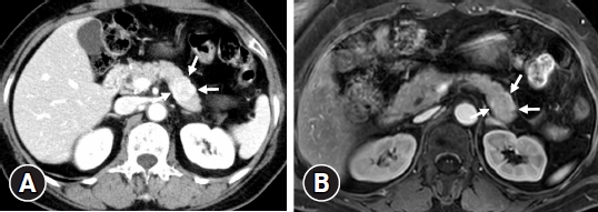
Fig.┬Ā2.
Endoscopic ultrasound. (A) B-mode endoscopic ultrasound revealed a well-defined, hypoechoic mass in the pancreatic body (arrowheads). (B) Contrast-enhanced harmonic imaging showing hyperenhancement inside the tumor. 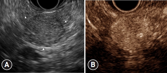
Fig.┬Ā3.
Histopathological findings. (A) A photograph of a gross specimen of the mass revealed a round, well-defined, and solid mass measuring 20 mm in size. (B) Low magnification of the microphotograph revealed that the tumor was composed of numerous microcysts with densely fibrocollagenous stroma (hematoxylin and eosin stain, ├Ś40). (C) Higher magnification of the microphotograph (hematoxylin-eosin stain, ├Ś200) showing a single layer of bland-looking cuboidal epithelium resting on the fibrous cyst wall (hematoxylin and eosin stain, ├Ś200). 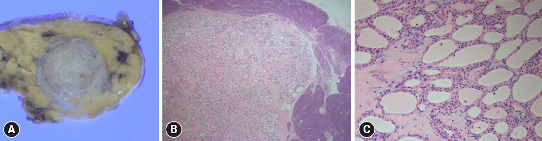
|
|


