Aortoduodenal fistula bleeding caused by an aortic stent graft
Article information

A 65-year-old female visited the emergency room due to sudden hematemesis and hematochezia. One year earlier, the patient underwent endovascular implantation of an aorto-bi-iliac bifurcation stent graft for a fusiform infrarenal aortic aneurysm that extended to the left common iliac artery. At the time of presentation, her blood pressure and heart rate were 70/40 mmHg and 105 beats/min, respectively. Laboratory findings showed a white blood cell count of 13,000/μL, hemoglobin level of 8.0 g/dL, and platelet count of 405,000/μL. Emergent endoscopy identified a foreign material (an aortic stent graft) with vascular pulsation in the third part of the duodenum (Fig. 1). Abdominal computed tomography revealed increased fat stranding around the aorta, which is indicative of inflammation, and a loss of the fat plane between the duodenum and aorta (Fig. 2). Therefore, the cause of bleeding was diagnosed as an aortoduodenal fistula caused by the previously placed aortic stent graft. The patient underwent primary closure of the duodenal perforation and omental wrapping and was discharged without any complications. However, the patient passed away 31 months later due to recurrence of the aortoduodenal fistula.
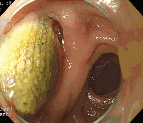
Upper gastrointestinal endoscopy reveals a foreign material (an aortic stent graft) and blood clot with vascular pulsation (asterisk) in the third part of the duodenum.
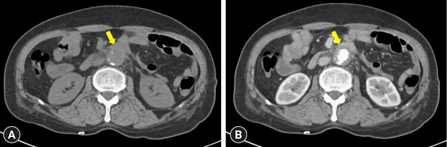
Abdominal computed tomography reveals increased fat stranding around the aorta, which is indicative of inflammation, and a loss of the fat plane between the duodenum and aorta (arrows). (A) Pre-enhanced phase. (B) Arterial phase.
Aortoenteric fistula can occur secondary to aortic reconstruction procedures1 and can develop anywhere in the gastrointestinal tract adjacent to the aorta; however, it is most frequently observed in the third part of the duodenum, which is anchored in the retroperitoneal space and positioned directly anterior to the aorta. The location of the duodenum in close proximity to the aorta makes it susceptible to fistula formation due to the constant transmission of pulsatile forces from the aorta.2 Therefore, aortoduodenal fistula should be considered a potential cause of hematemesis or hematochezia in patients with a history of aortic stent graft.3
Notes
Conflicts of Interest
Gwang Ha Kim is currently serving as a deputy editor for Clinical Endoscopy; however, he was not involved in the peer reviewer selection, evaluation, or decision process of this article. Seunghyun Hong has no potential conflicts of interest.
Funding
None.
Author Contributions
Conceptualization: GHK; Data curation: SH, GHK; Investigation: GHK; Methodology: SH; Writing–original draft: SH; Writing–review & editing: all authors.