Clin Endosc > Volume 57(2); 2024 > Article
Kim and Joo: A remnant cystic duct presenting as a duodenal subepithelial tumor
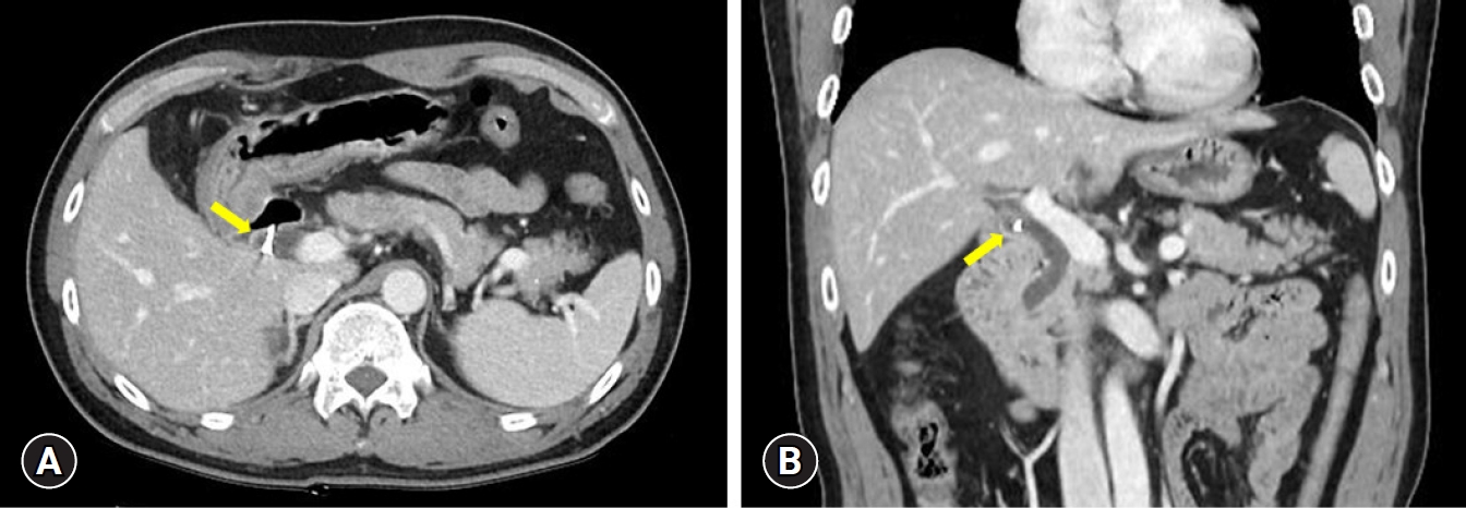
A 51-year-old man was referred to our institution because of a subepithelial tumor (SET) of the duodenum, which was detected incidentally during esophagogastroduodenoscopy (EGD). Twenty years earlier, he had undergone a cholecystectomy because of cholelithiasis. During EGD, an SET-like lesion, covered by normal duodenal mucosa, was observed in the anterior wall of the duodenal bulb (Fig. 1A). Endoscopic ultrasonography (EUS) using a 20-MHz catheter probe (UM3D-DP20-25R; Olympus) revealed a 0.8×0.6 cm-sized anechoic lesion with hyperechoic suture material outside the duodenal wall (Fig. 1B, C), which was connected to the common bile duct. No Doppler flow was detected by 7.5-MHz conventional EUS using a radial echoendoscope (GF-UE260-AL5; Olympus) (Fig. 1D, Video 1). Abdominal computed tomography scans also showed a remnant cystic duct with suture material (Fig. 2). Therefore, the lesion was diagnosed as a duodenal SET-like lesion caused by a remnant cystic duct.
A remnant cystic duct occurring after cholecystectomy can cause an SET-like lesion in the duodenum, particularly when the SET-like lesion is in the anterior side of the duodenal bulb in patients with a history of cholecystectomy. Although it is very rare, adenocarcinoma can arise from such a remnant cystic duct.1 Detailed examination of a remnant cystic duct by EUS is necessary to avoid missing malignant tumors or stones in the remnant duct.2,3

Video

Video 1. Endoscopic ultrasonography of a duodenal subepithelial lesion.
A video related to this article can be found online at https://doi.org/10.5946/ce.2023.275.

NOTES

Conflicts of Interest
Gwang Ha Kim is currently serving as a deputy editor in Clinical Endoscopy; however, he was not involved in the peer reviewer selection, evaluation, or decision process of this article. Dong Chan Joo has no potential conflicts of interest.
Funding
None.
Author Contributions
Conceptualization: GHK; Data curation: all authors; Investigation: GHK; Methodology: DCJ; Writing–original draft: GHK; Writing–review & editing: all authors.

Fig. 1.
Endoscopic ultrasonography (EUS) findings. (A) A 1-cm-sized subepithelial lesion covered by normal duodenal mucosa is observed in the anterior wall of the duodenal bulb. (B, C) EUS using a 20-MHz catheter probe reveals a 0.8×0.6 cm-sized anechoic lesion with hyperechoic suture material (arrow) outside the duodenal wall. (D) Conventional EUS (7.5 MHz) reveals that the anechoic lesion was connected to the common bile duct and had no Doppler flow (arrow).
ce-2023-275f1.jpg
Fig. 2.
Abdominal computed tomography reveals a remnant cystic duct with suture material (arrows). (A) Transverse section view. (B) Coronal section view.
ce-2023-275f2.jpg

REFERENCES

1. Eum JS, Kim GH, Park CH, et al. A remnant cystic duct cancer presenting as a duodenal submucosal tumor. Gastrointest Endosc 2008;67:975–976.
crossref pmid
2. Han SY, Chon HK, Kim SH, et al. Quality indicators of endoscopic ultrasound in the pancreatobiliary system: a brief review of current guidelines. Clin Endosc 2024;57:158–163.
crossref pmid
3. Ryou SH, Kim HJ. Successful removal of remnant cystic duct stump stone using single-operator cholangioscopy-guided electrohydraulic lithotripsy: two case reports. Clin Endosc 2023;56:375–380.
crossref pmid pmc pdf
TOOLS
PDF Links  PDF Links
PubReader  PubReader
ePub Link  ePub Link
XML Download  XML Download
Full text via DOI  Full text via DOI
Download Citation  Download Citation
Supplement  Supplement
  Print
Share:      
METRICS
0
Crossref
0
Scopus
1,712
View
143
Download
Related articles
Prevalence and natural course of incidental gastric subepithelial tumors  
A Rare Cause of Gastric Subepithelial Tumor  2020 May;53(3)
Contrast Enhanced Endoscopic Ultrasound Imaging for Gastrointestinal Subepithelial Tumors  2019 July;52(4)
Intramural Gastric Abscess Caused by a Toothpick Presenting as a Subepithelial Tumor  2014 May;47(3)
Endoscopic Resection of Subepithelial Tumors  2012 September;45(3)
Editorial Office
Korean Society of Gastrointestinal Endoscopy
#817, 156 Yanghwa-ro (LG Palace, Donggyo-dong), Mapo-gu, Seoul, 04050, Korea
TEL: +82-2-335-1552   FAX: +82-2-335-2690    E-mail: CE@gie.or.kr
Copyright © Korean Society of Gastrointestinal Endoscopy.                 Developed in M2PI
Close layer