

Search
- Page Path
- HOME > Search
Case Reports
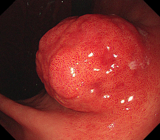
- Gastric Angiolipoma Resected with Endoscopic Submucosal Dissection
- Sang Myung Yeo, Jae Kwang Lee, Hyun Soo Kim, Chang Geun Park, Jae Kwon Jung, Dae Jin Kim, Yun Jin Chung, Han Jun Ryu
- Clin Endosc 2021;54(3):432-435. Published online March 15, 2021
- DOI: https://doi.org/10.5946/ce.2020.146
-
 Abstract
 PDF
PDF PubReader
 ePub
ePub - Angiolipoma is a benign fatty neoplasm that has components of proliferating blood vessels. These types of lesions commonly occur in the subcutaneous tissue of the limbs and trunk. Angiolipoma in the gastrointestinal tract is extremely rare, and the final diagnosis generally depends on histological examination of the excised biopsy. In most previously reported cases, the lesions were diagnosed and treated with surgical management. In this study, we report a case of gastric angiolipoma of approximately 4 cm in size that was diagnosed and treated with endoscopic submucosal dissection.
-
Citations
Citations to this article as recorded by
- A rare cause of upper gastrointestinal bleeding in an elderly female: Gastric Angiolipoma
Xiufan Ni, Sujun Gao, Lei Chen, Li Zhang, Jian Yin, Zhen Zhu
Revista Española de Enfermedades Digestivas.2023;[Epub] CrossRef - A Huge Gastric Angiolipoma Presenting with Acute Upper Gastrointestinal
Hemorrhage: A Case Report
Joo Hyeok Choi, Sung Bin Park, Jong Beum Lee, Tae-Jin Lee, Hyun Jeong Park, Eun Sun Lee
Current Medical Imaging Formerly Current Medical Imaging Reviews.2023;[Epub] CrossRef
- A rare cause of upper gastrointestinal bleeding in an elderly female: Gastric Angiolipoma
- 3,733 View
- 80 Download
- 2 Web of Science
- 2 Crossref

- A Case of a Bleeding Duodenal Lipoma Successfully Controlled by Endoscopic Resection
- Seo Yeon Gwak, Mi Kyung Lee, Yong Kang Lee
- Clin Endosc 2020;53(2):236-240. Published online July 24, 2019
- DOI: https://doi.org/10.5946/ce.2019.035
-
Abstract
PDFPubReaderePub
- This is a case report of successful endoscopic resection (ER) of a bleeding duodenal lipoma. An 85-year-old woman who was diagnosed with asymptomatic subepithelial tumor of the duodenum 3 years ago visited the emergency room with hematemesis and was admitted to our hospital. Emergent esophagogastroduodenoscopy revealed bleeding from an ulcer on the superior aspect of a subepithelial tumor measuring about 20 mm in diameter, at the superior duodenal angle. The ulcer was in the active stage (A1), with a visible vessel. The bleeding was controlled by ER of the tumor using a snare. The final pathological diagnosis was duodenal lipoma with mucosal ulceration. The patient showed no signs of bleeding for 10 days after the procedure; subsequently, she was discharged and followed up for regular checkups.
-
Citations
Citations to this article as recorded by- Endoscopically resected duodenal lipoma as an uncommon cause of upper gastrointestinal bleeding: a case report
Dong Chan Joo, Gwang Ha Kim, Bong Eun Lee, Moon Won Lee, Cheolung Kim
The Ewha Medical Journal.2024;[Epub] CrossRef
- Endoscopically resected duodenal lipoma as an uncommon cause of upper gastrointestinal bleeding: a case report
- 6,653 View
- 131 Download
- 3 Web of Science
- 1 Crossref
- Endoscopic Resection of Giant Colonic Lipoma: Case Series with Partial Resection
- Gun Woo Kim, Chang-Il Kwon, Sang Hee Song, Sun Mi Jin, Kyung Ho Kim, Jie Hye Moon, Sung Pyo Hong, Pil Won Park
- Clin Endosc 2013;46(5):586-590. Published online September 30, 2013
- DOI: https://doi.org/10.5946/ce.2013.46.5.586
-
Abstract
PDFPubReaderePub
Colonic lipoma, a very rare form of benign tumor, is typically detected incidentally in asymptomatic patients. The size of lipoma is reported variously from 2 mm to 30 cm, with higher likelihood of symptoms as the size is bigger. Cases with symptom or bigger lesion are surgically resected in principle; endoscopic resection, which has developed recently with groundbreaking advance of endoscopic excision technology, is being used more often but with rare report of success due to high chance of complications such as bowel perforation or bleeding. The authors report here, together with a literature review, our experiences of three cases of giant colonic lipomas showing complete remission after aggressive unroofing technique, at certain intervals, using snare catheter at the origin of the lipoma so that the remaining lipoma could be drained out of the exposed surface spontaneously, in order to reduce complications.
-
Citations
Citations to this article as recorded by- Colonic intussusception from pedunculated colonic lipoma at hepatic flexure: A case report and review of current literature
Richard Edmund Hogan, Ben Michael Murray, Michael Flanagan, Shane Brennan, Conor Shortt, Dara Kavanagh
Surgery Case Reports.2024; 1: 100008. CrossRef - A giant colonic lipoma
Tara M. Connelly, Cillian Clancy, Shaomin Hu, Joshua Sommovilla
ANZ Journal of Surgery.2023; 93(1-2): 428. CrossRef - Obscure gastrointestinal bleeding from a large jejunal lipoma treated using an endoscopic unroofing technique with double balloon enteroscopy: a case study
Reo Kobayashi, Ken Inoue, Ryohei Hirose, Toshifumi Doi, Akihito Harusato, Osamu Dohi, Naohisa Yoshida, Kazuhiko Uchiyama, Takeshi Ishikawa, Tomohisa Takagi, Hiroaki Yasuda, Hideyuki Konishi, Yukiko Morinaga, Yoshito Itoh
Clinical Journal of Gastroenterology.2023; 16(1): 32. CrossRef - Endoscopic debulking of a large colonic lipoma causing recurrent intussusception using endoscopic mucosotomy technique
Jenson Phung, Morgan Freeman, Mohammad Bilal
Endoscopy.2023; 55(S 01): E817. CrossRef - Large colonic lipoma with a laterally spreading tumor treated by endoscopic submucosal dissection: A case report
Jun Yong Bae, Hun Kyu Kim, Yee Jin Kim, Se Woong Kim, Youngeun Lee, Chang Beom Ryu, Moon Sung Lee
World Journal of Clinical Cases.2023; 11(26): 6194. CrossRef - Observation of the drainage process of the residual lipoma after endoscopic unroofing technique during colonoscopic evaluation of post-procedural hematochezia
Yi-Ling Ko, Hiroki Matsuoka, Ryohei Nomaru, So Imakiire, Hideto Sakisaka, Satoshi Matsuoka, Nobuaki Kuno, Koichi Abe, Sadahiro Funakoshi, Yusuke Ishida, Hideki Ishibashi, Kaori Koga, Tetsuhiro Saito, Morishige Takeshita, Fumihito Hirai
Clinical Journal of Gastroenterology.2022; 15(2): 407. CrossRef - Spontaneous expulsion of a duodenal lipoma after endoscopic biopsy: A case report
Zhi-Hao Chen, Li-Hong Lv, Wen-Sheng Pan, Yi-Miao Zhu
World Journal of Gastroenterology.2022; 28(34): 5086. CrossRef - Pedunculated sigmoid lipoma causing colo-colonic intussusception
Kenneth Ford, Samantha Lopez, Gaurav Synghal, Yomi Fayiga, Brittany Carter, Anuj Kandel, Kenneth Ford
Baylor University Medical Center Proceedings.2021; 34(3): 371. CrossRef - Colonoscopic resection of giant colonic lipoma causing subacute large bowel obstruction
Amy Donovan, Sandun Abeyasundara, Hajir Nabi
ANZ Journal of Surgery.2020;[Epub] CrossRef - COLON LIPOMA COMPLICATED BY COLON INVAGINATION
U. B. Urmonov, S. G. Afanasyev, A. Yu. Dobrodeev, A. V. Avgustinovich, M. Yu. Volkov, N. V. Vasiliev, E. N. Samtsov
Grekov's Bulletin of Surgery.2020; 178(6): 63. CrossRef - Endoscopic treatment of large symptomatic colon lipomas: A systematic review of efficacy and safety
Michiel Bronswijk, Anne‐Marie Vandenbroucke, Peter Bossuyt
United European Gastroenterology Journal.2020; 8(10): 1147. CrossRef - Endoscopic resection of giant colon lipomas: get rid of the roof!
Michiel Bronswijk
VideoGIE.2019; 4(7): 341. CrossRef - Submucosal lipoma of the sigmoid colon as a rare cause of mucoid diarrhea: a case report
S. U. B. Dassanayake, N. P. Dinamithra, N. M. M. Nawarathne
Journal of Medical Case Reports.2016;[Epub] CrossRef - Endoscopic resection of giant GI lipoma: a case series
Diane Lorenzo, Jean Michel Gonzalez, Alban Benezech, Marc Barthet
VideoGIE.2016; 1(2): 43. CrossRef - Two Patients with Large Colonic Lipomas for which Endoscopic Unroofing was Ineffective
Yuichi Tomiki, Koichiro Niwa, Kiichi Nagayasu, Yu Okazawa, Shingo Ito, Ryosuke Ichikawa, Hisashi Ro, Shun Ishiyama, Kiichi Sugimoto, Kazuhiro Sakamoto
Case Reports in Gastroenterology.2016; 10(3): 538. CrossRef - Colonoscopy‐assisted laparoscopic resection of an obstructing ‘giant’ lipoma of the transverse colon
B. Asantha De Silva, Raeed Deen, Wasantha K. Wijenayake
ANZ Journal of Surgery.2015; 85(10): 785. CrossRef - Unroofing Technique as an Option for the Endoscopic Treatment of Giant Gastrointestinal Lipomas
Marcela Kopáčová, Stanislav Rejchrt, Jan Bureš
Acta Medica (Hradec Kralove, Czech Republic).2015; 58(4): 115. CrossRef - Large “pedunculated” colonic lipoma: A word of caution while cutting into fat!
Syed Adnan Mohiuddin, Saad Al Kaabi, Ragesh Babu Thandassery, Khalid Mohsin Al Ejji, Nazeeh Al Dweik, Emran Amir, Manik Sharma
Indian Journal of Gastroenterology.2014; 33(6): 571. CrossRef
- Colonic intussusception from pedunculated colonic lipoma at hepatic flexure: A case report and review of current literature
- 7,514 View
- 95 Download
- 18 Crossref
- Endoscopic Treatment of a Symptomatic Ileal Lipoma with Recurrent Ileocolic Intussusceptions by Using Cap-Assisted Colonoscopy
- Eun Sung Lee, Kang Nyeong Lee, Kyung Soo Choi, Hang Lak Lee, Dae Won Jun, Oh Young Lee, Byung Chul Yoon, Ho Soon Choi
- Clin Endosc 2013;46(4):414-417. Published online July 31, 2013
- DOI: https://doi.org/10.5946/ce.2013.46.4.414
-
Abstract
PDFPubReaderePub
A 73-year-old woman presented with intermittent abdominal pain and weight loss of 15 kg for 2 years. Colonoscopy revealed an erythematous polypoid tumor with a long and wide stalk in the cecum, but with air inflation, it abruptly went away through the ileocecal valve (ICV). An abdominal computed tomography showed a well-demarcated pedunculated subepithelial mass of 2.6×2.7 cm size with fat attenuation in the terminal ileum. It was an intussusceptum of the ileal lipoma through the ICV. This ileal lipoma was causing her symptoms because repeated ileocolic intussusceptions resulted in intermittent intestinal obstructions. In order to avoid surgical sequelae of ileal resection, snare polypectomy using cap-assisted colonoscopy technique was performed within the ileum without complications. The histopathology report confirmed it as a subepithelial lipoma. After endoscopic resection of the ileal lipoma, the patient has been free of symptoms and was restored to the original weight.
-
Citations
Citations to this article as recorded by- Extraction of terminal ileal lipomas to cecum can facilitate endoscopic resection: A case series with video
Hiroshi Yamazaki, Yohei Minato, Deepak Madhu, Toshifumi Iida, Susumu Banjyoya, Tomoya Kimura, Koichi Furuta, Shinya Nagae, Yohei Itou, Nao Takeuchi, Shunya Takayanagi, Yoshiaki Kimoto, Yuki Kano, Takashi Sakuno, Kohei Ono, Ken Ohata
DEN Open.2025;[Epub] CrossRef - A Rare Case of Multiple Ileal Lipoma in A Young Male
Ramprashanth MP
Journal of Surgery Research and Practice.2024; : 1. CrossRef - Terminal Ileum Lipoma Causing Ileocolic Intussusception: A Case Report and Literature Review
Siddhant Dogra, Jason Wei, Benjamin Wadowski, Virginia Devi-Chou, Leandra Krowsoski, Rajiv R Shah
Cureus.2023;[Epub] CrossRef - Successful endoscopic management of adult ileocecal intussusception secondary to a large ileal lipoma
Akira Teramoto, Seiji Hamada, Takahiro Utsumi, Daizen Hirata, Yasushi Sano
VideoGIE.2021; 6(4): 187. CrossRef - Life‐threatening gastrointestinal bleeding from a giant ileal lipoma
Amy Donovan, Sandun Abeyasundara, Hajir Nabi
ANZ Journal of Surgery.2020;[Epub] CrossRef - Intususcepción íleo-cólica de lipoma ileal como causa de hemorragia digestiva baja
Eduardo Valdivielso Cortázar, María López Álvarez, Alberto Guerrero Montañes, Loreto Yañez González-Dopeso, Jesus Ángel Yañez López, Pedro Antonio Alonso Aguirre
Gastroenterología y Hepatología.2017; 40(7): 457. CrossRef - Ileocolic intussusception of ileal lipoma as a cause of lower gastrointestinal bleeding
Eduardo Valdivielso Cortázar, María López Álvarez, Alberto Guerrero Montañes, Loreto Yañez González-Dopeso, Jesus Ángel Yañez López, Pedro Antonio Alonso Aguirre
Gastroenterología y Hepatología (English Edition).2017; 40(7): 457. CrossRef - Unroofing Technique as an Option for the Endoscopic Treatment of Giant Gastrointestinal Lipomas
Marcela Kopáčová, Stanislav Rejchrt, Jan Bureš
Acta Medica (Hradec Kralove, Czech Republic).2015; 58(4): 115. CrossRef
- Extraction of terminal ileal lipomas to cecum can facilitate endoscopic resection: A case series with video
- 7,159 View
- 62 Download
- 8 Crossref
- A Case of Giant Lipoma Causing Chronic Recurrent Intussusception of the Colon
- Chang Seob Lee, Mi Jin Lee, Kyoung Lee Kim, Yeon Soo Kim, Gwang Ho Baik, Jin Bong Kim, Dong Joon Kim, Sang Hak Han
- Clin Endosc 2012;45(2):165-168. Published online June 30, 2012
- DOI: https://doi.org/10.5946/ce.2012.45.2.165
-
Abstract
PDFPubReaderePub
Colonic lipomas, which often occur in elderly women, usually have small size and occur mainly in the cecum and ascending colon. Most colonic lipomas are asymptomatic and identified incidentally at the time of endoscopy or surgery. However, they may cause symptoms such as bleeding, obstruction or intussusception as their size increases. Intermittent episodes of intussusception are uncommon but may be caused by large pedunculated lipoma. In a 68-year-old woman suffering intermittent abdominal pain, 5.5×4.5×3.8-cm huge mass was found by colonoscopy at proximal ascending colon, which was intussuscepted to proximal transverse colon on abdominal computed tomography. Segmental right colonic resection was conducted. We report a case of symptomatic giant pedunculated colonic lipoma causing intussusception requiring surgical intervention, with a successful recovery after surgery.
-
Citations
Citations to this article as recorded by- Large colonic lipoma with a laterally spreading tumor treated by endoscopic submucosal dissection: A case report
Jun Yong Bae, Hun Kyu Kim, Yee Jin Kim, Se Woong Kim, Youngeun Lee, Chang Beom Ryu, Moon Sung Lee
World Journal of Clinical Cases.2023; 11(26): 6194. CrossRef - Concomitant ileocecal intussusception due to cecal lipoma and paraduodenal hernia
Thomas A. O'Hara, Maeghan L. Ciampa, Constance L. Joel, Kay E. Bush, Robyn M. Hatley
Journal of Pediatric Surgery Case Reports.2022; 78: 102186. CrossRef - Colon lipoma causing intussusception in adults: literature review
Antonio LO CASTO, Marta FARINELLA, Crispino R. TOSTO, Emanuela FARINELLA, Alessandro MASSARA, Vito RODOLICO
Journal of Radiological Review.2022;[Epub] CrossRef - Kolon kanserini taklit eden kolon lipomu: Olgu sunumu
İlke Evrim SEÇİNTİ, Betül ŞİMŞEK, Salih ŞİMŞEK, Süleyman UYSAL, Ozan ÖZTÜRK
Mustafa Kemal Üniversitesi Tıp Dergisi.2022; 13(46): 230. CrossRef - Large lipoma of the ascending colon: a case report and review of literature
Aya N Farfour, Noor A AbuOmar, Fahad I Alsohaibani
Journal of Surgical Case Reports.2020;[Epub] CrossRef - Curative endoscopic treatment of intussusception due to a giant colonic lipoma using a wedged balloon and ligation with detachable snares
Masahiro Okada, Hirotsugu Sakamoto, Yoshikazu Hayashi, Tomonori Yano, Satoshi Shinozaki, Keijiro Sunada, Alan Kawarai Lefor, Hironori Yamamoto
Clinical Journal of Gastroenterology.2019; 12(4): 320. CrossRef - Lipome géant colique : à propos d’un cas à l’origine d’une invagination iléo-colique
A. S. Ouedraogo, K. S. Somda, M. Zida, A. Lamien-Sanou, W. N. Ramde, W. P. L. Guiguimde, V. Konsegre, F. A. H. A. Ido, O. M. Lompo-Goumbri
Journal Africain du Cancer / African Journal of Cancer.2015; 7(3): 140. CrossRef - A 4-cm lipoma of the transverse colon causing colonic intussusception: A case report and literature review
XIAO-CONG ZHOU, KE-QIONG HU, YI JIANG
Oncology Letters.2014; 8(3): 1090. CrossRef
- Large colonic lipoma with a laterally spreading tumor treated by endoscopic submucosal dissection: A case report
- 6,575 View
- 44 Download
- 8 Crossref
- A Giant Lipoma Incidentally Found in Massive Jejunal Diverticular Bleeding
- Seung Hye Jung, M.D., Woo Chul Chung, M.D., Kang Moon Lee, M.D., Chang Nyol Paik, M.D., Hyun Jin Kim, M.D., Sung Hoon Jung, M.D., Jae Wuk Kwak, M.D. and Ji Han Jung, M.D.*
- Korean J Gastrointest Endosc 2010;40(3):190-194. Published online March 30, 2010
-
Abstract
PDF
- Jejunal diverticulosis is a rare malady and it is often asymptomatic. It may lead to chronic non-specific or acute symptoms such as malabsorption, intussusception, obstruction, bleeding, perforation and abscess formation. It usually is seen as an incidental finding on computerized tomography, enteroclysis or during an emergency operation. Since the advent of double balloon enteroscopy and capsule endoscopy, several cases of small bowel diverticulosis with complications have recently been reported. Lipomas are the rare benign tumors of the small intestine with no malignant potential and they are mostly incidentally encountered during investigation of the gastrointestinal tract. We report here on a case of massive small bowel bleeding with jejunal diverticulosis, and a pedunculated elongated lipoma was incidentally found. (Korean J Gastrointest Endosc 2010;40:190-194)
- 2,112 View
- 8 Download
- A Case of Giant Gastric Lipoma Showing Upper Gastrointestinal Bleeding
- Hee Seok Moon, M.D., Jae Kyu Sung, M.D., Hyun Yong Jeong, M.D. and Dae Young Kang, M.D.*
- Korean J Gastrointest Endosc 2009;38(4):214-217. Published online April 30, 2009
-
Abstract
PDF
- Gastric lipoma is a typical benign submucosal tumor that is usually asymptomatic and it is generally detected incidentally when performing upper GI endoscopy. However, depending on its size and location, an atypical gastrointestinal lipoma can cause abdominal pain, diarrhea, constipation, intestinal obstruction, intussuception and life-threatening gastrointestinal bleeding. This tumor is diagnosed and differentiated from other malignant and submucosal tumors on the basis of its characteristic findings at endoscopy and on computed tomography, magnetic resonance imaging and endoscopic ultrasonography. We report here on the case of a 58-year-old female with epigastric discomfort and melena; a 4.5×4 cm ulcero-fungating mass was detected on the anterior wall of the gastric antrum. Surgical subtotal gastrectomy was performed and the lesion was diagnosed as gastric lipoma. (Korean J Gastrointest Endosc 2009;38:214-217)
- 2,176 View
- 10 Download
- Three Cases of Colonic Pseudolipomatosis Induced by Endoscope Disinfectant
- Jun Young Lee, M.D., Yong Sung Kim, M.D., Young Woo Sohn, M.D., Yong Reol Oh, M.D.,Jung Hyun Park, M.D., Hui Jung Kim, M.D., Weon Cheol Han, M.D.* and Byoung Kwan Son, M.D.†
- Korean J Gastrointest Endosc 2008;37(5):374-379. Published online November 30, 2008
-
Abstract
PDF
- Colonic pseudolipomatosis is a benign condition that is caused by mechanical trauma during an endoscopic procedure or by disinfectant colitis. It is characterized by empty vacuoles that are similar to the adipocyte in the lamina propria on histology and whitish plaques that are seen endoscopy. The prevalence of pseudolipomatosis is relatively low due to the lack of clinical experience and there have been no Korean reports about colonic pseudolipomatosis with the typical endoscopic findings. We report here on three cases of colonic pseudolipomatosis that was caused by endoscope disinfectant (paracetic acid). Typical whitish plaques were observed during the colonoscopic procedures in all 3 cases. In one case, whitish plaques appeared before our eyes immediately after the sudden appearance of whitish foamy fluid when the water button was depressed. H&E stain revealed empty vacuoles in the lamina propria and immunohistochemical staining showed no expression of CD31, CD34 and s-100. There were no symptoms related to these lesions in our cases. (Korean J Gastrointest Endosc 2008;37:374-379)
- 2,412 View
- 25 Download
- A Case of Fibrolipoma of the Colon
- Young Jae Lee, M.D., Jin Woong Cho, M.D., Gum Mo Jung, M.D., Ji Woong Kim, M.D., Yong Keun Cho, M.D., Myoung Jin Ju, M.D.* and Yong Ung Lee, M.D.
- Korean J Gastrointest Endosc 2008;37(2):142-145. Published online August 30, 2008
-
Abstract
PDF
- Lipomas of the gastrointestinal tract are rare, and most of them are frequently seen in the colon. This tumor is classified into subtypes by the proportion of the inner mesenchymal components. Fibrolipoma, as a variant type of lipoma, is rich in the fibrous component. It is generally detected incidentally, but sometimes symptoms such as bleeding, abdominal pain or anemia can be observed according to the size, shape and location of the tumor. It can be resected surgically or endoscopically, and then it can be confirmed by the pathologic diagnosis. Recurrence can occur, so follow-up evaluation is needed. We report here on a case of a fibrolipoma of the colon, and the tumor was endoscopically resected. (Korean J Gastrointest Endosc 2008;37:142-145)
- 2,746 View
- 33 Download
- Endoscopic Resection of a Large Colonic Lipoma
- Hye Suk Son, M.D., Young Seok Cho, M.D., Jin Soo Kim, M.D., Hyung Keun Kim, M.D., Chang Hyuk Ahn, M.D.*, Sung Soo Kim, M.D., Hiun Suk Chae, M.D. and Kyu Yong Choi, M.D.
- Korean J Gastrointest Endosc 2008;37(2):122-126. Published online August 30, 2008
-
Abstract
PDF
- Although colonic lipomas constitute the most common nonepithelial neoplasms of the gastrointestinal tract, colonic lipomas are rare benign tumors. Most colonic lipomas are asymptomatic and are incidentally identified at the time of endoscopy or surgery. Lipomas may cause symptoms such as bleeding, obstruction or intussusception when the size of a tumor exceeds 2 cm. Surgical resection is recommended for larger lipomas to relieve symptoms or exclude a malignancy. There are few published reports on the endoscopic removal of colonic lipomas. Endoscopic snare polypectomy has been used to treat clinically symptomatic colonic lipomas. However, removal of lipomas 2 cm or greater in diameter has been associated with a greater risk of perforation. Using a detachable snare or hemoclipping may reduce the risk of complications after a polypectomy. We report a case of a large colonic lipoma that was treated with endoscopic polypectomy using a detachable snare and hemoclipping. (Korean J Gastrointest Endosc 2008;37:122-126)
- 2,144 View
- 12 Download
- A Case of Giant Colonic Lipoma Endoscopically Removed Using an Unroofing Technique in Phases
- Young Kook Shin, M.D., Eun Young Kim, M.D., Seung Woon Jeon, M.D., Chang Jae Huh, M.D., Byung Seok Kim, M.D., Jae Uk Shin, M.D.†, Jin Tae Jung, M.D., Joong Goo Kwon, M.D. and Chang Ho Cho, M.D.*
- Korean J Gastrointest Endosc 2008;36(4):242-247. Published online April 30, 2008
-
Abstract
PDF
- Gastrointestinal lipomas are benign adipose tumors that are usually submucosal, and most commonly found in the colon. However, they have also been discovered in the small bowel, stomach and very rarely in the esophagus. Although most of gastrointestinal lipomas are asymptomatic and are found incidentally at time of endoscopy, surgery or autopsy, large lipomas can cause acute abdominal pain, bowel habit changes, gastrointestinal bleeding, intussusception or bowel obstruction. Lipomas can be diagnosed by colonoscopy, abdominal CT, barium series and endoscopic ultrasonography (EUS). Large lipomas need to be treated using various techniques. However, the best treatment modality for large lipomas has not yet been established. A surgical resection of lipomas should be considered for a giant lipoma >2 cm in diameter due to the risk of perforation or hemorrhage. Currently, endoscopic snare polypectomy or endo-loop ligation is used to treat symptomatic lipomas, which may reduce the risk of complications associated with endoscopic treatment. We report a case of giant colonic lipoma that was diagnosed successfully with EUS and treated safely using an endoscopic unroofing technique, endoloop ligation and snare polypectomy in phases. (Korean J Gastrointest Endosc 2008;36:242-247)
- 1,906 View
- 8 Download
- A Case of a Colonic Giant Lipoma Removed by Endoscopic Resection
- Hyun Chul Whang, M.D., Dong Han Im, M.D., Joon Seok Oh, M.D., Hyun Ju Kim, M.D.,Hwa Mock Lee, M.D., Youn Uk Ko, M.D., Won Il Park, M.D., Kwang Jin Kim, M.D., Jin Kwang An, M.D. and Ung Suk Yang, M.D.
- Korean J Gastrointest Endosc 2007;35(5):355-358. Published online November 30, 2007
-
Abstract
PDF
- A gastrointestinal lipoma, though rare, is a mesencymal tumor of the large bowel, and the second most common benign colonic tumor detected after an adenomatous polyp. The lesion may be asymoptomatic when small and may be detected incidentally, usually during a colonoscopic examination for another purpose. Lipomas of the large bowel that are not causing symptoms probably need no treatment, as malignant transformation has not been documented. If the mass is large, it can cause pain, anal bleeding due to intussusception, bowel obstruction and diarrhea, and thus resection should be considered. Due to the risk of perforation, endoscopic resection of large colonic lipomas has been discouraged. However, large colonic lipomas can be removed safely by endoscopic resection with the use of an endoscopic ultrasonogram and submucosal injection to elevate the lesion. (Korean J Gastrointest Endosc 2007;35:355-358)
- 1,983 View
- 9 Download
- A Case of Polypoid Angiolipoma of the Distal Descending Colon as Cause of Hematochezia
- Bong Hwan Kim, M.D., Won-Kyu Lee, M.D., Young Sam Kim, M.D., Yun-Hyoung Kim, M.D.,Yoo-Soon Ko, M.D., Tae Sik Won, M.D., Dae Jin Kim, M.D., Hyo Seung Kang, M.D., Dong Il Byun, M.D., In Sik Park, M.D.* and Soo Nam Lee, M.D.†
- Korean J Gastrointest Endosc 2007;34(2):115-118. Published online March 2, 2007
-
Abstract
PDF
- Angiolipoma is a benign tumor that is mainly observed in the subcutaneous tissue and is composed of mature adipose tissue and proliferative blood vessels. However, the condition is rare in the gastrointestinal tract including the colon. There was a case report of angiolipoma of the proximal ileum but there are no reports of angiolipoma of the colon in Korea. A 47-year-old man, who presented with intermittent left lower quadrant pain and hematochezia, underwent contrast enhancement CT, which revealed a huge mass with inhomogeneous density in the distal descending colon. The colonoscopy viewed a large polypoid mass with vascular engorgement, and a laparotomy was performed urgently due to the persistent abdominal pain, intussusception and hematochezia. The histology examination disclosed a benign angiolipoma. We report this case of symptomatic angiolipoma of the distal descending colon.
- 2,233 View
- 7 Download
- A Case of Angiomyolipoma of the Colon Manifested by Intussusception
- Seong Deuk Baek, M.D.
- Korean J Gastrointest Endosc 2006;33(6):377-380. Published online December 30, 2006
-
Abstract
PDF
- Angiomyolipomas are a form of mesenchymal hamartoma that consists of blood vessels, smooth muscle cells, and mature fat cells. The vast majority of these tumors occur in the kidney. Extrarenal angiomyolipomas are extremely rare and have been reported in the liver, nasal cavity, vagina, spermatic cord, skin, and mediastinum. We report a case of symptomatic angiomyolipoma manifestated caused by colonic intussusception. A 67-year-old male was admitted because of lower abdominal pain that began 10 days prior. Abdominal computed tomography showed intussusception, and colonofiberscopic finding showed a lumen filled with a smooth surfaced pedunculated mass in the left side colon. The patient underwent a partial segmental resection of the sigmoid colon. I report a case of angiomyolipoma that was confirmed by the pathology findings. (Korean J Gastrointest Endosc 2006;33:377380)
- 1,794 View
- 12 Download
- A Case of Pseudolipomatosis in the Gastric Mucosa
- Seung Kyun Song, M.D., Hae Won Han, M.D., Jin Ho Jeon, M.D., Jae Ha Maeng, M.D., Chang Jung Lee, M.D. and Suk Joon Park, M.D.
- Korean J Gastrointest Endosc 2006;33(6):361-363. Published online December 30, 2006
-
Abstract
PDF
- Mucosal pseudolipomatosis is a recently described endoscopic finding consisting of benign transient lesions. This condition, resembling fatty infiltration, is characterized by the presence of small gas voids in the gastrointestinal wall, particularly in the mucosa. The frequency of colonic pseudolipomatosis is higher than the frequency of pseudolipomatosis of the stomach. Both mechanical and chemical theories have been offered to explain the pathogenesis of colonic pseudolipomatosis. The mechanical theory pertains to an air pressure-related complication of the colonoscopy procedure. The chemical theory concerns a drug-related complication of the detergent used during colonoscopies. However, the pathogenesis of gastric pseudolipomatosis is still unclear. Recently, we had a patient who experienced gastric pseudolipomatosis after endoscopoy and biopsy procedures. In the following report we discuss this interesting case of gastric pseudolipomatosis. (Korean J Gastrointest Endosc 2006;33:361363)
- 2,267 View
- 22 Download
- Endoscopic Removal of Bleeding Duodenal Lipoma Using a Detachable Snare
- Gun-Min Kim, M.D., Woo-Chul Chung, M.D., Seong-Su Hwang, M.D.*, Kang-Moon Lee, M.D., Bo-In Lee, M.D., U-Im Chang, M.D., Jin-Mo Yang, M.D., Kyu-Yong Choi, M.D. and In-Sik Chung, M.D.
- Korean J Gastrointest Endosc 2006;33(2):100-104. Published online August 30, 2006
-
Abstract
PDF
- Duodenal lipomas are relatively uncommon and asymptomatic unless they are large. Tumors greater than 4 cm in diameter can cause obstructive symptoms as a result of intussusception necessitating a surgical resection. However, acute upper gastrointestinal bleeding is an extremely rare complication. Duodenal lipomas are most often submucosal but they can also be subserosal. Their shape can vary, and they can be either sessile or pedunculated. The overlying mucosa is usually normal but it may be ulcerated. Those that cause symptoms require treatment. Endoscopic snare polypectomy has been used to treat clinically symptomatic lipomas. A detachable snare may reduce the risk of complications after a polypectomy, including bleeding and perforation. We report a case of duodenal lipoma accompanied by massive upper GI bleeding that was treated by an endoscopic polypectomy using a detachable snare. (Korean J Gastrointest Endosc 2006;33:100104)
- 2,313 View
- 18 Download
- A Case of Giant Pedunculated Submucosal Lipoma Causing Intussusception of the Colon
- Joong Ho Bae, M.D., Dong Soo Han, M.D., Chang Hee Baek, M.D., Yong Woo Chung, M.D., Jong Pyo Kim, M.D., Joo Hyun Sohn, M.D., Yong Cheol Jeon, M.D. and Joon Soo Hahm, M.D.
- Korean J Gastrointest Endosc 2006;32(2):147-151. Published online February 27, 2006
-
Abstract
PDF
- Intussusception is a relatively common cause of intestinal obstruction in children. However, it is quite uncommon in adults, representing ≤1% of intestinal obstructions in this patient population. Colonic lipoma is rare, usually small, and occurs mainly in the right colon, particularly in the cecum. They often occur in elderly women. Intermittent episodes of intussusception are not uncommon in patients with colonic lipoma but they are usually caused by larger pedunculated lipomas. Almost all gastrointestinal lipomas are submucosal or subserosal, and most are asymptomatic, even though they can cause abdominal pain, bowel obstruction, and gastrointestinal bleeding. Colonic lipoma with a dramatic presentation requiring urgent surgery is rare. Symptomatic lipomas or complicated cases require surgical or endoscopical intervention. We report a case of symptomatic giant pedunculated colonic lipoma causing intussusception requiring surgical intervention, with a successful recovery after surgery. (Korean J Gastrointest Endosc 2006;32:147151)
- 2,044 View
- 7 Download
- A Case of Duodenal Lipoma with Upper Gastrointesinal Bleeding
- Jae Bum Park, M.D., Sang Won Park, M.D., Yun Sok Yang, M.D., Ho Sup Lee, M.D.,Byung Gu Yoon, M.D., Chang Goo Lee, M.D. and Sun Young Kim, M.D.*
- Korean J Gastrointest Endosc 2005;31(2):126-129. Published online August 30, 2005
-
Abstract
PDF
- Duodenal lipoma is relatively rare, and usually located in the second portion of the duodenum. Most lipomas of the small intestine are asymptomatic and incidentally found. However, epigastric pain, intussusception, ulcer, intestinal obstruction and rarely severe hemorrhage can occur depending on the size or location. Duodenal lipoma is mostly confirmed by surgical removal with histopathologic valuation because it is difficult to make a differential diagnosis of duodenal lipoma from malignant tumor or other submucosal tumor based only on the findings of radiology or endoscopy. Endoscopic ultrasonography can so help for the differential diagnosis of the submucosal tumor. We report a case of duodenal lipoma accompanied by upper gastrointesinal hemorrhage, treated by both endoscopic resection and surgical operation and confirmed by histopathologic diagnosis. (Korean J Gastrointest Endosc 2005;31:126129)
- 2,153 View
- 14 Download
- A Case of Strangulated Intussusception Caused by the Small Intestinal Lipoma in Adult
- Tae Hee Kim, M.D., Sung Yeun Yang, M.D., Soo Kyoung Kwon, M.D., Jeong Ha Pak, M.D., Kyung Im Bae, M.D., Sang Heon Lee, M.D., Sam Rong Jee, M.D., Eun Taek Pak, M.D., Sang Hyuk Lee, M.D., Sang Yong Seol, M.D., Jung Myung Chung, M.D., Woon Won Kim, M.D.*, Sa
- Korean J Gastrointest Endosc 2004;29(3):156-159. Published online September 30, 2004
-
Abstract
PDF
- An intussusception in adulthood is an unusual cause of bowel obstruction. It accounts for up to 5% of all intussusception. Approximately 90% of cases are secondary to a definite lesion such as malignancy or lipoma. Most patients are asymptomatic and the lesion is often detected incidentally at colonoscopy, operation and autopsy. Strangulated intussuscetion is a rare case and also requires emergency operation. A 32-year-old woman visited our emergency room because of severe epigastric pain. Abdominal CT revealed a low density mass in bowel loop and distended small bowel loops filled with fluid. Colonoscopic finding showed huge purple-colored coil-spring lesion in the ascending colon. From this findings, we diagnosed a strangulated intussusception. Surgically removed specimen revealed a small intestinal lipoma. (Korean J Gastrointest Endosc 2004;29:156159)
- 2,010 View
- 12 Download
- A Case of Gastric Lipoma with Early Gastric Cancer Removed by Subtotal Gastrectomy
- Ji Young Park, M.D., Jong Tae Baek, M.D., Dong Soo Lee, M.D., Soon Woo Nam, M.D.,Byung Min Ahn, M.D., Sun Jong Jeung, M.D., Hong Gern Bin, M.D.,In Sik Chung, M.D., Hee Sik Sun, M.D. and Eun Hee Lee, M.D.*
- Korean J Gastrointest Endosc 2004;28(6):312-316. Published online June 30, 2004
-
Abstract
PDF
- Gastric lipomas account for less than 3% of benign gastric tumor arising from the submucosal layer. Gastric lipomas are usually asymptomatic, but occasionally diagnosed from epigastric pain, obstruction and bleeding by their size and location. A 68-year-old female with melena was diagnosed as gastric lipoma, having a bleeding focus at endoscopy. Endoscopy with biopsy revealed a yellowish fat containing lipoma which was located prepyloric antrum and protruded to the duodenal lumen, and synchoronous adenocarcinoma which was located at the gastric angle and distal body. Adenocarcinoma was confined to the mucosa and seperated from lipoma by normal stomach tissue. The patient received Billroth-II subtotoal gastrectomy, and gastric lipoma and early gastric cancer were resected, completely. (Korean J Gastrointest Endosc 2004;28:312316)
- 2,090 View
- 9 Download
- A Case of Colonic Giant Lipoma Removed by Endoscopic Resection
- Bo Young Lee, M.D., Seung Won Jeong, M.D., Soon Hyo Kwon, M.D., Jae Young Jang, M.D., In Sub Jung, M.D., Chang Bum Ryu, M.D., Su Jin Hong, M.D., Jin Oh Kim, M.D., Joo Young Cho, M.D., Joon Seong Lee, M.D., Moon Sung Lee, M.D., Chan Sup Shim, M.D., Boo Sun
- Korean J Gastrointest Endosc 2003;26(2):99-102. Published online February 28, 2003
-
Abstract
PDF
- Colonic lipomas represent mesenchymal origin tumors that are second most common benign colonic tumor after hyperplastic polyps and adenomatous polyps. The patho- genesis of them is not clear. Most patients are asymptomatic and the lesion is often detected incidentally at colonoscopy, operation, and autopsy. According to the size and the location of lipoma, it may cause intestinal obstruction, perforation, intussusception, and life-threatening bleeding. There have been many reports of small colonic lipomas removed by endoscopic resection. Giant lipoma which is greater than 2 cm in size has been associated with higher risk of perforation, thus it has been removed by surgery until now. We report a case of colonic giant lipoma inducing intussusception which could be removed by endoscopic resection. (Korean J Gastrointest Endosc 2003;26: 99102)
- 2,013 View
- 4 Download
- 다발성 위 지방종증 1 예 ( A Case of Gastric Lipomatosis )
- Korean J Gastrointest Endosc 2001;23(6):489-493. Published online November 30, 2000
- 1,543 View
- 2 Download
- 출혈을 동반한 위지방종 1예 ( A Case of Gastric Lipoma with Hemorrhage )
- Korean J Gastrointest Endosc 2001;22(1):41-44. Published online November 30, 2000
-
Abstract
PDF
- Gastric lipoma is rare submucosal tumor, accounting for less than 3% of all be- nign gastric tumor. Most are usually asymptomatic, but on occasion, they may present with abdominal pain, obstruction, dyspepsia, intussuception and gastrointestinal bleeding. Surgical resection is definitive diagnostic and therapeutic procedure. Surgical removal of gastric lipoma should be considered in the following situations: 1) the lesion is large, 2) the lesion is difficult to differentiate from malignant tumor, 3) the patient is symptomatic or has recurrent bleeding or obstruction. We report a case of gastric lipoma with bleeding in a 67-year-old male. Gastroscopy showed active gastric ulcer with fresh blood clot. Although medical conservative treatment was done, bleeding was continued. We referred patient to general surgical department for open surgical procedure and subtotal gastrectomy was performed. Histopathological examination of surgical gastric segment showed 5 x 5.5 cm sized ulcerated mass. Microscopic finding of cross section showed uniform and mature adipose cell, finding consistent with lipoma. We confirmed it submucosal gastric lipoma with ulcer bleeding. (Korean J Gastrointest Endosc 2001;22:41 - 44)
- 1,561 View
- 2 Download
- 증례 : 식도위장관 ; 대장 지방종의 내시경적 치험 2예 ( Case Reports : Esophagus , Stomach & Intestine ; The Endoscopic Polypectomy in Two Cases of Colonic Lipoma )
- Korean J Gastrointest Endosc 1997;17(6):855-859. Published online November 30, 1996
-
Abstract
PDF
- Lipomas are one of the most common benign nonepithelial tumors of the colon and which are often detected incidentally at radiologic investigation or on operation without specific symptoms, Most of them look like submucosal tumor with yellowish-white color, smooth surface and solitary lesion. Sometimes they produce symptoms with large size, bleeding, constipation, diarrhea, abdominal pain, indigestion, intestinal obstruction and intussusception. Diagnosis can be made by colonoscopy, abdominal CT, MRI, barium enema and histologic confirmation achieves by the endoscopic polypectomy. Endoscopic polypectomy or mucosectomy can make it easy to remove them, as therapeutic method. We report 2 cases of lipomas successfully removed by endoseopic polypectomy-the one with two lipomas each at right descending colon and ileocecal valve and the other with one lipoma at the ileocecal valve-with the relevant literatures. (Korean J Gastrointest Endosc 17: 855-859, 1997)
- 1,321 View
- 0 Download
- 내시경적 박리생검술 ( Strip biopsy ) 을 이용한 위지방종의 치료 ( Endoscopic Enucleation of a Gastric Lipoma by Strip Biopsy )
- Korean J Gastrointest Endosc 1991;11(2):273-277. Published online November 30, 1990
-
Abstract
PDF
- Gastric lipomas are rare benign submucosal tumors which can present the diagnostic and therapeutic problems. The preferred treattment is observation or local excision. Since preoperative diagnosis and differentation from malignant tumors can be difficult by use of the conventional diagnostic modalities such as X-ray or endoscopic examination, patients are sometimes subjected to more extensive surgical procedures than warranted. As to the diagnosis of submucosal tumors of the upper digestive tract, a newly developed diagnostic modality of endoscopic ultrasonography(EUS) allows us to visuialize the structures underlying the gastrointestinal wall in a noninvasive maneuver, and can contribute to make differential diagnosis and decision of management. Strip biopsy is an endascopic tissue resection technique which permits resection of both mucosal and submucosal tissue regardless of the morphological type of the lesion, because the submucosal saline injections during the procedures make the flat or depressed lesions to be elevated. Now, we report a 56-yr-old female which had a Yamada type I polypoid gastric submucosal lipoma in the antrum, which could be performed the different diagnosis and successful endoscopic removal by endoscopic ultrasonography and strip biopsy.
- 1,270 View
- 6 Download
 First
First Prev
Prev


