Skip Navigation
Skip to contents

Clin Endosc : Clinical Endoscopy

OPEN ACCESS

Articles

Page Path
HOME > Clin Endosc > Volume 46(1); 2013 > Article
Focused Review Series: Photodynamic Therapy Photodynamic Therapy for Barrett's Esophagus and Esophageal Carcinoma
Bashar J. Qumseya, Waseem David, Herbert C. Wolfsen
Clinical Endoscopy 2013;46(1):30-37.
DOI: https://doi.org/10.5946/ce.2013.46.1.30
Published online: January 31, 2013

Division of Gastroenterology and Hepatology, Mayo Clinic, Jacksonville, FL, USA.

Correspondence: Herbert C. Wolfsen. Division of Gastroenterology and Hepatology, Mayo Clinic, 4500 San Pablo Road, Jacksonville, FL 32224, USA. Tel: +1-904-953-6319, Fax: +1-904-953-7366, pdt@mayo.edu
• Received: November 21, 2012   • Revised: January 14, 2013   • Accepted: January 14, 2013

Copyright © 2013 Korean Society of Gastrointestinal Endoscopy

This is an Open Access article distributed under the terms of the Creative Commons Attribution Non-Commercial License (http://creativecommons.org/licenses/by-nc/3.0/) which permits unrestricted non-commercial use, distribution, and reproduction in any medium, provided the original work is properly cited.

  • 10,356 Views
  • 83 Download
  • 55 Crossref
  • 59 Scopus
prev next
Photodynamic therapy (PDT) uses photosensitizer drugs that are activated by light energy and are then able to interact with oxygen or other vascular and cellular components to stimulate a photodynamic reaction often producing damage or necrosis to the target tissue. This treatment effect occurs in all sensitized cells (such as normal esophageal mucosa, as well as dysplastic or neoplastic mucosa) that are exposed to a light source, in proportion of the mucosal concentration of the photosensitizer drug (often determined by mucosal blood flow and oxygenation status) and dose of light energy. PDT has been used for decades to treat diseased esophageal mucosa in the setting of squamous dysplasia and carcinoma, as well as Barrett's mucosa with dysplasia or neoplasia.1
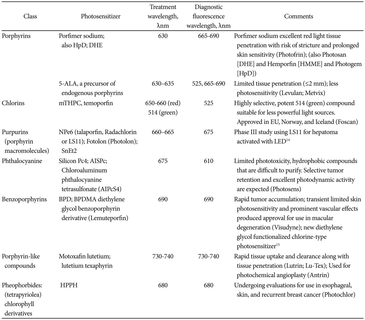
Many agents have been used as photosensitizers in esophageal disease. The initial studies were focused on patients with esophageal cancer and the use of porfimer sodium (Ps) PDT.2,3 Such research led to approval of PDT in the United States, Europe, and Japan. Later, Overholt et al.4,5 published a landmark study on the use of Ps PDT and patients with Barrett's esophagus (BE) with high grade dysplasia (HGD). This trial was a prospective, randomized, controlled, multicenter study and results of the study were published in three major research articles.4,5 The trial showed a lower progression rate to invasive esophageal carcinoma among patients who were treated with PDT. Since then, several studies demonstrated the clinical usefulness of Ps PDT on BE patient with dysplasia and neoplasia which has resulted in the development of other photosensitizers (Table 1).6-15
The initial description of PDT using porphyrin-based agents, hematoporphyrin derivative (HpD) and dihematoporphyrin ether (DHE) in combination with red light, became the predominant forms of PDT.16 After further purification of the photosensetizers, several agents were released: Ps (Photofrin; Pinnacle Biologics Inc., Bannockburn, IL, USA),17 DHE (available in Europe under the trade name Photosan [Seehof Laboratories, Wesselburen, Germany], a second-generation form of HpD available in China under the trade name Hemporfin [hematoporphyrin monomethyl ether; Funda-Zhangjiang Bio-Pharmaceutical, Shanghai, China]).18 Ps has recently undergone pharmacokinetic studies after repeated administration (demonstrating prolonged half-life and delayed excretion after repeated infusion).19 A recent review from Nara, Japan, has also described advances in the research and development of 5-aminolevulinic acid (ALA) for PDT, as well as the use of nonporphyrinoid compounds.20
When reviewing the literature about PDT, one needs to pay attention to three major points of interest. First of those points is the disease status of the study population. The results of a study on patient with BE and low grade dysplasia (LGD) should be interpreted differently from that of patients with HGD or early carcinoma (EC) and squamous dysplasia or carcinoma. Second, one requires a basic understanding of the physics and mechanics of the PDT technique. This includes how different photosensitizers are used, the various light sources, and the routes of administration. Equivalent doses of various agents may result in varying depth of tissue necrosis even if the source of energy is constant.21-25 Similarly, varying the light source, and wavelengths, will result in varying penetration into esophageal tissue. For example, red light (630 nm) seems to penetrate esophageal mucosa deeper than green light (532 nm).
Lastly, one needs to understand the ablative modalities used in a given study. Most regulatory trials allow the use of PDT only. On the other hand, clinical series seem to use additional ablative modalities, like radiofrequency energy or argon plasma coagulation (APC), to ablate residual BE after the initial PDT treatment. Such use, though appropriate,26 may introduce bias into the final conclusions of a study unless clearly accounted for by the authors.
Dr. Norman Barrett initially described BE, intestinal metaplasia of the esophagus, in the 1950s. Since then, the link between BE and esophageal adenocarcinoma has been extensively studied and well established.27,28 The incidence of esophageal cancer is on the rise.29,30 The development of esophageal adenocarcinoma among patients with BE seems to follow an established pathway through LGD, HGD, then adenocarcinoma. Therefore, detecting and treating dysplasia is of critical importance. Yet, dysplasia can be hard to detect in white light endoscopy (WLE). Therefore, a protocol was established to perform four-quadrant biopsy every 1 to 2 cm of BE (Seattle protocol). This, however, is cumbersome, and many clinicians do not adhere to such protocol resulting in many false negative screening. Therefore, many studies have evaluated the use of chromoendoscopy (CE) to detect dysplasia in BE. CE uses various dyes (indigo carmine, methylene blue, acetic acid, etc.) to highlight vascular patterns on surface mucosa. This enables the endoscopist to perform targeted biopsies of suspicious areas and increases the yield for dysplasia. Virtual chromoendoscopy (VE) uses various light sources and lenses on the endoscope in order to highlight the surface vasculature.
Both methods have been extensively studied in patients with BE. We recently assessed the increase in the diagnostic yield in detecting dysplasia using VE and CE compared to WLE with random biopsies in a meta-analysis. The study has been submitted for publication and an abstract form has been submitted to Digestive Disease Week 2013. We found that using either CE or VE increases the yield of detecting dysplasia by 34% (95% confidence interval, 20% to 56%, p=0.0001). This study, among others, highlights the importance of improved endoscopic imaging, which will be required to detect and treat patients with Barrett's dysplasia.
As discussed earlier, PDT offers different tissue penetration depending on the agent and the wavelength of the light source. In the esophagus, PDT offers clear advantages by allowing targeted therapy to the affected area using endoscopy. By controlling the depth of tissue necrosis, one can ensure eradication of Barrett's mucosa while minimizing the risk of esophageal perforation.31
In early clinical studies, surgeons and gastroenterologists used Ps combined with red light for palliation of obstructing lesions, as well as for curative treatment of early cancers. As clinicians became more comfortable with the technique, and with continued improvements in endoscopic light dosimetry, the uses of sodium porfimer PDT expanded to esophageal dysplasia (BE and squamous disease). As a result, Ps has been extensively studied in the United States and North America. The efficacy of this form of PDT in eradicating esophageal dysplasia has been documented in large prospective studies.5,12
Ps, however, is a first generation agent that has some shortcomings. Ps is not specific to mucosa. In addition, it causes prolonged photosensitivity, lasting up to 6 weeks, which can cause significant skin damage if a patient is exposed to light. Therefore, new photosensitizers have been produced to overcome those issues. One of such agents is metatetrahydroxy-phenyl chlorine (mTHPC, temoporfin, Foscan; Biolitec AG, Jena, Germany). The mTHPC is more potent compared to sodium porfimer, requires a smaller dosage and lower light energy. The mTHPC shortens the period of photosensitivity to 2 to 3 weeks. Yet, it seems to be associated with an increased risk of perforation and stricture formation compared to sodium porfimer.32 ALA is another photosensitizer which works on mucosal cells only.33 ALA allows targeted necrosis of mucosal cells only, thus preventing damage to submucosal tissue. In theory, this should lead to decreased risk of perforation and stricture formation.25 A recent trial from University College London sought to randomize 128 patients with Barrett's HGD to PDT using 5-ALA or Ps. Over the study period from 2006 to 2009, 64 patients were actually randomized to PDT with complete response for HGD in 16/34 patients in the ALA group (47%) and 12/30 patients in the Ps group (40%). The overall cancer incidence was 14% (nine/64 patients) that was usually treated with additional PDT or EMR plus chemo-radiation therapy. At a median 2 year follow-up, there was no statistically significant difference in outcome of PDT using either 5-ALA or Ps. A subgroup analysis suggested that patients with BE segments less than 6 cm length had improved outcomes with low risk of stricture or death after 5-ALA PDT.34 Several other agents have been studied as photosensitizers in PDT. Those are summarized in Table 1, after Yano et al.20
Porfimer sodium (Ps)
As mentioned earlier, Ps is the most widely used agent for esophageal PDT. It is administered by intravenous injection. This chemical is a HpD and was first approved in the United States in 1995 for use among patient with advanced esophageal carcinoma.35 The best study on sodium porfimer PDT came from Overholt et al.35 who introduced cylindrical inflatable balloon which is used to administer light to 101 patients with HGD. Patients were then followed up for 4 years showing a resolution of HGD in 54% complete remission of intestinal metaplasia (CRIM). Among patients with LGD, success rate was 93%. The success rate for HGD and cancer were noted to be 78% and 48%. In those patients, stenosis was noted in 30% of all patients. A larger (208 patients), multicenter study by Overholt et al.35 later showed complete eradication of HGD in 77% versus 39% among patients who had PDT and control respectively. This response was still present at 5 years of follow-up.
Since then, several other centers have reported clinical results of PDT. We reported our experience in 102 patients with BE and HGD/EC.11,36-38 After a mean follow-up of 18 months, a single session of Ps PDT showed CRIM in 56% of patients.38 We also reviewed the combined experience of Mayo Clinic in Jacksonville and Rochester. One hundred and forty-two patients with BE and HGD were treated with sodium porfimer PDT. At 19 months of follow-up, CRIM was noted in 50% of patients in Jacksonville compared to 35% in Rochester.39,41 HGD was eliminated in 100% in Jacksonville, and 80% in Rochester. Esophageal strictures were noted in 20% of patients in Jacksonville and 27% of patients in Rochester.
Stricture formation is one of the most common complications post PDT. Panjehpour et al.41,42 looked at the effect of oral steroids post PDT on stricture formation. Sixty patients with BE and HGD were randomized to be treated with either PDT alone or PDT combined with oral prednisone. Surprisingly, the results showed a trend towards more stricture formation in the prednisone group compared (29% vs. 16%); yet this did not reach statistical significance.41,42
ALA
As previously mentioned, ALA is another photosensitizing agent that has been used in PDT. ALA is a pro-drug which stimulates the endogenous production of protoporphyrin IX within the gut mucosa.43,44 ALA compounds have been used in Europe of several years. A commercially available form of ALA, Levulan, was approved by the US Food and Drug Administration.45 ALA is usually activated by red light and was expected to offer several advantages including a shorter photosensitivity period of 1 to 2 days and the preferential targeting of the superficial mucosal layer.46-49
The initial studies on ALA PDT were encouraging.50-53 Most notably, Ackroyd et al.54,55 conducted a double-blinded, randomized, placebo-controlled clinical trial among patients with BE and LGD using ALA PDT. After a mean follow-up of 24 months, complete remission of dysplasia was achieved in 98% of patients.54,55
Pech et al.56 used ALA PDT to treat 35 patients with BE with HGD and reported a very high complete response rate of 97% of patients after mean follow-up of 42 months. Since then, several other studies have been published with similar results.56-59
Despite the encouraging results of those studies, orally-administered ALA was found to be associated with significant side effects including elevated liver enzyme tests, chest pain, neuropathy, and sudden death.60 To avoid such toxicity, Ortner et al.61 administered ALA topically using a spray catheter at the time of endoscopy. This was used to treat seven patient with BE and seven patients with BE and LGD.61 The results of this study showed progression from BE to HGD in one patient and CRIM in only 21% of patients. Hence, this form of using ALA was abandoned. As discussed above, at a median 2 year follow-up, there was no statistically significant difference in outcome of PDT using either 5-ALA or Ps. However, a subgroup analysis suggested that patients with BE segments less than 6 cm length had improved outcomes with low risk of stricture or death after 5-ALA PDT.34
In addition to the above shortcomings, ALA PDT appears to perform poorly when compared to other ablation methods. In a study by Ragunath et al.,62 ALA PDT was compared to APC in 26 patients with BE with dysplasia. The authors found that APC was more effective, safer, and less expensive compared to ALA PDT. Other authors have reproduced these results.54,63,64 Therefore, the correct ALA dose and the optimal light dose have not been established and further studies need to be conducted before this form of PDT can be recommended as first line for Barrett's ablation.
mTHPC
As discussed earlier, mTHPC, a chlorine derivative, is a potent photosensitizer. It has a shorter period of photosensitivity compared to sodium porfimer and requires less light and lower drug doses to produce the effect. The use of mTHPC has been limited due to reported cases of tissue necrosis among patients with head and neck cancer.65 The use of mTHPC in patients with BE has been limited to a small number of studies (Table 2). The optimal light settings and drug dosimetry are not known. Further studies may be needed to optimize treatment parameters for mTHPC PDT in patients with BE.
At its core, PDT works by sensitizing esophageal mucosa to light. Therefore, the method by which light is delivered is of great importance to the success of this therapy. There is no easy way to determine the ideal dosage of light for any given patients. Too much light may lead to tissue necrosis and stricture formation. Too little light means incomplete eradication of dysplasia or BE. In addition to finding the right dosage, one has to overcome the uneven exposure to light caused by esophageal folds. Due to peristalsis and respiratory motion, maintaining central positioning of the light sources can be a challenge.50 The light delivery system is usually composed of a modified diffuser. One way to overcome this was the creation of an elastic catheter balloon.66 This allowed better centering the fiber and more equal distribution of light to the treatment area. However, over-distention of the esophageal wall may result in decreased mucosal perfusion leading to decreasing effectiveness.66,67 In addition to the balloon, another group of researchers used a large diameter rigid light distributor to improve light delivery.68 The use of advanced optical techniques, such as fluorescence spectroscopy or optical coherence tomography, may help assess drug levels at the level of the mucosa and the progression of the photodynamic reaction to improve PDT outcomes.69-71 The development of improved light dosimetry and delivery system will be crucial to the future of PDT and will determine if PDT can remain a viable endoscopic mucosal ablation treatment option.72-74
PDT has been particularly promising in patients with advanced esophageal carcinoma that was unsuitable for surgery, or for persistent/recurrent disease after chemoradiation therapy (CRT). Yano et al.20 performed Ps-PDT as salvage treatment in patients with recurrent disease after CRT with promising results. Subsequently, a phase I/II study is being conducted using PDT with talaporfin sodium in patients with recurrent disease after CRT for esophageal carcinoma in Japan.20 Lindenmann has also reported the salvage or palliative use of Ps PDT to prolong survival in patients with persistent/recurrent disease after CRT for esophageal cancer.75
EMR uses diathermy snare to resect nodular, dysplastic, or neoplastic esophageal lesions. EMR is used for staging of esophageal neoplasia but can also be used for curative intent.76 There are several EMR techniques. The most common includes using the multiband mucosectomy method with or without saline injection lift to remove the most suspicious lesions (DT-6-5F; Cook Medical, Bloomington, IN, USA). This involves placement of a rubber band around the suspicious lesion followed by resection of the area using a snare. For larger lesions, the area is lifted by injection of fluid, which can vary by different endoscopists. A cap is used to suction the mucosa while a snare is used to resect the affected area (K-008; Olympus America Inc., Center Valley, PA, USA). This is repeated multiple times until the whole lesion has been resected.
Several studies have established the utility of EMR, with or without ablation therapy, for treatment of EC and HGD in patients with BE.77-80 When EMR is done on focal lesions, Barrett's mucosa is still present and dysplasia or neoplasia can recur. There are two schools of thought regarding the eradication of BE. The traditional approach has been to use ablation after EMR with the goal of achieving CRIM.
Complete Barrett's eradication is a subset of EMR in which the whole BE mucosa is resected endoscopically. In doing so, there is no need for ablative therapies including PDT. Satodate et al.81 first described this approach as a case report in 2003. Since then, other investigators have studied and advocated for this approach. Using this approach, studies have shown the rate of CRIM to be 86% to 100%. The main limitation for this approach is the high rate of dysphagia due to stenosis (up to 50%). This approach continues to be a viable alternative to patients who do not undergo ablative therapies including PDT.80,82-84
Besides PDT, other ablative therapies are used for this purpose. This includes radiofrequency ablation (RFA), cryotherapy, and APC.85-87 The most commonly used of those is RFA using the HALO system (MARRX Medical Inc., Sunnyvale, CA, USA). RFA can be applied locally or circumferentially. Under standard settings, the depth of penetration is 500 to 1,000 µm. The most important study on RFA came by Shaheen et al.87 This was a multicentered, randomized, controlled clinical trial of 127 patients with Barrett's dysplasia. Patients were randomized to RFA or sham endoscopic therapy in a 2:1 ratio. The final results showed CRIM in 77.4% of RFA group compared to 2.3% in the control group (p<0.0001). Progression to cancer was seen in 1.2% in the RFA group compared to 9.3% in the control group (p<0.05). In addition, RFA has had a low rate of stenosis of less than 3% and perforation seems to occur in less than 0.2% of patients.88,89
For many years, PDT became an important method for mucosal ablation in patients with BE and squamous dysplasia and carcinoma. Ps PDT has been shown to be a safe and reliable treatment method for eliminating dysplasia and preventing the development of esophageal cancer. The technique seems to have the advantage of selectively targeting mucosal layer while minimizing stricturing and perforation. However, significant drawbacks to PDT have caused a decrease in the use of this method compared to RFA. These include the need for IV administration agents and extended periods of photosensitization. Newer photosensitizing agents and more sophisticated light dosimetry will be needed to bring PDT back to mainstream ablative therapy in gastroenterology.
  • 1. Hahn SM, Putt ME, Metz J, et al. Photofrin uptake in the tumor and normal tissues of patients receiving intraperitoneal photodynamic therapy. Clin Cancer Res 2006;12:5464–5470.ArticlePubMed
  • 2. McCaughan JS Jr, Ellison EC, Guy JT, et al. Photodynamic therapy for esophageal malignancy: a prospective twelve-year study. Ann Thorac Surg 1996;62:1005–1009.ArticlePubMed
  • 3. Litle VR, Luketich JD, Christie NA, et al. Photodynamic therapy as palliation for esophageal cancer: experience in 215 patients. Ann Thorac Surg 2003;76:1687–1692.ArticlePubMed
  • 4. Overholt BF, Lightdale CJ, Wang KK, et al. Photodynamic therapy with porfimer sodium for ablation of high-grade dysplasia in Barrett's esophagus: international, partially blinded, randomized phase III trial. Gastrointest Endosc 2005;62:488–498.ArticlePubMed
  • 5. Overholt BF, Wang KK, Burdick JS, et al. Five-year efficacy and safety of photodynamic therapy with Photofrin in Barrett's high-grade dysplasia. Gastrointest Endosc 2007;66:460–468.ArticlePubMed
  • 6. Wolfsen HC. Endoluminal therapy for Barrett's esophagus. Gastrointest Endosc Clin N Am 2007;17:59–82.ArticlePubMed
  • 7. Wolfsen HC. Present status of photodynamic therapy for high-grade dysplasia in Barrett's esophagus. J Clin Gastroenterol 2005;39:189–202.ArticlePubMed
  • 8. Wolfsen HC. Carpe luz-seize the light: endoprevention of esophageal adenocarcinoma when using photodynamic therapy with porfimer sodium. Gastrointest Endosc 2005;62:499–503.ArticlePubMed
  • 9. Wolfsen HC. Endoprevention of esophageal cancer: endoscopic ablation of Barrett's metaplasia and dysplasia. Expert Rev Med Devices 2005;2:713–723.ArticlePubMed
  • 10. Prosst RL, Wolfsen HC, Gahlen J. Photodynamic therapy for esophageal diseases: a clinical update. Endoscopy 2003;35:1059–1068.ArticlePubMed
  • 11. Wang KK. Current status of photodynamic therapy of Barrett's esophagus. Gastrointest Endosc 1999;49(3 Pt 2):S20–S23.ArticlePubMed
  • 12. Overholt BF, Panjehpour M, Halberg DL. Photodynamic therapy for Barrett's esophagus with dysplasia and/or early stage carcinoma: long-term results. Gastrointest Endosc 2003;58:183–188.ArticlePubMed
  • 13. Sampliner RE. Prevention of adenocarcinoma by reversing Barrett's esophagus with mucosal ablation. World J Surg 2003;27:1026–1029.ArticlePubMed
  • 14. Winship MJ, Wang S, Chen JC, Keltner L, Christophersen JS. Treatment of colorectal liver metastases using talaporfin sodium and intratumoral placement of LEDs [abstract]. J Clin Oncol 2005;23(16 Suppl):3663.ArticlePubMed
  • 15. Konan-Kouakou YN, Boch R, Gurny R, Allémann E. In vitro and in vivo activities of verteporfin-loaded nanoparticles. J Control Release 2005;103:83–91.ArticlePubMed
  • 16. Dougherty TJ. An update on photodynamic therapy applications. J Clin Laser Med Surg 2002;20:3–7.ArticlePubMed
  • 17. Allison SP, Lobo DN. Fluid and electrolytes in the elderly. Curr Opin Clin Nutr Metab Care 2004;7:27–33.ArticlePubMed
  • 18. Qiang YG, Zhang XP, Li J, Huang Z. Photodynamic therapy for malignant and non-malignant diseases: clinical investigation and application. Chin Med J (Engl) 2006;119:845–857.ArticlePubMed
  • 19. Pereira SP, Ayaru L, Ackroyd R, et al. The pharmacokinetics and safety of porfimer after repeated administration 30-45 days apart to patients undergoing photodynamic therapy. Aliment Pharmacol Ther 2010;32:821–827.ArticlePubMedPMC
  • 20. Yano S, Hirohara S, Obata M, et al. Current states and future views in photodynamic therapy. J Photochem Photobiol C 2011;12:46–67.Article
  • 21. Perry Y, Epperly MW, Fernando HC, et al. Photodynamic therapy induced esophageal stricture: an animal model: from mouse to pig. J Surg Res 2005;123:67–74.ArticlePubMed
  • 22. Cramers P, Ruevekamp M, Oppelaar H, Dalesio O, Baas P, Stewart FA. Foscan uptake and tissue distribution in relation to photodynamic efficacy. Br J Cancer 2003;88:283–290.ArticlePubMedPMCPDF
  • 23. Wagnieres G, Hadjur C, Grosjean P, et al. Clinical evaluation of the cutaneous phototoxicity of 5,10,15,20-tetra(m-hydroxyphenyl)chlorin. Photochem Photobiol 1998;68:382–387.ArticlePubMed
  • 24. Barr H, Kendall C, Stone N. Photodynamic therapy for esophageal cancer: a useful and realistic option. Technol Cancer Res Treat 2003;2:65–76.ArticlePubMed
  • 25. Bown SG, Rogowska AZ. New photosensitizers for photodynamic therapy in gastroenterology. Can J Gastroenterol 1999;13:389–392.ArticlePubMedPDF
  • 26. Paskowitz DM, Donohue-Rolfe KM, Yang H, et al. Neurotrophic factors minimize the retinal toxicity of verteporfin photodynamic therapy. Invest Ophthalmol Vis Sci 2007;48:430–437.ArticlePubMed
  • 27. Montgomery E, Bronner MP, Goldblum JR, et al. Reproducibility of the diagnosis of dysplasia in Barrett esophagus: a reaffirmation. Hum Pathol 2001;32:368–378.ArticlePubMed
  • 28. Zaninotto G, Minnei F, Guirroli E, et al. The Veneto region's Barrett's oesophagus registry: aims, methods, preliminary results. Dig Liver Dis 2007;39:18–25.ArticlePubMed
  • 29. Falk GW. Barrett's esophagus. Gastroenterology 2002;122:1569–1591.ArticlePubMed
  • 30. Devesa SS, Blot WJ, Fraumeni JF Jr. Changing patterns in the incidence of esophageal and gastric carcinoma in the United States. Cancer 1998;83:2049–2053.ArticlePubMed
  • 31. Tokar JL, Haluszka O, Weinberg DS. Endoscopic therapy of dysplasia and early-stage cancers of the esophagus. Semin Radiat Oncol 2007;17:10–21.ArticlePubMed
  • 32. Andrejevic Blant S, Grosjean P, Ballini JP, et al. Localization of tetra(m-hydroxyphenyl) chlorin (Foscan) in human healthy tissues and squamous cell carcinomas of the upper aero-digestive tract, the esophagus and the bronchi: a fluorescence microscopy study. J Photochem Photobiol B 2001;61:1–9.ArticlePubMed
  • 33. Ackroyd R, Kelty C, Brown N, Reed M. The history of photodetection and photodynamic therapy. Photochem Photobiol 2001;74:656–669.ArticlePubMed
  • 34. Dunn JM, Mackenzie GD, Banks MR. A randomised controlled trial of ALA vs. Photofrin photodynamic therapy for high-grade dysplasia arising in Barrett's oesophagus. Lasers Med Sci; Epub 2012 Jun 15. DOI: http://dx.doi.org/10.1007/s10103-012-1132-1
  • 35. Overholt B, Panjehpour M, Tefftellar E, Rose M. Photodynamic therapy for treatment of early adenocarcinoma in Barrett's esophagus. Gastrointest Endosc 1993;39:73–76.ArticlePubMed
  • 36. Wang KK, Kim JY. Photodynamic therapy in Barrett's esophagus. Gastrointest Endosc Clin N Am 2003;13:483–489.ArticlePubMed
  • 37. Wolfsen HC, Woodward TA, Raimondo M. Photodynamic therapy for dysplastic Barrett esophagus and early esophageal adenocarcinoma. Mayo Clin Proc 2002;77:1176–1181.ArticlePubMed
  • 38. Wolfsen HC, Hemminger LL, Wallace MB, Devault KR. Clinical experience of patients undergoing photodynamic therapy for Barrett's dysplasia or cancer. Aliment Pharmacol Ther 2004;20:1125–1131.ArticlePubMed
  • 39. Wang KK, Wong Kee, Buttar N, Papenfuss S, Lutzke L. Barrett's esophagus after photodynamic therapy: risk of cancer development during long term follow-up [abstract]. Gastroenterology 2004;126(5 Suppl 2):A50.Article
  • 40. Wolfsen HC, Hemminger LL. Photodynamic therapy for dysplastic Barrett's esophagus and mucosal adenocarcinoma [abstract]. Gastrointest Endosc 2004;59:AB251.Article
  • 41. Panjehpour M, Overholt BF, Haydek JM, Lee SG. Results of photodynamic therapy for ablation of dysplasia and early cancer in Barrett's esophagus and effect of oral steroids on stricture formation. Am J Gastroenterol 2000;95:2177–2184.ArticlePubMed
  • 42. Panjehpour M, Overholt BF, Phan MN, Haydek JM. Optimization of light dosimetry for photodynamic therapy of Barrett's esophagus: efficacy vs. incidence of stricture after treatment. Gastrointest Endosc 2005;61:13–18.ArticlePubMed
  • 43. Peng Q, Warloe T, Moan J, et al. Antitumor effect of 5-aminolevulinic acid-mediated photodynamic therapy can be enhanced by the use of a low dose of photofrin in human tumor xenografts. Cancer Res 2001;61:5824–5832.PubMed
  • 44. Peng Q, Warloe T, Berg K, et al. 5-Aminolevulinic acid-based photodynamic therapy. Clinical research and future challenges. Cancer 1997;79:2282–2308.ArticlePubMed
  • 45. Godal A, Nilsen NO, Klaveness J, Branden JE, Nesland JM, Peng Q. New derivatives of 5-aminolevulinic acid for photodynamic therapy: chemical synthesis and porphyrin production in in vitro and in vivo biological systems. J Environ Pathol Toxicol Oncol 2006;25:109–126.ArticlePubMed
  • 46. Bedwell J, MacRobert AJ, Phillips D, Bown SG. Fluorescence distribution and photodynamic effect of ALA-induced PP IX in the DMH rat colonic tumour model. Br J Cancer 1992;65:818–824.ArticlePubMedPMCPDF
  • 47. Kennedy JC, Pottier RH. Endogenous protoporphyrin IX, a clinically useful photosensitizer for photodynamic therapy. J Photochem Photobiol B 1992;14:275–292.ArticlePubMed
  • 48. Peng Q, Berg K, Moan J, Kongshaug M, Nesland JM. 5-Aminolevulinic acid-based photodynamic therapy: principles and experimental research. Photochem Photobiol 1997;65:235–251.ArticlePubMed
  • 49. Webber J, Kessel D, Fromm D. Side effects and photosensitization of human tissues after aminolevulinic acid. J Surg Res 1997;68:31–37.ArticlePubMed
  • 50. Gossner L, May A, Sroka R, Ell C. A new long-range through-the-scope balloon applicator for photodynamic therapy in the esophagus and cardia. Endoscopy 1999;31:370–376.ArticlePubMedPDF
  • 51. Gossner L, Stolte M, Sroka R, et al. Photodynamic ablation of high-grade dysplasia and early cancer in Barrett's esophagus by means of 5-aminolevulinic acid. Gastroenterology 1998;114:448–455.ArticlePubMed
  • 52. Barr H, Shepherd NA, Dix A, Roberts DJ, Tan WC, Krasner N. Eradication of high-grade dysplasia in columnar-lined (Barrett's) oesophagus by photodynamic therapy with endogenously generated protoporphyrin IX. Lancet 1996;348:584–585.ArticlePubMed
  • 53. Orth K, Stanescu A, Rück A, Russ D, Beger HG. Photodynamic ablation and argon-plasma coagulation of premalignant and early-stage malignant lesions of the oesophagus: an alternative to surgery? Chirurg 1999;70:431–438.ArticlePubMedPDF
  • 54. Ackroyd R, Brown NJ, Davis MF, et al. Photodynamic therapy for dysplastic Barrett's oesophagus: a prospective, double blind, randomised, placebo controlled trial. Gut 2000;47:612–617.ArticlePubMedPMC
  • 55. Ackroyd R, Brown NJ, Davis MF, Stephenson TJ, Stoddard CJ, Reed MW. Aminolevulinic acid-induced photodynamic therapy: safe and effective ablation of dysplasia in Barrett's esophagus. Dis Esophagus 2000;13:18–22.ArticlePubMedPDF
  • 56. Pech O, Gossner L, May AD, Stolte M, Ell C. Long term results of PDT for early neoplasia in Barrett's esophagus [abstract]. Gastrointest Endosc 2004;59:P257.Article
  • 57. Guelrud M, Herrera I, Essenfeld H, Castro J. Enhanced magnification endoscopy: a new technique to identify specialized intestinal metaplasia in Barrett's esophagus. Gastrointest Endosc 2001;53:559–565.ArticlePubMed
  • 58. Behrens A, May A, Gossner L, et al. Curative treatment for high-grade intraepithelial neoplasia in Barrett's esophagus. Endoscopy 2005;37:999–1005.ArticlePubMedPDF
  • 59. May A, Gossner L, Pech O, et al. Intraepithelial high-grade neoplasia and early adenocarcinoma in short-segment Barrett's esophagus (SSBE): curative treatment using local endoscopic treatment techniques. Endoscopy 2002;34:604–610.ArticlePubMedPDF
  • 60. Sylantiev C, Schoenfeld N, Mamet R, Groozman GB, Drory VE. Acute neuropathy mimicking porphyria induced by aminolevulinic acid during photodynamic therapy. Muscle Nerve 2005;31:390–393.ArticlePubMed
  • 61. Ortner MA, Zumbusch K, Liebetruth J, et al. Is topical delta-aminolevulinic acid adequate for photodynamic therapy in Barrett's esophagus? A pilot study. Endoscopy 2002;34:611–616.ArticlePubMedPDF
  • 62. Ragunath K, Krasner N, Raman VS, Haqqani MT, Phillips CJ, Cheung I. Endoscopic ablation of dysplastic Barrett's oesophagus comparing argon plasma coagulation and photodynamic therapy: a randomized prospective trial assessing efficacy and cost-effectiveness. Scand J Gastroenterol 2005;40:750–758.ArticlePubMed
  • 63. Kelty CJ, Ackroyd R, Brown NJ, Brown SB, Reed MW. Comparison of high- vs low-dose 5-aminolevulinic acid for photodynamic therapy of Barrett's esophagus. Surg Endosc 2004;18:452–458.ArticlePubMed
  • 64. Kelty CJ, Ackroyd R, Brown NJ, Stephenson TJ, Stoddard CJ, Reed MW. Endoscopic ablation of Barrett's oesophagus: a randomized-controlled trial of photodynamic therapy vs. argon plasma coagulation. Aliment Pharmacol Ther 2004;20:1289–1296.ArticlePubMed
  • 65. Andrejevic-Blant S, Hadjur C, Ballini JP, et al. Photodynamic therapy of early squamous cell carcinoma with tetra(m-hydroxyphenyl)chlorin: optimal drug-light interval. Br J Cancer 1997;76:1021–1028.ArticlePubMedPMCPDF
  • 66. van den Bergh H. On the evolution of some endoscopic light delivery systems for photodynamic therapy. Endoscopy 1998;30:392–407.ArticlePubMedPDF
  • 67. Overholt BF, Panjehpour M, DeNovo RC, Peterson MG, Jenkins C. Balloon photodynamic therapy of esophageal cancer: effect of increasing balloon size. Lasers Surg Med 1996;18:248–252.ArticlePubMed
  • 68. Stepinac T, Grosjean P, Woodtli A, Monnier P, van den Bergh H, Wagnieres G. Optimization of the diameter of a radial irradiation device for photodynamic therapy in the esophagus. Endoscopy 2002;34:411–415.ArticlePubMedPDF
  • 69. Braichotte DR, Savary JF, Monnier P, van den Bergh HE. Optimizing light dosimetry in photodynamic therapy of early stage carcinomas of the esophagus using fluorescence spectroscopy. Lasers Surg Med 1996;19:340–346.ArticlePubMed
  • 70. Zellweger M, Grosjean P, Monnier P, van den Bergh H, Wagnières G. Stability of the fluorescence measurement of Foscan in the normal human oral cavity as an indicator of its content in early cancers of the esophagus and the bronchi. Photochem Photobiol 1999;69:605–610.ArticlePubMed
  • 71. Standish BA, Yang VX, Munce NR, et al. Doppler optical coherence tomography monitoring of microvascular tissue response during photodynamic therapy in an animal model of Barrett's esophagus. Gastrointest Endosc 2007;66:326–333.ArticlePubMed
  • 72. Boere IA, Robinson DJ, de Bruijn HS, et al. Monitoring in situ dosimetry and protoporphyrin IX fluorescence photobleaching in the normal rat esophagus during 5-aminolevulinic acid photodynamic therapy. Photochem Photobiol 2003;78:271–277.ArticlePubMed
  • 73. Cheung R, Solonenko M, Busch TM, et al. Correlation of in vivo photosensitizer fluorescence and photodynamic-therapy-induced depth of necrosis in a murine tumor model. J Biomed Opt 2003;8:248–252.ArticlePubMed
  • 74. Radu A, Conde R, Fontolliet C, Wagnieres G, Van den Bergh H, Monnier P. Mucosal ablation with photodynamic therapy in the esophagus: optimization of light dosimetry in the sheep model. Gastrointest Endosc 2003;57:897–905.ArticlePubMed
  • 75. Lindenmann J, Matzi V, Neuboeck N, et al. Individualized, multimodal palliative treatment of inoperable esophageal cancer: clinical impact of photodynamic therapy resulting in prolonged survival. Lasers Surg Med 2012;44:189–198.ArticlePubMed
  • 76. Namasivayam V, Wang KK, Prasad GA. Endoscopic mucosal resection in the management of esophageal neoplasia: current status and future directions. Clin Gastroenterol Hepatol 2010;8:743–754.ArticlePubMedPMC
  • 77. Ell C, May A, Gossner L, et al. Endoscopic mucosal resection of early cancer and high-grade dysplasia in Barrett's esophagus. Gastroenterology 2000;118:670–677.ArticlePubMed
  • 78. Pech O, Bollschweiler E, Manner H, Leers J, Ell C, Hölscher AH. Comparison between endoscopic and surgical resection of mucosal esophageal adenocarcinoma in Barrett's esophagus at two high-volume centers. Ann Surg 2011;254:67–72.ArticlePubMed
  • 79. Moss A, Bourke MJ, Hourigan LF, et al. Endoscopic resection for Barrett's high-grade dysplasia and early esophageal adenocarcinoma: an essential staging procedure with long-term therapeutic benefit. Am J Gastroenterol 2010;105:1276–1283.ArticlePubMedPDF
  • 80. Larghi A, Lightdale CJ, Ross AS, et al. Long-term follow-up of complete Barrett's eradication endoscopic mucosal resection (CBE-EMR) for the treatment of high grade dysplasia and intramucosal carcinoma. Endoscopy 2007;39:1086–1091.ArticlePubMed
  • 81. Satodate H, Inoue H, Yoshida T, et al. Circumferential EMR of carcinoma arising in Barrett's esophagus: case report. Gastrointest Endosc 2003;58:288–292.ArticlePubMed
  • 82. Chung A, Bourke MJ, Hourigan LF, et al. Complete Barrett's excision by stepwise endoscopic resection in short-segment disease: long term outcomes and predictors of stricture. Endoscopy 2011;43:1025–1032.ArticlePubMedPDF
  • 83. Chennat J, Konda VJ, Ross AS, et al. Complete Barrett's eradication endoscopic mucosal resection: an effective treatment modality for high-grade dysplasia and intramucosal carcinoma: an American single-center experience. Am J Gastroenterol 2009;104:2684–2692.ArticlePubMedPDF
  • 84. Peters FP, Krishnadath KK, Rygiel AM, et al. Stepwise radical endoscopic resection of the complete Barrett's esophagus with early neoplasia successfully eradicates pre-existing genetic abnormalities. Am J Gastroenterol 2007;102:1853–1861.ArticlePubMed
  • 85. Ell C, May A, Pech O, et al. Curative endoscopic resection of early esophageal adenocarcinomas (Barrett's cancer). Gastrointest Endosc 2007;65:3–10.ArticlePubMed
  • 86. Sampliner RE. Endoscopic therapy for Barrett's esophagus. Clin Gastroenterol Hepatol 2009;7:716–720.ArticlePubMed
  • 87. Shaheen NJ, Overholt BF, Sampliner RE, et al. Durability of radiofrequency ablation in Barrett's esophagus with dysplasia. Gastroenterology 2011;141:460–468.ArticlePubMedPMC
  • 88. Fleischer DE, Overholt BF, Sharma VK, et al. Endoscopic radiofrequency ablation for Barrett's esophagus: 5-year outcomes from a prospective multicenter trial. Endoscopy 2010;42:781–789.ArticlePubMed
  • 89. Lyday WD, Corbett FS, Kuperman DA, et al. Radiofrequency ablation of Barrett's esophagus: outcomes of 429 patients from a multicenter community practice registry. Endoscopy 2010;42:272–278.ArticlePubMed
Table 1
Photosensitizers for Esophageal Photodynamic Therapy
ce-46-30-i001.jpg

HpD, hematoporphyrin derivative; DHE, dihematoporphyrin ether; HMME, hematoporphyrin monomethyl ether; ALA, aminolevulinic acid; mTHPC, meso-tetrahydroxyphenyl chlorine; NPe6, mono-L-aspartyl chlorine e6; SnEt2, tin-etio-purpurin; LED, light emitting diode; Pc4, phthalocyanine; AISPc, aluminum disulphonated phthalocyanine; AIPcS4, aluminum phthalocyanine tetrasulfonate; BPD, benzoporphyrin derivative; BPDMA, benzoporphyrin derivative monoacid; HPPH, 2-[1-hexyloxyethyl]2-devinyl-pyropheophorbide-a.

Table 2
Studies of Meta-Tetrahydroxy-Phenyl Chlorine Photodynamic Therapy in Patients with BE
ce-46-30-i002.jpg

BE, Barrett's esophagus; HGD, high grade dysplasia; AC, adenocarcinoma.

Figure & Data

REFERENCES

    Citations

    Citations to this article as recorded by  
    • Localized Photodynamic Therapy Using a Chlorin e6-Embedded Silicone-Covered Self-Expandable Metallic Stent as a Palliative Treatment for Malignant Esophageal Strictures
      Dae Sung Ryu, Ji Won Kim, Hyeonseung Lee, Seong Jin Eo, Song Hee Kim, Jin Hee Noh, Yuri Kim, Seokin Kang, Kun Na, Jung-Hoon Park, Do Hoon Kim
      ACS Biomaterials Science & Engineering.2024; 10(3): 1869.     CrossRef
    • Patients' values and preferences regarding endoscopic therapy for Barrett's esophagus: A cross‐sectional survey study
      Bashar J. Qumseya, David Estores, Venkata Gorripati, Nanlog Liu, Amira Qumseya
      DEN Open.2024;[Epub]     CrossRef
    • Multifunctional Ablative Gastrointestinal Imaging Capsule (MAGIC) for Esophagus Surveillance and Interventions
      Hyeon-Cheol Park, Dawei Li, Rongguang Liang, Gina Adrales, Xingde Li
      BME Frontiers.2024;[Epub]     CrossRef
    • Polyamine Derived Photosensitizer: A Novel Approach for Photodynamic Therapy of Cancer
      Hao Deng, Ke Xie, Liling Hu, Xiaowen Liu, Qingyun Li, Donghui Xie, Fengyi Xiang, Wei Liu, Weihong Zheng, Shuzhang Xiao, Jun Zheng, Xiao Tan
      Molecules.2024; 29(17): 4277.     CrossRef
    • Applications of Microwaves in Medicine
      J.-C. Chiao, Changzhi Li, Jenshan Lin, Robert H. Caverly, James C. M. Hwang, Harel Rosen, Arye Rosen
      IEEE Journal of Microwaves.2023; 3(1): 134.     CrossRef
    • Current Status and Future Prospects for Esophageal Cancer
      Mahdi Sheikh, Gholamreza Roshandel, Valerie McCormack, Reza Malekzadeh
      Cancers.2023; 15(3): 765.     CrossRef
    • A View on Drug Development for Cancer Prevention
      Andrew R. Reynolds, Michele Moschetta, Alula R. Yohannes, Farzana Walcott, Marianne Ashford, Zoltan Szucs, Tina Sarbajna, James Hadfield, Eden Harrison, Benjamin G. Challis, Amaya Gasco Hernandez, Gaia Schiavon, Caroline Germa, Gyorgy Zorenyi, Barry Crean
      Cancer Discovery.2023; 13(5): 1058.     CrossRef
    • Management of Long-Segment Barrett's Esophagus
      Sarah E. Kim, Francisco Schlottmann, Mario A. Masrur
      Journal of Laparoendoscopic & Advanced Surgical Techniques.2023; 33(12): 1201.     CrossRef
    • Recent developments in photodynamic therapy and its application against multidrug resistant cancers
      Debalina Bhattacharya, Mainak Mukhopadhyay, Kumar Shivam, Satyajit Tripathy, Ranjan Patra, Arindam Pramanik
      Biomedical Materials.2023; 18(6): 062005.     CrossRef
    • Endoscope Capsules: The Present Situation and Future Outlooks
      Rodrigo Gounella, Talita Conte Granado, Oswaldo Hideo Ando Junior, Daniel Luís Luporini, Mario Gazziro, João Paulo Carmo
      Bioengineering.2023; 10(12): 1347.     CrossRef
    • Photodynamic Therapy and Immunological View in Gastrointestinal Tumors
      David Aebisher, Paweł Woźnicki, Klaudia Dynarowicz, Aleksandra Kawczyk-Krupka, Grzegorz Cieślar, Dorota Bartusik-Aebisher
      Cancers.2023; 16(1): 66.     CrossRef
    • Synthesis of N-aryl-2,6-diphenyl-2H-pyrazolo[4,3-c]pyridin-7-amines and their photodynamic properties in the human skin melanoma cell line G361
      Beatričė Razmienė, Veronika Vojáčková, Eva Řezníčková, Lukáš Malina, Vaida Dambrauskienė, Martin Kubala, Robert Bajgar, Hana Kolářová, Asta Žukauskaitė, Eglė Arbačiauskienė, Algirdas Šačkus, Vladimír Kryštof
      Bioorganic Chemistry.2022; 119: 105570.     CrossRef
    • Some Natural Photosensitizers and Their Medicinal Properties for Use in Photodynamic Therapy
      Tomasz Piotr Kubrak, Przemysław Kołodziej, Jan Sawicki, Anna Mazur, Katarzyna Koziorowska, David Aebisher
      Molecules.2022; 27(4): 1192.     CrossRef
    • The emerging role of photoacoustic imaging in clinical oncology
      Li Lin, Lihong V. Wang
      Nature Reviews Clinical Oncology.2022; 19(6): 365.     CrossRef
    • Singlet Oxygen, Photodynamic Therapy, and Mechanisms of Cancer Cell Death
      Prabal Singh Maharjan, Hitesh Kumar Bhattarai, Zhen-Jian Zhuo
      Journal of Oncology.2022; 2022: 1.     CrossRef
    • Endoscopic Management of Esophageal Cancer
      Christopher Paiji, Alireza Sedarat
      Cancers.2022; 14(15): 3583.     CrossRef
    • Oxygen‐Generated Hierarchical‐Structured AuNRs@MnO2@SiO2 Nanocarrier for Enhanced NIR‐ and H2O2‐Responsive Mild‐Hyperthermia Photodynamic/Photothermal Combined Tumor Therapy
      Wen‐Jie Zheng, Ze‐Lei Zhou, Qi‐Yan Lv, Xiaojie Song, Wen‐Xing Zhang, Hui‐Fang Cui
      Advanced Therapeutics.2022;[Epub]     CrossRef
    • Advancements in photodynamic therapy of esophageal cancer
      Dorota Bartusik-Aebisher, Michał Osuchowski, Marta Adamczyk, Joanna Stopa, Grzegorz Cieślar, Aleksandra Kawczyk-Krupka, David Aebisher
      Frontiers in Oncology.2022;[Epub]     CrossRef
    • Barrett's esophagus: What do we need to know?
      Nabeeha Mohy-ud-din, Timothy S. Krill, Aun Raza Shah, Ahmed T. Chatila, Shailendra Singh, Mohammad Bilal, Sreeram Parupudi
      Disease-a-Month.2020; 66(1): 100850.     CrossRef
    • Evolving role of biomaterials in diagnostic and therapeutic radiation oncology
      Siyu Shi, Ravi Vissapragada, Joseph Abi Jaoude, Caroline Huang, Anmol Mittal, Elisa Liu, Jim Zhong, Vivek Kumar
      Bioactive Materials.2020; 5(2): 233.     CrossRef
    • Light Sources and Dosimetry Techniques for Photodynamic Therapy
      Michele M. Kim, Arash Darafsheh
      Photochemistry and Photobiology.2020; 96(2): 280.     CrossRef
    • Recent advances in understanding prodrug transport through the SLC15 family of proton-coupled transporters
      Gurdeep S. Minhas, Simon Newstead
      Biochemical Society Transactions.2020; 48(2): 337.     CrossRef
    • Radiodynamic therapy using 15-MV radiation combined with 5-aminolevulinic acid and carbamide peroxide for prostate cancer in vivo
      Joseph V Panetta, Dusica Cvetkovic, Xiaoming Chen, Lili Chen, C-M Charlie Ma
      Physics in Medicine & Biology.2020; 65(16): 165008.     CrossRef
    • Clinical development and potential of photothermal and photodynamic therapies for cancer
      Xingshu Li, Jonathan F. Lovell, Juyoung Yoon, Xiaoyuan Chen
      Nature Reviews Clinical Oncology.2020; 17(11): 657.     CrossRef
    • A nucleus-directed bombesin derivative for targeted delivery of metallodrugs to cancer cells
      Sílvia Barrabés, Iteng Ng-Choi, María Ángeles Martínez, Blanca R. Manzano, Félix A. Jalón, Gustavo Espino, Lidia Feliu, Marta Planas, Rafael de Llorens, Anna Massaguer
      Journal of Inorganic Biochemistry.2020; 212: 111214.     CrossRef
    • Applications of Minimally Invasive Nanomedicine-Based Therapies in 3D in vitro Cancer Platforms
      Layla Mohammad-Hadi, Marym Mohammad-Hadi
      Synthesis Lectures on Materials and Optics.2020; 1(5): 1.     CrossRef
    • Challenges in Endoscopic Therapy of Dysplastic Barrett’s Esophagus
      Aurada Cholapranee, Arvind J Trindade
      Current Treatment Options in Gastroenterology.2019; 17(1): 32.     CrossRef
    • A Basic Study of Photodynamic Therapy with Glucose-Conjugated Chlorin e6 Using Mammary Carcinoma Xenografts
      Tomohiro Osaki, Shota Hibino, Inoru Yokoe, Hiroaki Yamaguchi, Akihiro Nomoto, Shigenobu Yano, Yuji Mikata, Mamoru Tanaka, Hiromi Kataoka, Yoshiharu Okamoto
      Cancers.2019; 11(5): 636.     CrossRef
    • Ring-Fused Diphenylchlorins as Potent Photosensitizers for Photodynamic Therapy Applications: In Vitro Tumor Cell Biology and in Vivo Chick Embryo Chorioallantoic Membrane Studies
      Bruno F. O. Nascimento, Mafalda Laranjo, Nelson A. M. Pereira, João Dias-Ferreira, Marta Piñeiro, Maria Filomena Botelho, Teresa M. V. D. Pinho e Melo
      ACS Omega.2019; 4(17): 17244.     CrossRef
    • Photodynamic therapy using pheophorbide and 670 nm LEDs exhibits anti-cancer effects in-vitro in androgen dependent prostate cancer
      Taher Gheewala, Troy Skwor, Gnanasekar Munirathinam
      Photodiagnosis and Photodynamic Therapy.2018; 21: 130.     CrossRef
    • Photodynamic therapy in 3D cancer models and the utilisation of nanodelivery systems
      Layla Mohammad-Hadi, Alexander J. MacRobert, Marilena Loizidou, Elnaz Yaghini
      Nanoscale.2018; 10(4): 1570.     CrossRef
    • Evidence of photoinduced lipid hydroperoxidation in Langmuir monolayers containing Eosin Y
      Lucas S.A. Pereira, Sabrina A. Camacho, Antonio A. Malfatti-Gasperini, Karen Jochelavicius, Thatyane M. Nobre, Osvaldo N. Oliveira, Pedro H.B. Aoki
      Colloids and Surfaces B: Biointerfaces.2018; 171: 682.     CrossRef
    • Preclinical Study of Antineoplastic Sinoporphyrin Sodium-PDT via In Vitro and In Vivo Models
      Rui Shi, Chao Li, Zhihuan Jiang, Wanfang Li, Aiping Wang, Jinfeng Wei
      Molecules.2017; 22(1): 112.     CrossRef
    • Novel zinc phthalocyanine as a promising photosensitizer for photodynamic treatment of esophageal cancer
      Weronika Kuzyniak, Jacob Schmidt, Wojciech Glac, Janine Berkholz, Gustav Steinemann, Björn Hoffmann, Eugeny A. Ermilov, Ayşe Gül Gürek, Vefa Ahsen, Bianca Nitzsche, Michael Höpfner
      International Journal of Oncology.2017; 50(3): 953.     CrossRef
    • Photosensitizers for Two‐Photon Excited Photodynamic Therapy
      Zhiyuan Sun, Li‐Peng Zhang, Feipeng Wu, Yuxia Zhao
      Advanced Functional Materials.2017;[Epub]     CrossRef
    • The effect of aloe-emodin-induced photodynamic activity on the apoptosis of human gastric cancer cells: A pilot study
      Hai-Dan Lin, Kai-Ting Li, Qin-Qin Duan, Qing Chen, Shi Tian, Ellie Shihng Meir Chu, Ding-Qun Bai
      Oncology Letters.2017; 13(5): 3431.     CrossRef
    • Photodynamic Therapy with Aminolevulinic Acid: a novel therapeutic Target in the Management of Gastrointestinal Malignancies
      Emel Canbay
      Journal of Cancer Prevention & Current Research.2017;[Epub]     CrossRef
    • Photosensitizers in prostate cancer therapy
      Taher Gheewala, Troy Skwor, Gnanasekar Munirathinam
      Oncotarget.2017; 8(18): 30524.     CrossRef
    • The effect of aloe emodin–encapsulated nanoliposome‐mediated r‐caspase‐3 gene transfection and photodynamic therapy on human gastric cancer cells
      Kai‐Ting Li, Qin‐Qin Duan, Qing Chen, Juan‐Wen He, Si Tian, Hai‐Dan Lin, Qing Gao, Ding‐Qun Bai
      Cancer Medicine.2016; 5(2): 361.     CrossRef
    • Red Blood Cell‐Facilitated Photodynamic Therapy for Cancer Treatment
      Wei Tang, Zipeng Zhen, Mengzhe Wang, Hui Wang, Yen‐Jun Chuang, Weizhong Zhang, Geoffrey D Wang, Trever Todd, Taku Cowger, Hongmin Chen, Lin Liu, Zibo Li, Jin Xie
      Advanced Functional Materials.2016; 26(11): 1757.     CrossRef
    • Nonthermal Ablation by Using Intravascular Oxygen Radical Generation with WST11: Dynamic Tissue Effects and Implications for Focal Therapy
      Simon Y. Kimm, Tatum V. Tarin, Sébastien Monette, Govindarajan Srimathveeravalli, Daniel Gerber, Jeremy C. Durack, Stephen B. Solomon, Peter T. Scardino, Avigdor Scherz, Jonathan Coleman
      Radiology.2016; 281(1): 109.     CrossRef
    • Treatment Effects of WST11 Vascular Targeted Photodynamic Therapy for Urothelial Cell Carcinoma in Swine
      Katie S. Murray, Ashley G. Winter, Renato Beluco Corradi, Stephen LaRosa, Sylvia Jebiwott, Alexander Somma, Haruyuki Takaki, Govindarajan Srimathveeravalli, Michelle Lepherd, Sebastien Monette, Kwanghee Kim, Avigdor Scherz, Jonathan A. Coleman
      Journal of Urology.2016; 196(1): 236.     CrossRef
    • The potential of photodynamic therapy (PDT)—Experimental investigations and clinical use
      Anna Oniszczuk, Karolina A. Wojtunik-Kulesza, Tomasz Oniszczuk, Kamila Kasprzak
      Biomedicine & Pharmacotherapy.2016; 83: 912.     CrossRef
    • Safety evaluation of repeated intravenous infusion of sinoporphyrin with and without PDT in rats
      Rui Shi, Xiaoqi Lin, Jingxuan Zhang, Hongtao Jin, Aiping Wanga, Jinfeng Wei
      Photochemical & Photobiological Sciences.2016; 15(11): 1366.     CrossRef
    • Flexible synthesis of cationic peptide–porphyrin derivatives for light-triggered drug delivery and photodynamic therapy
      R. Dondi, E. Yaghini, K. M. Tewari, L. Wang, F. Giuntini, M. Loizidou, A. J. MacRobert, I. M. Eggleston
      Organic & Biomolecular Chemistry.2016; 14(48): 11488.     CrossRef
    • Inhibition of hypoxia inducible factor 1 and topoisomerase with acriflavine sensitizes perihilar cholangiocarcinomas to photodynamic therapy
      Ruud Weijer, Mans Broekgaarden, Massis Krekorian, Lindy K. Alles, Albert C. van Wijk, Claire Mackaaij, Joanne Verheij, Allard C. van der Wal, Thomas M. van Gulik, Gert Storm, Michal Heger
      Oncotarget.2016; 7(3): 3341.     CrossRef
    • Interventional Therapy of Esophageal Cancer
      Aiwu Mao
      Gastrointestinal Tumors.2016; 3(2): 59.     CrossRef
    • Tumor cell survival pathways activated by photodynamic therapy: a molecular basis for pharmacological inhibition strategies
      Mans Broekgaarden, Ruud Weijer, Thomas M. van Gulik, Michael R. Hamblin, Michal Heger
      Cancer and Metastasis Reviews.2015; 34(4): 643.     CrossRef
    • Endoscopic Treatments for Dysplastic Barrett’s Esophagus: Resection, Ablation, What Else?
      Charumathi Raghu Subramanian, George Triadafilopoulos
      World Journal of Surgery.2015; 39(3): 597.     CrossRef
    • Photodynamic therapy in esophageal cancer
      Richard A. Kozarek
      International Journal of Gastrointestinal Intervention.2015; 4(2): 95.     CrossRef
    • Treatment of Near-Infrared Photodynamic Therapy Using a Liposomally Formulated Indocyanine Green Derivative for Squamous Cell Carcinoma
      Tetsuro Maruyama, Yasunori Akutsu, Akiko Suganami, Yutaka Tamura, Hiromichi Fujito, Tomoki Ouchi, Naoki Akanuma, Yuka Isozaki, Nobuyoshi Takeshita, Isamu Hoshino, Masaya Uesato, Taro Toyota, Hideki Hayashi, Hisahiro Matsubara, A R M Ruhul Amin
      PLOS ONE.2015; 10(4): e0122849.     CrossRef
    • WITHDRAWN: Photodynamic therapy in esophageal cancer
      Richard A. Kozarek
      Gastrointestinal Intervention.2014;[Epub]     CrossRef
    • Ablative Therapy for Esophageal Dysplasia and Early Malignancy: Focus on RFA
      Rebecca Tuttle, Steven J. Nurkin, Steven N. Hochwald
      BioMed Research International.2014; 2014: 1.     CrossRef
    • Clinical outcome of photodynamic therapy in esophageal squamous cell carcinoma
      Eunjue Yi, Chan Kyu Yang, Chosun Leem, Youngsoo Park, Ji-Eun Chang, Sukki Cho, Sanghoon Jheon
      Journal of Photochemistry and Photobiology B: Biology.2014; 141: 20.     CrossRef
    • Barrett's oesophagus: diagnosis, surveillance and treatment
      Jan Bornschein, Rebecca C Fitzgerald
      British Journal of Hospital Medicine.2013; 74(8): 444.     CrossRef

    • PubReader PubReader
    • ePub LinkePub Link
    • Cite
      CITE
      export Copy Download
      Close
      Download Citation
      Download a citation file in RIS format that can be imported by all major citation management software, including EndNote, ProCite, RefWorks, and Reference Manager.

      Format:
      • RIS — For EndNote, ProCite, RefWorks, and most other reference management software
      • BibTeX — For JabRef, BibDesk, and other BibTeX-specific software
      Include:
      • Citation for the content below
      Photodynamic Therapy for Barrett's Esophagus and Esophageal Carcinoma
      Clin Endosc. 2013;46(1):30-37.   Published online January 31, 2013
      Close
    • XML DownloadXML Download
    Related articles
    Photodynamic Therapy for Barrett's Esophagus and Esophageal Carcinoma
    Photodynamic Therapy for Barrett's Esophagus and Esophageal Carcinoma

    Photosensitizers for Esophageal Photodynamic Therapy

    HpD, hematoporphyrin derivative; DHE, dihematoporphyrin ether; HMME, hematoporphyrin monomethyl ether; ALA, aminolevulinic acid; mTHPC, meso-tetrahydroxyphenyl chlorine; NPe6, mono-L-aspartyl chlorine e6; SnEt2, tin-etio-purpurin; LED, light emitting diode; Pc4, phthalocyanine; AISPc, aluminum disulphonated phthalocyanine; AIPcS4, aluminum phthalocyanine tetrasulfonate; BPD, benzoporphyrin derivative; BPDMA, benzoporphyrin derivative monoacid; HPPH, 2-[1-hexyloxyethyl]2-devinyl-pyropheophorbide-a.

    Studies of Meta-Tetrahydroxy-Phenyl Chlorine Photodynamic Therapy in Patients with BE

    BE, Barrett's esophagus; HGD, high grade dysplasia; AC, adenocarcinoma.

    Table 1 Photosensitizers for Esophageal Photodynamic Therapy

    HpD, hematoporphyrin derivative; DHE, dihematoporphyrin ether; HMME, hematoporphyrin monomethyl ether; ALA, aminolevulinic acid; mTHPC, meso-tetrahydroxyphenyl chlorine; NPe6, mono-L-aspartyl chlorine e6; SnEt2, tin-etio-purpurin; LED, light emitting diode; Pc4, phthalocyanine; AISPc, aluminum disulphonated phthalocyanine; AIPcS4, aluminum phthalocyanine tetrasulfonate; BPD, benzoporphyrin derivative; BPDMA, benzoporphyrin derivative monoacid; HPPH, 2-[1-hexyloxyethyl]2-devinyl-pyropheophorbide-a.

    Table 2 Studies of Meta-Tetrahydroxy-Phenyl Chlorine Photodynamic Therapy in Patients with BE

    BE, Barrett's esophagus; HGD, high grade dysplasia; AC, adenocarcinoma.


    Clin Endosc : Clinical Endoscopy Twitter Facebook
    Close layer
    TOP