

Search
- Page Path
- HOME > Search
Original Article
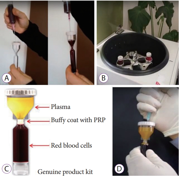
- Effectiveness of Autologous Platelet-Rich Plasma for the Healing of Ulcers after Endoscopic Submucosal Dissection
- Eunju Jeong, In kyung Yoo, Ozlem Ozer Cakir, Hee Kyung Kim, Won Hee Kim, Sung Pyo Hong, Joo Young Cho
- Clin Endosc 2019;52(5):472-478. Published online May 17, 2019
- DOI: https://doi.org/10.5946/ce.2018.152
-
 Abstract
 PDF
PDF PubReader
 ePub
ePub - Background
/Aims: Platelet-rich plasma (PRP) has been used for wound healing in various medical fields. The aim of this study was to evaluate the clinical efficacy and safety of local PRP injections after endoscopic submucosal dissection (ESD).
Methods
Patients were non-randomly divided into the following two groups: (1) control group in which patients were administered only an intravenous proton pump inhibitor (PPI), and (2) a study group in which patients were administered an intravenous PPI and a topical PRP injection. We assessed the reduction in the ulcer area and stage of the ulcer after the procedure (24 hours, 48 hours, and 28 days after endoscopic surgery).
Results
We enrolled 7 study and 7 control patients. In the study group, the rate of ulcer reduction was 59% compared to 52% in the control group (p=0.372), 28 days after ESD. There were 5 patients in the S stage and 2 patients in the H stage in the study group compared to no patient in the S stage and 7 patients in the H stage in the control group (p=0.05), 28 days after ESD. There were no serious complications in either group.
Conclusions
The local injection of PRP is a safe and effective procedure for ulcer healing after ESD. -
Citations
Citations to this article as recorded by
- Clinical efficacy of blood derivatives on wound healing: A systematic review and network meta‐analysis
Yanhong Wu, Guang Peng, Yuzhi Wang, Jianwu Chen, Bin Zhang, Jianbing Tang, Biao Cheng
International Wound Journal.2024;[Epub] CrossRef - Endoscopic Shielding With Platelet-rich Plasma After Resection Of Large Colorectal Lesions
Vicente Lorenzo-Zúñiga, Vicente Moreno de Vega, Ramón Bartolí
Surgical Laparoscopy, Endoscopy & Percutaneous Techniques.2021; 31(3): 376. CrossRef - The Additive Effect of Platelet-Rich Plasma in the Treatment of Actively Bleeding Peptic Ulcer
Waseem M. Seleem, Amr Shaaban Hanafy
Clinical Endoscopy.2021; 54(6): 864. CrossRef - Endless Challenges in Overcoming Complications Associated with Endoscopic Submucosal Dissection
Satoshi Ono, Shun Ito, Kenji Ogata
Clinical Endoscopy.2019; 52(5): 395. CrossRef
- Clinical efficacy of blood derivatives on wound healing: A systematic review and network meta‐analysis
- 6,535 View
- 139 Download
- 4 Web of Science
- 4 Crossref

Brief Report
- Immediate Endoscopic Management of an Intramural Hematoma Developed during Colonoscopy
- Chang-Il Kwon, Duck Hwan Kim, Sung Pyo Hong
- Clin Endosc 2017;50(5):508-509. Published online August 3, 2017
- DOI: https://doi.org/10.5946/ce.2017.037
-
PDFPubReaderePub
-
Citations
Citations to this article as recorded by- Systemic AL amyloidosis with multiple submucosal hematomas of the colon: a case report and literature review
Makomo Makazu, Akiko Sasaki, Chikamasa Ichita, Chihiro Sumida, Takashi Nishino, Miki Nagayama, Shinichi Teshima
Clinical Journal of Gastroenterology.2024; 17(1): 69. CrossRef - Perspectives and Management Strategies for Acute Colonic Intramural Hematoma
Reham Samir, Mohamed B Hashem, Hedy A Badary, Ahmed Bahaa, Nader Bakheet
International Journal of General Medicine.2022; Volume 15: 2861. CrossRef - Colonic Intramural Hematoma in a Cat: A Case Report
Ti-Chiu Hsu, Lee-Shuan Lin, Cheng-Shu Chung, Chuan Chiang, Hsien-Chieh Chiu, Ping-Hsun Huang
Frontiers in Veterinary Science.2022;[Epub] CrossRef - Traumatic Acute Colonic Intramural Hematoma: A Rare Entity and Successful Expectant Approach
Devarajan Jebin Aaron, Sandeep Bhattarai, Oseen Shaikh, Sarath Chandra Sistla
Cureus.2020;[Epub] CrossRef
- Systemic AL amyloidosis with multiple submucosal hematomas of the colon: a case report and literature review
- 5,899 View
- 101 Download
- 4 Web of Science
- 4 Crossref
Case Report
- Double-Scope Peroral Endoscopic Myotomy (POEM) for Esophageal Achalasia: The First Trial of a New Double-Scope POEM
- Hee Jin Hong, Ga Won Song, Weon Jin Ko, Won Hee Kim, Ki Baik Hahm, Sung Pyo Hong, Joo Young Cho
- Clin Endosc 2016;49(4):383-386. Published online March 15, 2016
- DOI: https://doi.org/10.5946/ce.2015.108
-
Abstract
PDFPubReaderePub
- With the accumulation of clinical trials demonstrating its efficacy and safety, peroral endoscopic myotomy (POEM) has emerged as a less invasive treatment option for esophageal achalasia compared with laparoscopic Heller myotomy. However, the difficulty in determining the exact extent of myotomy, a critical factor associated with the success and safety of the procedure, remains a limitation. Although the various endoscopic landmarks and ancillary techniques have been applied, none of these has been proven sufficient. As a solution for this limitation, the double-scope POEM technique with a second endoscope to assure the exact length of the submucosal tunnel has been applied since 2014. Before double-scope POEM was introduced, the second endoscope was applied only to confirm the accuracy of the procedure. In the present study, we performed double-scope POEM in the treatment of esophageal achalasia through a novel procedure of simultaneous application of the second endoscope to assist in the conventional POEM procedure.
-
Citations
Citations to this article as recorded by- Peroral Endoscopic Myotomy (POEM) in Children: A State of the Art Review
Ali A. Mencin, Amrita Sethi, Monique T. Barakat, Diana G. Lerner
Journal of Pediatric Gastroenterology & Nutrition.2022; 75(3): 231. CrossRef - Per-oral endoscopic myotomy (POEM) for a sigmoid type of achalasia: short-term outcomes and changes in the esophageal angle
Shota Maruyama, Yusuke Taniyama, Tadashi Sakurai, Makoto Hikage, Chiaki Sato, Kai Takaya, Takuro Konno, Takeshi Naitoh, Michiaki Unno, Takashi Kamei
Surgical Endoscopy.2020; 34(9): 4124. CrossRef - Characteristics of a Subset of Achalasia With Normal Integrated Relaxation Pressure
Eunju Kim, In Kyung Yoo, Dong Keon Yon, Joo Young Cho, Sung Pyo Hong
Journal of Neurogastroenterology and Motility.2020; 26(2): 274. CrossRef - Feasibility of using an led-probe in third-space endoscopy: a clinical study
Oscar Víctor Hernández Mondragón, Raúl Zamarripa Mottú, Omar Solórzano Pineda, Raúl Alberto Gutierrez Aguilar, Luís Fernando García Contreras
BMC Gastroenterology.2020;[Epub] CrossRef - 2007–2019: a “Third”-Space Odyssey in the Endoscopic Management of Gastrointestinal Tract Diseases
Anastassios C. Manolakis, Haruhiro Inoue, Akiko Ueno, Yuto Shimamura
Current Treatment Options in Gastroenterology.2019; 17(2): 202. CrossRef - Treatment of Achalasia with Per-Oral Endoscopic Myotomy: Analysis of 50 Consecutive Patients
Erica D. Kane, David J. Desilets, Donna Wilson, Marc Leduc, Vikram Budhraja, John R. Romanelli
Journal of Laparoendoscopic & Advanced Surgical Techniques.2018; 28(5): 514. CrossRef - Two penetrating vessels as a novel indicator of the appropriate distal end of peroral endoscopic myotomy
Shinwa Tanaka, Fumiaki Kawara, Takashi Toyonaga, Haruhiro Inoue, Robert Bechara, Namiko Hoshi, Hirohumi Abe, Yoshiko Ohara, Tsukasa Ishida, Yoshinori Morita, Eiji Umegaki
Digestive Endoscopy.2018; 30(2): 206. CrossRef
- Peroral Endoscopic Myotomy (POEM) in Children: A State of the Art Review
- 7,732 View
- 109 Download
- 9 Web of Science
- 7 Crossref
Original Article
- Optimal Methods for the Management of Iatrogenic Colonoscopic Perforation
- Dae Kyu Shin, Sun Young Shin, Chi Young Park, Sun Mi Jin, Yang Hyun Cho, Won Hee Kim, Chang-Il Kwon, Kwang Hyun Ko, Ki Baik Hahm, Pil Won Park, Jong Woo Kim, Sung Pyo Hong
- Clin Endosc 2016;49(3):282-288. Published online February 18, 2016
- DOI: https://doi.org/10.5946/ce.2015.046
-
Abstract
PDFPubReaderePub
- Background
/Aims: Colonoscopic perforations have been managed with exploratory laparotomy, and have resulted in some morbidity and mortality. Recently, laparoscopic surgery is commonly performed for this purpose. The aim of this study was to compare the outcomes of several management strategies for iatrogenic colonoscopic perforations.
Methods
We retrospectively reviewed the medical records of patients who had been treated for colonoscopic perforation between January 2004 and April 2013 at CHA Bundang Medical Center in Korea.
Results
A total of 41 patients with colonoscopic perforation were enrolled. Twenty patients underwent conservative management with a success rate of 90%. Surgical management was performed in 23 patients including two patients who were converted to surgical management after the failure of the initial conservative management. Among 14 patients who underwent surgery at 8 hours after the perforation, there was no considerable difference in adverse outcomes between the laparotomy group and the laparoscopic surgery group. The medical costs and claim rate were 1.45 and 1.87 times greater in the exploratory laparotomy group, respectively.
Conclusions
Conservative management of colonoscopic perforation could be an option for patients without overt symptoms of peritonitis or with a small defect size. If surgical management is required, laparoscopic surgery may be considered as the initial procedure even with a delayed diagnosis. -
Citations
Citations to this article as recorded by- Surgical repair of endoscopy-induced colonic perforations: a case-matched study of short-term morbidity and mortality
Fady DANIEL, Suha JABAK, Mohammad HOSNI, Hani TAMIM, Aurelie MAILHAC, Ayman ALRAZIM, Noura AL-ALI, Robert CHURCH, Mohammad KHALIFE, Shafik SIDANI, Faek JAMALI
Minerva Surgery.2024;[Epub] CrossRef - Laparoscopic versus open surgery for colonoscopic perforation: A systematic review and meta-analysis
Wu Zhong, Chuanyuan Liu, Chuanfa Fang, Lei Zhang, Xianping He, Weiquan Zhu, Xueyun Guan
Medicine.2023; 102(24): e34057. CrossRef - Elastography for Pediatric Chronic Liver Disease
Giovanna Ferraioli, Richard G. Barr, Jonathan R. Dillman
Journal of Ultrasound in Medicine.2021; 40(5): 909. CrossRef - Clinical outcomes of laparoscopic versus open surgery for repairing colonoscopic perforation: a multicenter study
Jae Seok Lee, Jeong Yeon Kim, Byung Mo Kang, Sang Nam Yoon, Jun Ho Park, Bo Young Oh, Jong Wan Kim
Surgery Today.2021; 51(2): 285. CrossRef - The analysis of outcomes of surgical management for colonoscopic perforations: A 16-years experiences at a single institution
Dae Ro Lim, Jung Kul Kuk, Taehyung Kim, Eung Jin Shin
Asian Journal of Surgery.2020; 43(5): 577. CrossRef - Multicenter retrospective evaluation of ileocecocolic perforations associated with diagnostic lower gastrointestinal endoscopy in dogs and cats
Vanessa L. Woolhead, Jacqueline C. Whittemore, Sarah A. Stewart
Journal of Veterinary Internal Medicine.2020; 34(2): 684. CrossRef - Endoscopic Management of the Ascending Colon Perforation Secondary to a Rare-Earth Magnets Ingestion in a Pediatric Patient
Sandra Mabel Camacho-Gomez, James Meredith Noel, Robert Adam Noel
ACG Case Reports Journal.2020; 7(8): e00436. CrossRef - Pseudo‐obstruction But a Real Perforation
AORN Journal.2019; 109(1): 142. CrossRef - Treatment of colonoscopic perforation: outcomes from a major single tertiary institution
Carolyn R. Chew, Justin M. C. Yeung, Ian G. Faragher
ANZ Journal of Surgery.2019; 89(5): 546. CrossRef - Management of colonoscopic perforations: A systematic review
Alexander T. Hawkins, Kenneth W. Sharp, Molly M. Ford, Roberta L. Muldoon, M. Benjamin Hopkins, Timothy M. Geiger
The American Journal of Surgery.2018; 215(4): 712. CrossRef - 2017 WSES guidelines for the management of iatrogenic colonoscopy perforation
Nicola de’Angelis, Salomone Di Saverio, Osvaldo Chiara, Massimo Sartelli, Aleix Martínez-Pérez, Franca Patrizi, Dieter G. Weber, Luca Ansaloni, Walter Biffl, Offir Ben-Ishay, Miklosh Bala, Francesco Brunetti, Federica Gaiani, Solafah Abdalla, Aurelien Ami
World Journal of Emergency Surgery.2018;[Epub] CrossRef - Management Outcomes of Colonoscopic Perforations Are Affected by the General Condition of the Patients
Jae Ho Park, Kyung Jong Kim
Annals of Coloproctology.2018; 34(1): 16. CrossRef - Abdominal Sepsis: An Update
Mircea Gabriel Mureșan, Ioan Alexandru Balmoș, Iudita Badea, Ario Santini
The Journal of Critical Care Medicine.2018; 4(4): 120. CrossRef - Laparoscopic vs. open surgery for the treatment of iatrogenic colonoscopic perforations: a systematic review and meta-analysis
Aleix Martínez-Pérez, Nicola de’Angelis, Francesco Brunetti, Yann Le Baleur, Carmen Payá-Llorente, Riccardo Memeo, Federica Gaiani, Marco Manfredi, Paschalis Gavriilidis, Giorgio Nervi, Federico Coccolini, Aurelien Amiot, Iradj Sobhani, Fausto Catena, Gia
World Journal of Emergency Surgery.2017;[Epub] CrossRef - Urinary Bladder Injury During Colonoscopy Without Colon Perforation
Jung Wook Suh, Jun Won Min, Hwan Namgung, Dong-Guk Park
Annals of Coloproctology.2017; 33(3): 112. CrossRef - The management of intra-abdominal infections from a global perspective: 2017 WSES guidelines for management of intra-abdominal infections
Massimo Sartelli, Alain Chichom-Mefire, Francesco M. Labricciosa, Timothy Hardcastle, Fikri M. Abu-Zidan, Abdulrashid K. Adesunkanmi, Luca Ansaloni, Miklosh Bala, Zsolt J. Balogh, Marcelo A. Beltrán, Offir Ben-Ishay, Walter L. Biffl, Arianna Birindelli, M
World Journal of Emergency Surgery.2017;[Epub] CrossRef - How Should We Manage Iatrogenic Perforation Caused by Colonoscopy?
Eun Sun Kim
Clinical Endoscopy.2016; 49(3): 214. CrossRef
- Surgical repair of endoscopy-induced colonic perforations: a case-matched study of short-term morbidity and mortality
- 8,245 View
- 151 Download
- 20 Web of Science
- 17 Crossref
Review
- Image Quality Analysis of Various Gastrointestinal Endoscopes: Why Image Quality Is a Prerequisite for Proper Diagnostic and Therapeutic Endoscopy
- Weon Jin Ko, Pyeong An, Kwang Hyun Ko, Ki Baik Hahm, Sung Pyo Hong, Joo Young Cho
- Clin Endosc 2015;48(5):374-379. Published online September 30, 2015
- DOI: https://doi.org/10.5946/ce.2015.48.5.374
-
Abstract
PDFPubReaderePub
Arising from human curiosity in terms of the desire to look within the human body, endoscopy has undergone significant advances in modern medicine. Direct visualization of the gastrointestinal (GI) tract by traditional endoscopy was first introduced over 50 years ago, after which fairly rapid advancement from rigid esophagogastric scopes to flexible scopes and high definition videoscopes has occurred. In an effort towards early detection of precancerous lesions in the GI tract, several high-technology imaging scopes have been developed, including narrow band imaging, autofocus imaging, magnified endoscopy, and confocal microendoscopy. However, these modern developments have resulted in fundamental imaging technology being skewed towards red-green-blue and this technology has obscured the advantages of other endoscope techniques. In this review article, we have described the importance of image quality analysis using a survey to consider the diversity of endoscope system selection in order to better achieve diagnostic and therapeutic goals. The ultimate aims can be achieved through the adoption of modern endoscopy systems that obtain high image quality.
-
Citations
Citations to this article as recorded by- Colonoscopy Quality, Innovation, and the Assessment of New Technology
Sanjay R.V. Gadi, Sriya S. Muralidharan, Jeremy R. Glissen Brown
Techniques and Innovations in Gastrointestinal Endoscopy.2024; 26(2): 177. CrossRef - Endoscopy image enhancement method by generalized imaging defect models based adversarial training
Wenjie Li, Jingfan Fan, Yating Li, Pengcheng Hao, Yucong Lin, Tianyu Fu, Danni Ai, Hong Song, Jian Yang
Physics in Medicine & Biology.2022; 67(9): 095016. CrossRef - Reduced detection rate of artificial intelligence in images obtained from untrained endoscope models and improvement using domain adaptation algorithm
Junseok Park, Youngbae Hwang, Hyun Gun Kim, Joon Seong Lee, Jin-Oh Kim, Tae Hee Lee, Seong Ran Jeon, Su Jin Hong, Bong Min Ko, Seokmin Kim
Frontiers in Medicine.2022;[Epub] CrossRef - Diagnosis of Early Gastric Cancer Using Image-enhanced Endoscopy
Weon Jin Ko
The Korean Journal of Medicine.2017; 92(3): 264. CrossRef
- Colonoscopy Quality, Innovation, and the Assessment of New Technology
- 8,137 View
- 104 Download
- 5 Web of Science
- 4 Crossref
Case Report
- Extragastroesophageal Malignancy-Associated Secondary Achalasia: A Rare Association of Pancreatic Cancer Rendering Alarm Manifestation
- Hong Min Kim, Ji Min Chu, Won Hee Kim, Sung Pyo Hong, Ki Baik Hahm, Kwang Hyun Ko
- Clin Endosc 2015;48(4):328-331. Published online July 24, 2015
- DOI: https://doi.org/10.5946/ce.2015.48.4.328
-
Abstract
PDFPubReaderePub
Secondary achalasia or pseudoachalasia is a rare esophageal motor abnormality, which mimics primary achalasia; it is not easily distinguishable from idiopathic achalasia by manometry, radiological examination, or endoscopy. Although the majority of reported pseudoachalasia cases are associated with neoplasms at or near the esophagogastric (EG) junction, other neoplastic processes or even chronic illnesses such as rheumatoid arthritis can lead to the development of pseudoachalasia, for example, mediastinal masses, gastrointestinal (GI) tumors of the liver and biliary tract, and non-GI malignancies. Therefore, even if a patient presents with the typical findings of achalasia, we should be alert to the possibility of other GI malignancies besides EG tumors. For instance, pancreatic cancer was found in the case reported here; only four such cases have been reported in the literature. A 47-year-old man was admitted to our center with a 3-month history of dysphagia. His endoscopic and esophageal manometric findings were compatible with primary achalasia. However, unresponsiveness to diverse conventional achalasia treatments led us to suspect secondary achalasia. An active search led to a diagnosis of pancreatic mucinous cystadenocarcinoma invading the gastric fundus and EG junction. This rare case of pseudoachalasia caused by pancreatic carcinoma emphasizes the need for suspecting GI malignancies other than EG tumors in patients refractory to conventional achalasia treatment.
-
Citations
Citations to this article as recorded by- Delayed Presentation of Malignancy-Associated Pseudoachalasia of the Gastric Cardia
Clive J Miranda, Farhan Azad, Ross R Moyer, Sasikanth N Ravi, Gina M Sparacino
Cureus.2024;[Epub] CrossRef - Is it necessary to perform a morphological assessment for an esophageal motility disorder? A retrospective descriptive study
Sofya Latrache, Chloe Melchior, Charlotte Desprez, Sabrina Sidali, Julien Recton, Olivier Touchais, Elise van der Eecken, Fabien Wuestenberghs, Cloe Charpentier, Anne Marie Leroi, Guillaume Gourcerol
Clinics and Research in Hepatology and Gastroenterology.2021; 45(6): 101633. CrossRef - When a Late Metastasis Is Hard to Swallow
Catarina Negrão, Rita Sismeiro, Margarida Monteiro, Filipa G Pereira, Marta Jonet
Cureus.2021;[Epub] CrossRef - Development of pseudoachalasia following magnetic sphincter augmentation (MSA) with restoration of peristalsis after endoscopic dilation
Katrin Schwameis, Shahin Ayazi, Ali H. Zaidi, Toshitaka Hoppo, Blair A. Jobe
Clinical Journal of Gastroenterology.2020; 13(5): 697. CrossRef - Burkitt’s Lymphoma of the Gastrohepatic Omentum: A Malignant Presentation of Pseudoachalasia
Eric Omar Then, Andrew Ofosu, Prashanth Rawla, Tagore Sunkara, Sriharsha Dadana, Andrea Culliford, Vinaya Gaduputi
Case Reports in Gastrointestinal Medicine.2019; 2019: 1. CrossRef
- Delayed Presentation of Malignancy-Associated Pseudoachalasia of the Gastric Cardia
- 7,128 View
- 57 Download
- 6 Web of Science
- 5 Crossref
Original Article
-
Endoscopic Submucosal Dissection Using a Novel Versatile Knife: An Animal Feasibility Study (with Video)

- Chang-Il Kwon, Gwangil Kim, Il-Kwun Chung, Won Hee Kim, Kwang Hyun Ko, Sung Pyo Hong, Seok Jeong, Don Haeng Lee
- Clin Endosc 2014;47(6):544-554. Published online November 30, 2014
- DOI: https://doi.org/10.5946/ce.2014.47.6.544
-
Abstract
PDF
 Supplementary MaterialPubReaderePub
 Background/Aims In order to reduce the procedure time and the number of accessory changes during endoscopic submucosal dissection (ESD), we developed a novel versatile knife, which has the combined advantages of several conventional knives. The aim of this study was to compare the efficacy, safety, and histological quality of ESD performed using this novel versatile knife and a combination of several conventional knives.
Methods This was an
in vivo animal study comparing two different modalities of ESD in mini-pigs. Completion time of each resection was documented, and the resected specimens were retrieved and evaluated for completeness. To assess the quality control of the procedures and adverse events, detailed histopathological examinations were performed.Results A total of 18 specimens were dissected by ESD safely and easily (nine specimens using the new versatile knife; nine specimens by mixing conventional knives). All resections were completed as
en bloc resections. There was no significant difference in procedure time between the 2 modalities (456 seconds vs. 355 seconds,p =0.258) and cutting speed (1.983 mm2/sec vs. 1.57 mm2/sec,p =1.000). The rate of adverse events and histological quality did not statistically differ between the modalities.Conclusions ESD with a versatile knife appeared to be an easy, safe, and technically efficient method.
-
Citations
Citations to this article as recorded by- Comparison of synchronous dual wavelength diode laser versus conventional endo-knives for esophageal endoscopic submucosal dissection: an animal study
Jian Tang, Shufang Ye, Xueliang Ji, Jun Li, Feng Liu
Surgical Endoscopy.2018; 32(12): 5037. CrossRef - Technological review on endoscopic submucosal dissection: available equipment, recent developments and emerging techniques
Georgios Mavrogenis, Juergen Hochberger, Pierre Deprez, Morteza Shafazand, Dimitri Coumaros, Katsumi Yamamoto
Scandinavian Journal of Gastroenterology.2017; 52(4): 486. CrossRef
- Comparison of synchronous dual wavelength diode laser versus conventional endo-knives for esophageal endoscopic submucosal dissection: an animal study
- 7,188 View
- 60 Download
- 3 Web of Science
- 2 Crossref
Case Reports
- Endoscopic Resection of Giant Colonic Lipoma: Case Series with Partial Resection
- Gun Woo Kim, Chang-Il Kwon, Sang Hee Song, Sun Mi Jin, Kyung Ho Kim, Jie Hye Moon, Sung Pyo Hong, Pil Won Park
- Clin Endosc 2013;46(5):586-590. Published online September 30, 2013
- DOI: https://doi.org/10.5946/ce.2013.46.5.586
-
Abstract
PDFPubReaderePub
Colonic lipoma, a very rare form of benign tumor, is typically detected incidentally in asymptomatic patients. The size of lipoma is reported variously from 2 mm to 30 cm, with higher likelihood of symptoms as the size is bigger. Cases with symptom or bigger lesion are surgically resected in principle; endoscopic resection, which has developed recently with groundbreaking advance of endoscopic excision technology, is being used more often but with rare report of success due to high chance of complications such as bowel perforation or bleeding. The authors report here, together with a literature review, our experiences of three cases of giant colonic lipomas showing complete remission after aggressive unroofing technique, at certain intervals, using snare catheter at the origin of the lipoma so that the remaining lipoma could be drained out of the exposed surface spontaneously, in order to reduce complications.
-
Citations
Citations to this article as recorded by- Colonic intussusception from pedunculated colonic lipoma at hepatic flexure: A case report and review of current literature
Richard Edmund Hogan, Ben Michael Murray, Michael Flanagan, Shane Brennan, Conor Shortt, Dara Kavanagh
Surgery Case Reports.2024; 1: 100008. CrossRef - A giant colonic lipoma
Tara M. Connelly, Cillian Clancy, Shaomin Hu, Joshua Sommovilla
ANZ Journal of Surgery.2023; 93(1-2): 428. CrossRef - Obscure gastrointestinal bleeding from a large jejunal lipoma treated using an endoscopic unroofing technique with double balloon enteroscopy: a case study
Reo Kobayashi, Ken Inoue, Ryohei Hirose, Toshifumi Doi, Akihito Harusato, Osamu Dohi, Naohisa Yoshida, Kazuhiko Uchiyama, Takeshi Ishikawa, Tomohisa Takagi, Hiroaki Yasuda, Hideyuki Konishi, Yukiko Morinaga, Yoshito Itoh
Clinical Journal of Gastroenterology.2023; 16(1): 32. CrossRef - Endoscopic debulking of a large colonic lipoma causing recurrent intussusception using endoscopic mucosotomy technique
Jenson Phung, Morgan Freeman, Mohammad Bilal
Endoscopy.2023; 55(S 01): E817. CrossRef - Large colonic lipoma with a laterally spreading tumor treated by endoscopic submucosal dissection: A case report
Jun Yong Bae, Hun Kyu Kim, Yee Jin Kim, Se Woong Kim, Youngeun Lee, Chang Beom Ryu, Moon Sung Lee
World Journal of Clinical Cases.2023; 11(26): 6194. CrossRef - Observation of the drainage process of the residual lipoma after endoscopic unroofing technique during colonoscopic evaluation of post-procedural hematochezia
Yi-Ling Ko, Hiroki Matsuoka, Ryohei Nomaru, So Imakiire, Hideto Sakisaka, Satoshi Matsuoka, Nobuaki Kuno, Koichi Abe, Sadahiro Funakoshi, Yusuke Ishida, Hideki Ishibashi, Kaori Koga, Tetsuhiro Saito, Morishige Takeshita, Fumihito Hirai
Clinical Journal of Gastroenterology.2022; 15(2): 407. CrossRef - Spontaneous expulsion of a duodenal lipoma after endoscopic biopsy: A case report
Zhi-Hao Chen, Li-Hong Lv, Wen-Sheng Pan, Yi-Miao Zhu
World Journal of Gastroenterology.2022; 28(34): 5086. CrossRef - Pedunculated sigmoid lipoma causing colo-colonic intussusception
Kenneth Ford, Samantha Lopez, Gaurav Synghal, Yomi Fayiga, Brittany Carter, Anuj Kandel, Kenneth Ford
Baylor University Medical Center Proceedings.2021; 34(3): 371. CrossRef - Colonoscopic resection of giant colonic lipoma causing subacute large bowel obstruction
Amy Donovan, Sandun Abeyasundara, Hajir Nabi
ANZ Journal of Surgery.2020;[Epub] CrossRef - COLON LIPOMA COMPLICATED BY COLON INVAGINATION
U. B. Urmonov, S. G. Afanasyev, A. Yu. Dobrodeev, A. V. Avgustinovich, M. Yu. Volkov, N. V. Vasiliev, E. N. Samtsov
Grekov's Bulletin of Surgery.2020; 178(6): 63. CrossRef - Endoscopic treatment of large symptomatic colon lipomas: A systematic review of efficacy and safety
Michiel Bronswijk, Anne‐Marie Vandenbroucke, Peter Bossuyt
United European Gastroenterology Journal.2020; 8(10): 1147. CrossRef - Endoscopic resection of giant colon lipomas: get rid of the roof!
Michiel Bronswijk
VideoGIE.2019; 4(7): 341. CrossRef - Submucosal lipoma of the sigmoid colon as a rare cause of mucoid diarrhea: a case report
S. U. B. Dassanayake, N. P. Dinamithra, N. M. M. Nawarathne
Journal of Medical Case Reports.2016;[Epub] CrossRef - Endoscopic resection of giant GI lipoma: a case series
Diane Lorenzo, Jean Michel Gonzalez, Alban Benezech, Marc Barthet
VideoGIE.2016; 1(2): 43. CrossRef - Two Patients with Large Colonic Lipomas for which Endoscopic Unroofing was Ineffective
Yuichi Tomiki, Koichiro Niwa, Kiichi Nagayasu, Yu Okazawa, Shingo Ito, Ryosuke Ichikawa, Hisashi Ro, Shun Ishiyama, Kiichi Sugimoto, Kazuhiro Sakamoto
Case Reports in Gastroenterology.2016; 10(3): 538. CrossRef - Colonoscopy‐assisted laparoscopic resection of an obstructing ‘giant’ lipoma of the transverse colon
B. Asantha De Silva, Raeed Deen, Wasantha K. Wijenayake
ANZ Journal of Surgery.2015; 85(10): 785. CrossRef - Unroofing Technique as an Option for the Endoscopic Treatment of Giant Gastrointestinal Lipomas
Marcela Kopáčová, Stanislav Rejchrt, Jan Bureš
Acta Medica (Hradec Kralove, Czech Republic).2015; 58(4): 115. CrossRef - Large “pedunculated” colonic lipoma: A word of caution while cutting into fat!
Syed Adnan Mohiuddin, Saad Al Kaabi, Ragesh Babu Thandassery, Khalid Mohsin Al Ejji, Nazeeh Al Dweik, Emran Amir, Manik Sharma
Indian Journal of Gastroenterology.2014; 33(6): 571. CrossRef
- Colonic intussusception from pedunculated colonic lipoma at hepatic flexure: A case report and review of current literature
- 7,513 View
- 95 Download
- 18 Crossref
- Gastrointestinal Cancers in a Peutz-Jeghers Syndrome Family: A Case Report
- Sang Hee Song, Kun Woo Kim, Won Hee Kim, Chang Il Kwon, Kwang Hyun Ko, Ki Baik Hahm, Pil Won Park, Sung Pyo Hong
- Clin Endosc 2013;46(5):572-575. Published online September 30, 2013
- DOI: https://doi.org/10.5946/ce.2013.46.5.572
-
Abstract
PDFPubReaderePub
A 17-year-old man was diagnosed as Peutz-Jeghers syndrome (PJS) because of pigmented lip and multiple gastrointestinal polyps. He had anemia and underwent polypectomy on the duodenum and colon. His maternal family members were patients with PJS. His mother used to be screened with endoscopy to remove large polyps. One and half years later, he underwent jejunal segmental resection due to intussusceptions. He underwent endoscopic polypectomy every 2 to 3 years. When he was 23 years old, high-grade dysplasia was found in colonic polyp and his mother underwent partial pancreatectomy due to intraductal papillary mucinous carcinoma. When he was 27 years old, diffuse gastric polyps on the greater curvature of corpus expanded and grew. Therefore, wide endoscopic polypectomy was done. Histological examination revealed focal intramucosal carcinoma and low-grade dysplasia in hamartomatous polyps. We report cases of cancers occurred in first-degree relatives with PJS.
-
Citations
Citations to this article as recorded by- Familial and hereditary gastric cancer, an overview
Fátima Carneiro
Best Practice & Research Clinical Gastroenterology.2022; 58-59: 101800. CrossRef - Small bowel intussusception and concurrent sigmoid polyp with malignant transformation in Peutz–Jeghers syndrome
Maidah Algarni, Enas Raml, Nora Trabulsi, Mohammed Nassif
Journal of Surgical Case Reports.2019;[Epub] CrossRef - The first European family with gastric adenocarcinoma and proximal polyposis of the stomach: case report and review of the literature
Rudolf Repak, Darina Kohoutova, Miroslav Podhola, Stanislav Rejchrt, Marek Minarik, Lucie Benesova, Michal Lesko, Jan Bures
Gastrointestinal Endoscopy.2016; 84(4): 718. CrossRef - Gastric Hamartomatous Polyps—Review and Update
Monika Vyas, Xiu Yang, Xuchen Zhang
Clinical Medicine Insights: Gastroenterology.2016; 9: CGast.S38452. CrossRef - Giant rectal polyp prolapse in an adult patient with the Peutz-Jeghers syndrome
Ana Delfina Cano-Contreras, Arturo Meixueiro-Daza, Peter Grube-Pagola, Jose Maria Remes-Troche
BMJ Case Reports.2016; : bcr2016215629. CrossRef - Prevention Strategies for Gastric Cancer: A Global Perspective
Jin Young Park, Lawrence von Karsa, Rolando Herrero
Clinical Endoscopy.2014; 47(6): 478. CrossRef
- Familial and hereditary gastric cancer, an overview
- 6,557 View
- 58 Download
- 6 Crossref
Original Article
- Angular Positioning of Stent Increases Bowel Perforation after Self-Expandable Metal Stent Placement for Malignant Colorectal Obstruction
- Jeong Guil Lee, Kwang Ho Yoo, Chang-Il Kwon, Kwang Hyun Ko, Sung Pyo Hong
- Clin Endosc 2013;46(4):384-389. Published online July 31, 2013
- DOI: https://doi.org/10.5946/ce.2013.46.4.384
-
Abstract
PDFPubReaderePub
Background/Aims Some factors like stent wires, balloon dilatation and use of guide wires seems to increase perforation after self-expandable metal stent (SEMS) placement, but few studies mentioned about the relationship between angulation of malignant stricture and perforation. The present study aimed to confirm that more angular positioning of stents increases perforation.
Methods This study was conducted with retrospectively evaluation at Digestive Disease Center, CHA Bundang Medical Center, CHA University. Between January 2002 and August 2011, SEMS was inserted in 130 patients with malignant colorectal obstruction. We studied the difference in the angle of stenosis between perforation and non-perforation groups using fluorography images.
Results SEMS insertion was performed in 130 cases of obstruction due to colon cancer. Perforation occurred in eight patients (6.2%) of them. Thirteen cases were excluded from the analysis due to poor fluoroscopic images. Among the eight patients with perforation, the mean stenosis angle was 109.9° compared to 153.1° in the nonperforation group, indicating that the angle was more acute in the perforation group (
p =0.016).Conclusions This study shows that more angular positioning of stent increases bowel perforation after SEMS placement for malignant colorectal obstruction.
-
Citations
Citations to this article as recorded by- Analysis of Reported Adverse Events With Colonic Stents: An FDA MAUDE Database Study
Chong-Chi Chiu, Kuo-Chuan Hung, Li-Ren Yeh
Journal of Clinical Gastroenterology.2023; 57(6): 640. CrossRef - Clinical outcomes of colorectal stenting
S.V. Davydova, A.G. Fedorov, O.A. Yun, A.E. Klimov
Endoskopicheskaya khirurgiya.2023; 29(6): 12. CrossRef - Colonic stenting for malignant obstructions-A review of current indications and outcomes
Amelie Lueders, Gabie Ong, Peter Davis, Jonathan Weyerbacher, Jonathan Saxe
The American Journal of Surgery.2022; 224(1): 217. CrossRef - Comparison of different types of covered self-expandable metal stents for malignant colorectal obstruction
Joon Seop Lee, Hyun Seok Lee, Eun Soo Kim, Min Kyu Jung, Jin Tae Jung, Ho Gak Kim, Dong Wook Lee, Dae Jin Kim, Yoo Jin Lee, Chang Heon Yang
Surgical Endoscopy.2021; 35(8): 4124. CrossRef - Self‐expanding metallic stent improves histopathological edema compared with transanal drainage tube for malignant colorectal obstruction
Hiroshi Takeyama, Kotaro Kitani, Tomoko Wakasa, Masanori Tsujie, Yoshinori Fujiwara, Shigeto Mizuno, Masao Yukawa, Yoshio Ohta, Masatoshi Inoue
Digestive Endoscopy.2016; 28(4): 456. CrossRef - Oncologic safety of self-expanded metal stent insertion as a bridge to elective surgery in malignant colorectal obstruction
Se Hui Oh, Nak Jun Choi, Sang Hyuk Seo, Min Sung An, Kwang Hee Kim, Ki Beom Bae, Jin Won Hwang, Sang Heon Lee, Ji Hyun Kim, Sam Ryong Jee, Mi Seon Kang, Kwan Hee Hong
Korean Journal of Clinical Oncology.2016; 12(1): 48. CrossRef - Enteral stent construction: Current principles
Hans-Ulrich Laasch, Derek W. Edwards, Ho-Young Song
International Journal of Gastrointestinal Intervention.2016; 5(2): 85. CrossRef - Comparison of through-the-scope stent insertion with standard stent insertion for the management of malignant colorectal obstruction: a prospective study
Y. Wan, Y.-Q. Zhu, N.-W. Chen, Z.-G. Wang, Y.-S. Cheng, J. Shi
Techniques in Coloproctology.2016; 20(10): 707. CrossRef - Intestinal Stricture in Crohn's Disease
Chen-Wang Chang, Jau-Min Wong, Chien-Chih Tung, I-Lun Shih, Horng-Yuan Wang, Shu-Chen Wei
Intestinal Research.2015; 13(1): 19. CrossRef - Predictive Factors for Successful Colonic Stenting in Acute Large-Bowel Obstruction
Derek J. Boyle, Christopher Thorn, Ashish Saini, Colin Elton, Gary K. Atkin, Ian C. Mitchell, Kevin Lotzof, Adrian Marcus, Pawan Mathur
Diseases of the Colon & Rectum.2015; 58(3): 358. CrossRef - Stents for colonic strictures: Materials, designs, and more
Nandakumar Srinivasan, Richard A. Kozarek
Techniques in Gastrointestinal Endoscopy.2014; 16(3): 100. CrossRef - Patients who failed endoscopic stenting for left-sided malignant colorectal obstruction suffered the worst outcomes
Tian-Zhi Lim, Dedrick Chan, Ker-Kan Tan
International Journal of Colorectal Disease.2014; 29(10): 1267. CrossRef
- Analysis of Reported Adverse Events With Colonic Stents: An FDA MAUDE Database Study
- 7,008 View
- 59 Download
- 12 Crossref
Review
- Endoscopic Treatment of Refractory Gastroesohageal Reflux Disease
- Won Hee Kim, Pil Won Park, Ki Baik Hahm, Sung Pyo Hong
- Clin Endosc 2013;46(3):230-234. Published online May 31, 2013
- DOI: https://doi.org/10.5946/ce.2013.46.3.230
-
Abstract
PDFPubReaderePub
Though efficient acid suppression with proton pump inhibitors (PPIs) remains the mainstay of treatment of gastroesophageal reflux disease (GERD), some of the patients showed refractory response to PPIs, necessitating further intervention. After increasing dose of PPIs and other kinds of pharmacological intervention adopting prokinetics or others, variable endoscopic treatments are introduced for the treatment of these refractory cases. The detailed introduction regarding endoscopic treatment for GERD is forwarded in this review article. Implantation of reabsorbable or synthetic materials in the distal esophagus was tried in vain and is expelled from the market due to limited efficacy and serious complication. Radiofrequency energy delivery (Stretta) and transoral incisionless fundoplication (EsophyX) are actively tried currently.
-
Citations
Citations to this article as recorded by- Refractory gastroesophageal reflux disease
C. R. Subramanian, G. Triadafilopoulos
Gastroenterology Report.2015; 3(1): 41. CrossRef - Long-term outcomes of patients with refractory gastroesophageal reflux disease following a minimally invasive endoscopic procedure: a prospective observational study
Wei-Tao Liang, Zhong-Gao Wang, Feng Wang, Yue Yang, Zhi-Wei Hu, Jian-Jun Liu, Guang-Chang Zhu, Chao Zhang, Ji-Min Wu
BMC Gastroenterology.2014;[Epub] CrossRef
- Refractory gastroesophageal reflux disease
- 7,725 View
- 73 Download
- 2 Crossref
Case Report
- An Impacted Pancreatic Stone in the Papilla Induced Acute Obstructive Cholangitis in a Patient with Chronic Pancreatitis
- Kwang-Ho Yoo, Chang-Il Kwon, Sang-Wook Yoon, Won Hee Kim, Jung Min Lee, Kwang Hyun Ko, Sung Pyo Hong, Pil Won Park
- Clin Endosc 2012;45(1):99-102. Published online March 31, 2012
- DOI: https://doi.org/10.5946/ce.2012.45.1.99
-
Abstract
PDFPubReaderePub
Obstructive jaundice is very rarely caused by impaction of a pancreatic stone in the papilla. We report here on a case of obstructive jaundice with acute cholangitis that was caused by an impacted pancreatic stone in the papilla in a patient with chronic pancreatitis. A 48-year-old man presented with acute obstructive cholangitis. Abdominal computed tomography with the reconstructed image revealed distal biliary obstruction that was caused by a pancreatic stone in the pancreatic head, and there was also pancreatic ductal dilatation and parenchymal atrophy of the pancreatic body and tail with multiple calcifications. Emergency duodenoscopy revealed an impacted pancreatic stone in the papilla. Precut papillotomy using a needle knife was performed, followed by removal of the pancreatic stone using grasping forceps. After additional sphincterotomy, a large amount of dark-greenish bile juice gushed out. The patient rapidly improved and he has remained well.
-
Citations
Citations to this article as recorded by- Pancreatic stones causing secondary biliary obstruction: An uncommon presentation of chronic pancreatitis
Wesley C. Judy, Tom K. Lin
JPGN Reports.2024;[Epub] CrossRef - The “squeezing with forceps” method for emergency endoscopic removal of an impacted pancreatic stone in the papilla of a patient on antithrombotic therapy
Sho Kitagawa, Shori Ishikawa, Keiya Okamura
Endoscopy.2023; 55(S 01): E454. CrossRef - Biliary Outlet Obstruction Due to Pancreatic Calculi in a Post-cholecystectomy Patient
Joey Almaguer, Dylan Murray, Matthew Murray, Richard Murray
Cureus.2023;[Epub] CrossRef - Ampullary stone in chronic pancreatitis causing obstructive jaundice and cholangitis
Sandheep Janardhanan, Allwin James, Alagammai Palaniappan, Ramesh Ardhanari
Gastroenterology, Hepatology and Endoscopy Practice.2021; 1(2): 69. CrossRef - Ursodeoxycholic acid attenuates 5‑fluorouracil‑induced mucositis in a rat model
Seung Kim, Hoon Chun, Hyuk Choi, Eun Kim, Bora Keum, Yeon Seo, Yoon Jeen, Hong Lee, Soon Um, Chang Kim
Oncology Letters.2018;[Epub] CrossRef - Pancreatic Calculus Causing Biliary Obstruction: Endoscopic Therapy for a Rare Initial Presentation of Chronic Pancreatitis
Anurag J. Shetty, C. Ganesh Pai, Shiran Shetty, Girisha Balaraju
Digestive Diseases and Sciences.2015; 60(9): 2840. CrossRef
- Pancreatic stones causing secondary biliary obstruction: An uncommon presentation of chronic pancreatitis
- 5,951 View
- 50 Download
- 6 Crossref
 First
First Prev
Prev


