Skip Navigation
Skip to contents

Clin Endosc : Clinical Endoscopy

OPEN ACCESS

Articles

Page Path
HOME > Clin Endosc > Volume 46(2); 2013 > Article
Case Report A Case of Esophageal Squamous Cell Carcinoma with Pancreatic Metastasis
Choulki Park1, Jae Young Jang1, Youn Hwa Kim2, Eun Jung Hwang1, Ki Yong Na2, Kyung-Yup Kim1, Jae Hyun Park1, Young Woon Chang1
Clinical Endoscopy 2013;46(2):197-200.
DOI: https://doi.org/10.5946/ce.2013.46.2.197
Published online: March 31, 2013

1Division of Gastroenterology and Hepatology, Department of Internal Medicine, Kyung Hee University Medical Center, Kyung Hee University School of Medicine, Seoul, Korea.

2Department of Pathology, Kyung Hee University Medical Center, Kyung Hee University School of Medicine, Seoul, Korea.

Correspondence: Jae Young Jang. Division of Gastroenterology and Hepatology, Department of Internal Medicine, Kyung Hee University Medical Center, Kyung Hee University School of Medicine, 23 Kyungheedae-ro, Dongdaemun-gu, Seoul 130-872, Korea. Tel: +82-2-958-8200, Fax: +82-2-968-1848, jyjang@khu.ac.kr
• Received: May 30, 2012   • Revised: August 29, 2012   • Accepted: October 23, 2012

Copyright © 2013 Korean Society of Gastrointestinal Endoscopy

This is an Open Access article distributed under the terms of the Creative Commons Attribution Non-Commercial License (http://creativecommons.org/licenses/by-nc/3.0/) which permits unrestricted non-commercial use, distribution, and reproduction in any medium, provided the original work is properly cited.

  • 6,745 Views
  • 51 Download
  • 11 Crossref
  • 11 Scopus
prev next
  • Solitary pancreatic metastasis of esophageal cancer is extremely rare. We report the case of a 58-year-old male admitted with esophageal cancer. Additional asymptomatic solitary hepatic and pancreatic masses were observed in the staging work-up for esophageal cancer. The hepatic mass was confirmed as a primary hepatocellular carcinoma with an ultrasound-guided needle biopsy. An esophagectomy with a distal pancreatectomy and radiofrequency ablation for hepatocellular carcinoma were performed. Histologically, the pancreatic mass was confirmed to be a metastasis from the esophageal cancer. The patient has been followed up with chemotherapy.
Tumors of the pancreas are divided into exocrine and endocrine tumors. Ductal carcinoma is the most common exocrine tumor of the pancreas and is subdivided into adenocarcinoma and squamous cell carcinoma. Adenocarcinoma, the most common subtype, comprises 80% of all malignant tumors of the pancreas,1,2 while primary squamous cell carcinoma is an extremely rare subtype of ductal carcinoma in the pancreas because the cell line does not exist in a normal pancreas.3,4 Consequently, if the presence of squamous cell carcinoma is confirmed in a biopsy, the possibility of metastatic disease must be considered first, and an extensive search for the primary lesion should be undertaken.5,6
Recently, we confirmed a case of synchronous esophageal cancer with pancreatic metastasis and hepatocellular carcinoma (HCC). This is reported here along with a literature review.
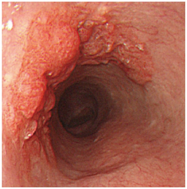
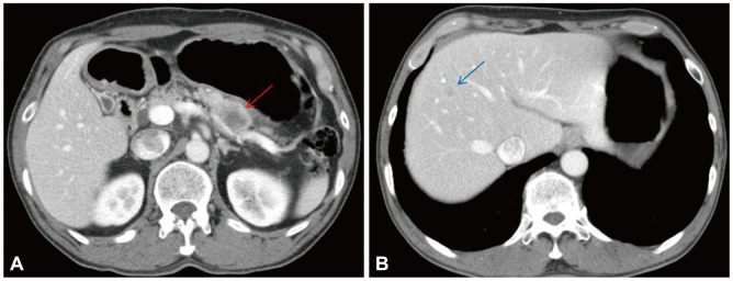
A 58-year-old male with a 1-month history of abdominal bloating and indigestion visited our hospital for esophagogastroduodenoscopy. This detected a 3×4-cm erythematous lesion with an irregular nodular surface extending between 38 and 41 cm from the incisors in the 9 to 1 o,clock position (Fig. 1). With Iodine staining, it was shown as an iodine-void area with a well-defined boundary. A biopsy revealed squamous cell carcinoma, and he was admitted for further evaluation and management of this esophageal cancer. He was an alcoholic who had been drinking one bottle of Soju daily for the past 30 years and had a 30-pack-year smoking history. He was a hepatitis B carrier without regular follow-up. At admission, the patient's vitals were stable and he was alert mentally. No abnormalities were detected in the physical examination. The peripheral blood tests were normal, with hemoglobin 13.6 g/dL, white blood cell count 7,330/mm3, platelets 238,000/mm3, as were the blood chemistry and electrolytes. Additional studies for hepatitis B showed hepatitis B e antigen (-) and anti-hepatitis B e antibody (+), and no virus was detected with hepatitis B virus real-time polymerase chain reaction. The results for tumor markers were as follows: carcinoembryonic antigen 2.39 ng/mL (reference range, <4.1), CA19-9 2 U/mL (reference range, <37), and alpha-fetoprotein (AFP) 8.58 ng/mL (reference range, <4.0). Abdominal computed tomography (CT) showed a poorly demarcated and poorly enhancing mass at the pancreatic tail (Fig. 2A). Magnetic resonance imaging (MRI) showed that the pancreatic mass appeared poorly demarcated and hyperintense on T2-weighted image and hypointense on T1-weighted image. Dynamic enhancing MRI showed that the mass was poorly enhanced without rim enhancement. So we were highly suspicious that the pancreatic mass was a primary adenocarcinoma rather than metastasis, because of tumor character and extremely low incidence of pancreatic metastasis from esophageal cancer. CT and MRI showed 1.2 cm-sized single nodule in the liver (Fig. 2B). MRI showed that the hepatic mass was hypointense on T1-weighted image and hyperintense on T2-weighted image, and poorly enhanced on dynamic enhancing study. However, serum AFP was elevated (8.52 ng/mL) and we had to undergo ultrasound-guided liver biopsy. The hepatic mass was confirmed as a primary HCC based on the loss of ductular reaction and CD34 positivity (Fig. 3). The patient underwent surgery for the esophagus and pancreas lesions and radiofrequency ablation for the hepatic cancer. Histologically, the partial esophagectomy and distal pancreatectomy specimens showed squamous cell carcinoma and local lymph node invasion and the patient was diagnosed with stage IV esophageal cancer with pancreatic metastasis (Fig. 4). In the esophagus, the tumor invaded to submucosal layer with lymphatic invasion and metastasis to 3 of 31 regional lymph nodes. In the pancreas, the size was 5×4.5×4 cm with clean resection margins but there was lymphatic invasion involving 2 of 20 regional lymph nodes. The patient had received 5-fluorouracil/cisplatin combination therapy six times for 4 months after the surgery. He has been followed up without any evidence of recurrence on abdomen CT and gastroscopy until this month.
Epithelial tumors of the pancreas are divided into exocrine tumors, which are derived from ductal and acinar cells, and endocrine tumors, which are derived from islet cells.1,2 Most malignant tumors in the pancreas are primary, and approximately 80% are adenocarcinomas. The rest include metastatic lesions, squamous cell carcinoma, small cell carcinoma, and acinar cell carcinoma.7 Primary squamous cell carcinoma of the pancreas is extremely rare, accounting for only 0.7% of 1,300 pancreatic cancer in a 1992 report from Japan because the normal pancreas does not contain squamous epithelium.8 Typically, primary squamous cell carcinoma of the pancreas is confirmed only after other primary sources have been excluded using diagnostic tests like CT of the head and chest, gastrointestinal endoscopy, a pelvic exam with a pap smear, and a thorough examination of the skin. Once there is no evidence of a primary lesion elsewhere, the diagnosis of primary pancreatic cancer can be made.5,6 Isolated pancreatic metastases from nonpancreatic primary tumors are also very rare, accounting for about 2% of all pancreatic tumors. Autopsy studies of cancer patients with pancreatic metastasis have shown that the lungs and liver are the most common sites of primary lesions, followed by the colon and kidney. Primary esophageal cancer compromises only 0% to 4.8% of pancreatic metastasis, and there has been no reported case in South Korea.8-10 Generally, it is difficult to distinguish a solitary pancreatic metastasis from a primary cancer and a double or triple primary pancreatic cancer is difficult to diagnose in almost all cases. A tissue diagnosis can be made before surgery with a fine needle aspiration biopsy, guided by ultrasonography, CT, or another modality. In the case of a pancreatic mass, resectability should be considered ahead of a preoperative biopsy.
In our case, both primary pancreatic cancer and pancreatic metastasis from the esophageal cancer or HCC were possible. But the pancreatic mass was considered as primary cancer on abdomen CT and MRI. So we decided surgical resection because the tumors were thought to be triple primary cancer arising from liver, esophagus, and pancreas. After the operation, the pancreatic mass was unexpectedly confirmed as metastasis from the esophageal cancer.
In the treatment of cancer patients with an isolated distant organ metastasis and no widespread disease, resection of the metastasis is beneficial for some types of tumor. Hiotis et al.11 reported that the resection of pancreatic metastatic lesions in 16 patients with different primary tumors led to a longer disease-free interval and improved overall survival. In addition, the surgical resection of solitary metastatic lesions from colorectal cancer to the pancreas increases patient survival.10 Furthermore, the resection of isolated extrahepatic recurrences of HCC prolonged the survival in selected patients.12 A review of the surgical outcomes of reported cases, including our patient, indicated that the surgical mortality rate in pancreatic surgery is less than 5%.13 In our case, surgical resection was reasonable for treatment and obtaining a definite diagnosis, as well as for improving patient survival, although there are few guidelines for managing cases such as ours.
However, these previous reports are based on metastatic lesions that developed after the diagnosis of well-controlled primary tumors. There are some limitations to applying these reported cases to our case, in which a primary lesion and an isolated metastatic lesion were found simultaneously and the metastatic lesion was considered as primary tumor on imaging studies. Further observations of the prognosis are required in cases diagnosed with synchronous double primary cancer with an isolated metastatic lesion. We reported this case because isolated pancreatic metastasis of esophageal cancer is rare, and careful treatment decisions are required. Moreover, established guidelines for treating double primary cancer with an isolated metastatic lesion are needed.
  • 1. Sears HF, Kim Y, Strawitz J. Squamous cell carcinoma of the pancreas. J Surg Oncol 1980;14:261–265. 7392648.ArticlePubMed
  • 2. Beyer KL, Marshall JB, Metzler MH, Poulter JS, Seger RM, Díaz-Arias AA. Squamous cell carcinoma of the pancreas. Report of an unusual case and review of the literature. Dig Dis Sci 1992;37:312–318. 1735353.ArticlePubMed
  • 3. Itani KM, Karni A, Green L. Squamous cell carcinoma of the pancreas. J Gastrointest Surg 1999;3:512–515. 10482708.ArticlePubMed
  • 4. Brown HA, Dotto J, Robert M, Salem RR. Squamous cell carcinoma of the pancreas. J Clin Gastroenterol 2005;39:915–919. 16208119.ArticlePubMed
  • 5. Al-Shehri A, Silverman S, King KM. Squamous cell carcinoma of the pancreas. Curr Oncol 2008;15:293–297. 19079631.ArticlePubMedPMC
  • 6. Mulkeen AL, Yoo PS, Cha C. Less common neoplasms of the pancreas. World J Gastroenterol 2006;12:3180–3185. 16718837.ArticlePubMedPMC
  • 7. Nakashima H, Hayakawa T, Hoshino M, et al. Squamous cell carcinoma of the pancreas with massive invasion of the retroperitoneum. Intern Med 1995;34:61–64. 7718985.ArticlePubMed
  • 8. Masetti M, Zanini N, Martuzzi F, et al. Analysis of prognostic factors in metastatic tumors of the pancreas: a single-center experience and review of the literature. Pancreas 2010;39:135–143. 19820422.ArticlePubMed
  • 9. Adsay NV, Andea A, Basturk O, Kilinc N, Nassar H, Cheng JD. Secondary tumors of the pancreas: an analysis of a surgical and autopsy database and review of the literature. Virchows Arch 2004;444:527–535. 15057558.ArticlePubMed
  • 10. Torres-Villalobos G, Podgaetz E, Anthon FJ, Remes-Troche JM, Robles-Diaz G, Nuñez CC. Single pancreatic metastasis from a previously resected carcinoma of the cecum: a case report. Curr Surg 2004;61:328–330. 15165777.ArticlePubMed
  • 11. Hiotis SP, Klimstra DS, Conlon KC, Brennan MF. Results after pancreatic resection for metastatic lesions. Ann Surg Oncol 2002;9:675–679. 12167582.ArticlePubMed
  • 12. Woo SM, Park JW, Han SS, et al. Isolated pancreatic metastasis of hepatocellular carcinoma after curative resection. World J Gastrointest Oncol 2010;2:209–212. 21160600.ArticlePubMedPMC
  • 13. Varker KA, Muscarella P, Wall K, Ellison C, Bloomston M. Pancreatectomy for non-pancreatic malignancies results in improved survival after R0 resection. World J Surg Oncol 2007;5:145. 18162131.ArticlePubMedPMC
Fig. 1
Endoscopic finding shows a 3×4-cm erythematous lesion with an irregular nodular surface extending between 38 and 41 cm from the incisors in the 9 to 1 o'clock position.
ce-46-197-g001.jpg
Fig. 2
(A, B) Abdomen computed tomography (CT) scans demonstrate about 3 cm-sized hypodense lesion on the inferior of the pancreas (red arrow) and another hypodense lesion approximately 1.4 cm in size on the right hepatic lobe (blue arrow) were detected on abdominal CT.
ce-46-197-g002.jpg
Fig. 3
Histological examination of liver biopsy specimen. Irregularly arranged, thickened liver cell plates, suggesting hepatocellular carcinoma (H&E stain, ×200).
ce-46-197-g003.jpg
Fig. 4
Histological examination of the mass in (A) esophagus and (B) pancreas showing squamous cell carcinoma (H&E stain, ×200).
ce-46-197-g004.jpg

Figure & Data

REFERENCES

    Citations

    Citations to this article as recorded by  
    • Pancreatic Metastases of Esophageal Squamous Cell Carcinoma: A Case Report and Review of the Literature
      Tivadar Bara, Alexandra Georgiana Scurtu, Tivadar Bara, Zsolt Zoltan Fulop, Renata Moriczi, Patricia Simu, Paul Borz, Simona Gurzu
      Diagnostics.2024; 14(19): 2164.     CrossRef
    • A case of esophageal squamous cell carcinoma accompanied by pancreatic and bile duct metastases: Successful treatment starts with an accurate diagnosis
      Yuki Ito, Yoshihiro Ikura, Hiroaki Sawai, Tatsuya Osuga
      Journal of Cancer Research and Therapeutics.2023; 19(7): 2060.     CrossRef
    • Simultaneous presentation and resection of esophageal cancer and metastasis to the pancreas: Α case report and literature review
      Yuki Denda, Yoichi Matsuo, Keisuke Nonoyama, Hiromichi Murase, Tomokatsu Kato, Yuichi Hayashi, Hiroyuki Imafuji, Kenta Saito, Mamoru Morimoto, Hiroyuki Kato, Michihiro Yoshida, Itaru Naitoh, Kazuki Hayashi, Ryo Ogawa, Hiroki Takahashi, Shuji Takiguchi
      Molecular and Clinical Oncology.2023;[Epub]     CrossRef
    • Malignant biliary obstruction due to metastatic non-hepato-pancreato-biliary cancer
      Takeshi Okamoto
      World Journal of Gastroenterology.2022; 28(10): 985.     CrossRef
    • An extremely atypical presentation of esophageal squamous cell carcinoma with pancreatic and hepatic metastases
      Lei Zhang, Xin Long, Zheng-Nan Hu, Yu Wu, Jia Song, Bi-Xiang Zhang, Wei-Xun Chen
      Medicine.2021; 100(20): e25785.     CrossRef
    • Primary squamous cell carcinoma of head of the pancreas
      Supratim Bhattacharyya, Padmalaya Devi, Subrat Samantara, KP Kunhi Mohammed
      Oncology Journal of India.2020; 4(3): 145.     CrossRef
    • Successful resection of pancreatic metastasis from oesophageal squamous cell carcinoma: a case report and review of the literature
      Wataru Koizumi, Minoru Kitago, Masahiro Shinoda, Hiroshi Yagi, Yuta Abe, Go Oshima, Shutaro Hori, Kenta Inomata, Hirofumi Kawakubo, Miho Kawaida, Yuko Kitagawa
      BMC Cancer.2019;[Epub]     CrossRef
    • Engineered Gold Nanoshells Killing Tumor Cells: New Perspectives
      Valeria De Matteis, Mariafrancesca Cascione, Chiara C. Toma, Rosaria Rinaldi
      Current Pharmaceutical Design.2019; 25(13): 1477.     CrossRef
    • An Entirely Atypical Presentation of Esophageal Squamous Cell Cancer with Pancreatic and Bone Metastases
      Eric Yoon, Yousef Nassar, Juan Tejada-Almonte, Muhammad Sohail Mansoor, Kavita Umrau, Sven Hida
      Case Reports in Gastroenterology.2019; 13(3): 423.     CrossRef
    • Pancreatic Squamous Cell Carcinoma
      Seyed Hassan Abedi, Alireza Ahmadzadeh, Amir Houshang Mohammad Alizadeh
      Case Reports in Gastroenterology.2017; 11(1): 219.     CrossRef
    • Esophageal Cancer Metastases to Unexpected Sites: A Systematic Review
      Osama Shaheen, Abdulaziz Ghibour, Bayan Alsaid
      Gastroenterology Research and Practice.2017; 2017: 1.     CrossRef

    • PubReader PubReader
    • ePub LinkePub Link
    • Cite
      CITE
      export Copy Download
      Close
      Download Citation
      Download a citation file in RIS format that can be imported by all major citation management software, including EndNote, ProCite, RefWorks, and Reference Manager.

      Format:
      • RIS — For EndNote, ProCite, RefWorks, and most other reference management software
      • BibTeX — For JabRef, BibDesk, and other BibTeX-specific software
      Include:
      • Citation for the content below
      A Case of Esophageal Squamous Cell Carcinoma with Pancreatic Metastasis
      Clin Endosc. 2013;46(2):197-200.   Published online March 31, 2013
      Close
    • XML DownloadXML Download
    Figure
    • 0
    • 1
    • 2
    • 3
    A Case of Esophageal Squamous Cell Carcinoma with Pancreatic Metastasis
    Image Image Image Image
    Fig. 1 Endoscopic finding shows a 3×4-cm erythematous lesion with an irregular nodular surface extending between 38 and 41 cm from the incisors in the 9 to 1 o'clock position.
    Fig. 2 (A, B) Abdomen computed tomography (CT) scans demonstrate about 3 cm-sized hypodense lesion on the inferior of the pancreas (red arrow) and another hypodense lesion approximately 1.4 cm in size on the right hepatic lobe (blue arrow) were detected on abdominal CT.
    Fig. 3 Histological examination of liver biopsy specimen. Irregularly arranged, thickened liver cell plates, suggesting hepatocellular carcinoma (H&E stain, ×200).
    Fig. 4 Histological examination of the mass in (A) esophagus and (B) pancreas showing squamous cell carcinoma (H&E stain, ×200).
    A Case of Esophageal Squamous Cell Carcinoma with Pancreatic Metastasis

    Clin Endosc : Clinical Endoscopy Twitter Facebook
    Close layer
    TOP