

Search
- Page Path
- HOME > Search
Review
- Submucosal endoscopy: the present and future
- Zaheer Nabi, Duvvur Nageshwar Reddy
- Clin Endosc 2023;56(1):23-37. Published online January 9, 2023
- DOI: https://doi.org/10.5946/ce.2022.139

-
 Abstract
 PDF
PDF PubReader
 ePub
ePub - Submucosal endoscopy or third-space endoscopy utilizes the potential space between the mucosal and muscularis layers of the gastrointestinal tract to execute therapeutic interventions for various diseases. Over the last decade, endoscopic access to the submucosal space has revolutionized the field of therapeutic endoscopy. Submucosal endoscopy was originally used to perform endoscopic myotomy in patients with achalasia cardia, and its use has grown exponentially since. Currently, submucosal endoscopy is widely used to resect subepithelial tumors and to manage refractory gastroparesis and Zenker’s diverticulum. While the utility of submucosal endoscopy has stood the test of time in esophageal motility disorders and subepithelial tumors, its durability remains to be established in conditions such as Zenker’s diverticulum and refractory gastroparesis. Other emerging indications for submucosal endoscopy include esophageal epiphrenic diverticulum, Hirschsprung’s disease, and esophageal strictures not amenable to conventional endoscopic treatment. The potential of submucosal endoscopy to provide easy and safe access to the mediastinum and peritoneal spaces may open doors to novel indications and rejuvenate the interest of endoscopists in natural orifice transluminal endoscopic surgery in the future. This review focuses on the current spectrum, recent updates, and future direction of submucosal endoscopy in the gastrointestinal tract.
-
Citations
Citations to this article as recorded by
- Emerging indications for third space endoscopy
Rahil H. Shah, Sunil Amin
Best Practice & Research Clinical Gastroenterology.2024; : 101911. CrossRef - Therapeutic endoscopy: Recent updates and future directions
Zaheer Nabi, D. Nageshwar Reddy
Digestive and Liver Disease.2024;[Epub] CrossRef - Endoscopic full thickness resection: techniques, applications, outcomes
Zaheer Nabi, D. Nageshwar Reddy
Expert Review of Gastroenterology & Hepatology.2024; : 1. CrossRef - The role of cap-assisted endoscopy and its future implications
Sol Kim, Bo-In Lee
Clinical Endoscopy.2024; 57(3): 293. CrossRef - Precision Endoscopy in Peroral Myotomies for Motility Disorders of the Upper Gastrointestinal Tract: Current Insights and Prospective Avenues—A Comprehensive Review
Francesco Vito Mandarino, Edoardo Vespa, Alberto Barchi, Ernesto Fasulo, Emanuele Sinagra, Francesco Azzolini, Silvio Danese
Life.2023; 13(11): 2143. CrossRef - An Esophageal Leiomyoma with Cystic Degeneration Mimicking a Malignant Neoplasm
Gwang Ha Kim, Dong Chan Joo, Moon Won Lee, Bong Eun Lee, Kyungbin Kim
The Ewha Medical Journal.2023;[Epub] CrossRef - Prevalence, natural progression, and clinical practices of upper gastrointestinal subepithelial lesions in Korea: a multicenter study
Younghee Choe, Yu Kyung Cho, Gwang Ha Kim, Jun-Ho Choi, Eun Soo Kim, Ji Hyun Kim, Eun Kwang Choi, Tae Hyeon Kim, Seong-Hun Kim, Do Hoon Kim
Clinical Endoscopy.2023; 56(6): 744. CrossRef
- Emerging indications for third space endoscopy
- 3,148 View
- 232 Download
- 5 Web of Science
- 7 Crossref

Original Articles
-
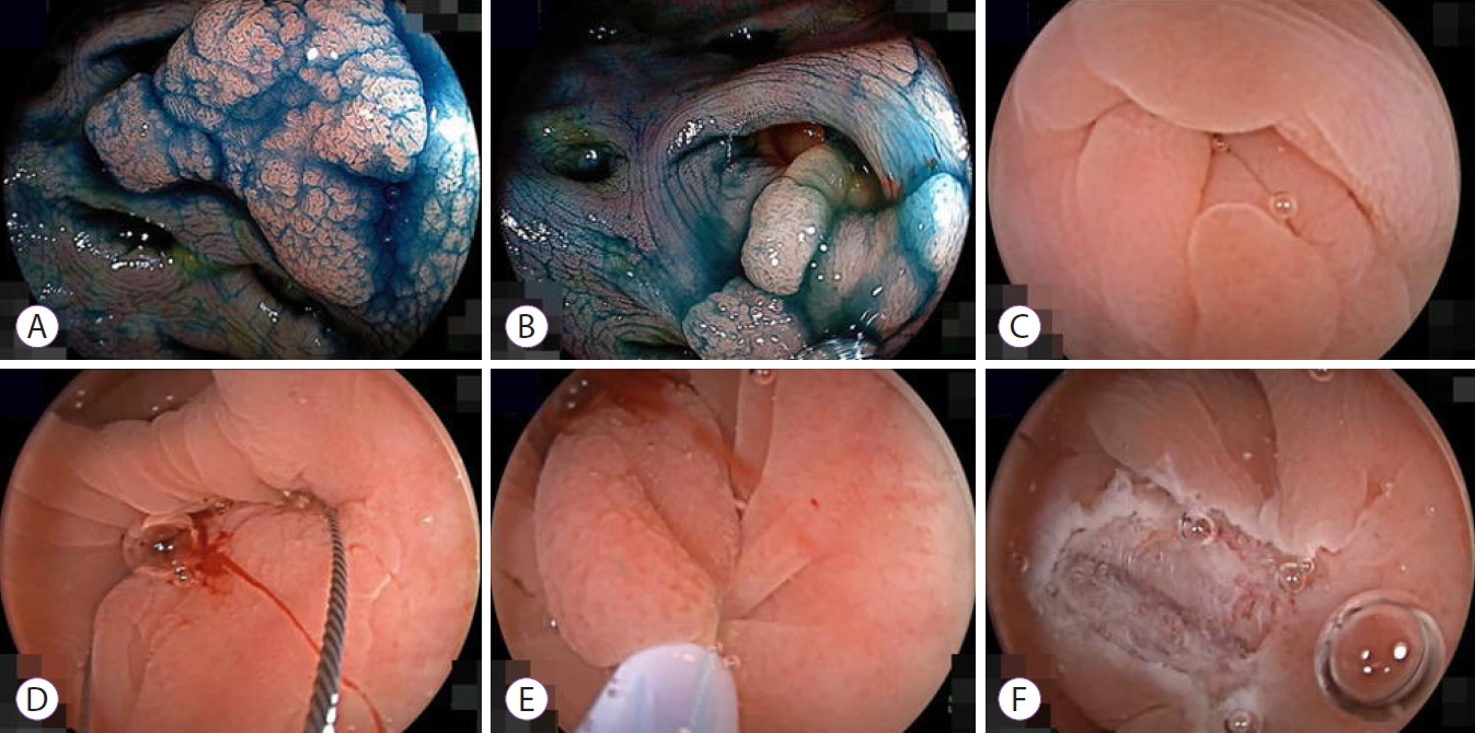
Feasibility and safety of endoscopic submucosal dissection for lesions in proximity to a colonic diverticulum

- Nobuaki Ikezawa, Takashi Toyonaga, Shinwa Tanaka, Tetsuya Yoshizaki, Toshitatsu Takao, Hirofumi Abe, Hiroya Sakaguchi, Kazunori Tsuda, Satoshi Urakami, Tatsuya Nakai, Taku Harada, Kou Miura, Takahisa Yamasaki, Stuart Kostalas, Yoshinori Morita, Yuzo Kodama
- Clin Endosc 2022;55(3):417-425. Published online May 12, 2022
- DOI: https://doi.org/10.5946/ce.2021.245
-
Abstract
PDF
 Supplementary MaterialPubReaderePub
 - Background
/Aims: Endoscopic submucosal dissection (ESD) for diverticulum-associated colorectal lesions is generally contraindicated because of the high risk of perforation. Several studies on patients with such lesions treated with ESD have been reported recently. However, the feasibility and safety of ESD for lesions in proximity to a colonic diverticulum (D-ESD) have not been fully clarified. The aim of this study was to evaluate the feasibility and safety of D-ESD.
Methods
D-ESD was defined as ESD for lesions within approximately 3 mm of a diverticulum. Twenty-six consecutive patients who underwent D-ESD were included. Two strategic approaches were used depending on whether submucosal dissection of the diverticulum-related part was required (strategy B) or not (strategy A). Treatment outcomes and adverse events associated with each strategy were analyzed.
Results
The en bloc resection rate was 96.2%. The rates of R0 and curative resection in strategies A and B were 80.8%, 73.1%, 84.6%, and 70.6%, respectively. Two cases of intraoperative perforation and one case of delayed perforation occurred. The delayed perforation case required emergency surgery, but the other cases were managed conservatively.
Conclusions
D-ESD may be a feasible treatment option. However, it should be performed in a high-volume center by expert hands because it requires highly skilled endoscopic techniques. -
Citations
Citations to this article as recorded by- Endoscopic submucosal dissection for diverticulum using combination of countertraction and circumferential-inversion method
Hiroshi Takayama, Yoshinori Morita, Toshitatsu Takao, Douglas Motomura, Madoka Takao, Takashi Toyonaga, Yuzo Kodama
Endoscopy.2024; 56(S 01): E91. CrossRef - Traction-assisted endoscopic submucosal dissection for resection of ileocecal valve neoplasia: a French retrospective multicenter case series
Clara Yzet, Timothée Wallenhorst, Jérémie Jacques, Mariana Figueiredo Ferreira, Jérôme Rivory, Florian Rostain, Louis-Jean Masgnaux, Jean Grimaldi, Romain Legros, Pierre Lafeuille, Jérémie Albouys, Fabien Subtil, Marion Schaefer, Mathieu Pioche
Endoscopy.2024;[Epub] CrossRef - The role of cap-assisted endoscopy and its future implications
Sol Kim, Bo-In Lee
Clinical Endoscopy.2024; 57(3): 293. CrossRef - Successful planned piecemeal endoscopic resection using gel immersion and an over-the-scope clip for a lesion extensively extended into the colonic diverticulum
Tomoaki Tashima, Takahiro Muramatsu, Tomonori Kawasaki, Tsubasa Ishikawa, Shomei Ryozawa
VideoGIE.2023; 8(4): 167. CrossRef - Future therapeutic implications of new molecular mechanism of colorectal cancer
Sen Lu, Cheng-You Jia, Jian-She Yang
World Journal of Gastroenterology.2023; 29(16): 2359. CrossRef - Iatrogenic colorectal perforation caused by a clip
Hirotaka Oura, Yasuki Hatayama, Erika Nomura, Harutoshi Sugiyama, Daisuke Murakami, Makoto Arai, Takayoshi Nishino
Endoscopy.2023; 55(S 01): E1091. CrossRef
- Endoscopic submucosal dissection for diverticulum using combination of countertraction and circumferential-inversion method
- 3,732 View
- 171 Download
- 4 Web of Science
- 6 Crossref
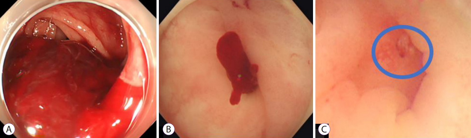
- Comparison of conventional and new endoscopic band ligation devices for colonic diverticular bleeding
- Ayaka Takasu, Takashi Ikeya, Yasutoshi Shiratori
- Clin Endosc 2022;55(3):408-416. Published online February 18, 2022
- DOI: https://doi.org/10.5946/ce.2021.200

-
Abstract
PDFPubReaderePub
- Background
/Aims: Endoscopic band ligation (EBL) is used to treat colonic diverticular bleeding (CDB). An endoscopic variceal ligation device for esophageal varices is used as a conventional EBL device (C-EBL). A new EBL device (N-EBL) was developed by Sumitomo Bakelite Co. in August 2018. We aimed to evaluate the clinical outcomes of N-EBL compared with those of C-EBL.
Methods
Seventy-nine patients who underwent EBL for CDB at St. Luke’s International Hospital, Japan, between 2017 and 2020 were included in this retrospective study. Patients were divided into the C-EBL and N-EBL groups. Their clinical outcomes, including achieving initial hemostasis, early rebleeding, procedure time, and EBL-associated adverse events, were evaluated.
Results
Of the 79 patients, 36 (45.6%) were in the C-EBL group and 43 (54.4%) were in the N-EBL group. The rate of achieving initial hemostasis was 100% in the C-EBL group and 93.0% in the N-EBL group. No significant difference was noted in the early rebleeding rate between the groups (p=0.24). The N-EBL group achieved a shorter median EBL procedure time than the C-EBL group (18.2 minutes vs. 14.2 minutes, p=0.02). No adverse events were observed in either group.
Conclusions
The N-EBL device is safe and useful and may reduce EBL procedure time. -
Citations
Citations to this article as recorded by- Advances in endoscopic management of colonic diverticular bleeding
Yasutoshi Shiratori, Syed Matthew Kodilinye, Ahmed E. Salem
Current Opinion in Gastroenterology.2024;[Epub] CrossRef - Management of Patients With Acute Lower Gastrointestinal Bleeding: An Updated ACG Guideline
Neil Sengupta, Joseph D. Feuerstein, Vipul Jairath, Amandeep K. Shergill, Lisa L. Strate, Robert J. Wong, David Wan
American Journal of Gastroenterology.2023; 118(2): 208. CrossRef - Effective endoscopic band ligation for diverticular perforation with a refractory pelvic abscess
Koichi Soga, Atsushi Majima
Clinical Endoscopy.2023; 56(2): 252. CrossRef - A new band ligation device to treat colonic diverticular bleeding
Yunho Jung
Clinical Endoscopy.2022; 55(3): 367. CrossRef
- Advances in endoscopic management of colonic diverticular bleeding
- 3,847 View
- 237 Download
- 2 Web of Science
- 4 Crossref
Case Reports
- Endoscopic Treatment of Iatrogenic Perforation of Sigmoid Diverticulum: A Case Report of Multidisciplinary Management
- Giacomo Emanuele Maria Rizzo, Giuseppina Ferro, Giovanna Rizzo, Giovanni Di Carlo, Alessandro Cantone, Gaetano Giuseppe Di Vita, Carmelo Sciumè
- Clin Endosc 2022;55(2):292-296. Published online June 7, 2021
- DOI: https://doi.org/10.5946/ce.2021.005
-
Abstract
PDFPubReaderePub
- Iatrogenic perforations are severe complications of gastrointestinal endoscopy; therefore, their management should be adequately planned. A 77-year-old man with a history of diverticulosis underwent a colonoscopy for anemia. During the procedure, an iatrogenic perforation occurred suddenly in the sigmoid colon, near a severe angle among the numerous diverticula. Through-the-scope clips were immediately applied to treat it and close mucosal edges. Laboratory tests showed increased levels of inflammation and infection, and although there were no complaints of abdominal pain, the patient had an extremely distended abdomen. A multidisciplinary board began management based on a conservative approach. Pneumoperitoneum was treated with computed tomography-assisted drainage. After 72 hours, his intestinal canalization and laboratory tests were normal. Though this adverse event is rare, a multidisciplinary board should be promptly gathered upon occurrence, even if the patient appears clinically stable, to consider a conservative approach and avoid surgical treatment.
- 3,552 View
- 161 Download
- Parapneumonic Effusion and Tension Pneumothorax after Diverticular Peroral Endoscopic Myotomy in a Woman with Large Epiphrenic Diverticulum: A Case Report and Literature Review
- Sz-Iuan Shiu
- Clin Endosc 2021;54(2):275-279. Published online January 15, 2021
- DOI: https://doi.org/10.5946/ce.2020.093
-
Abstract
PDFSupplementary MaterialPubReaderePub
- Esophageal diverticula (ED) represents a group of rare conditions that warrant intervention when they are symptomatic or coexisting with pulmonary disorders. Few literature reviews have described this patient entity or discussed the postoperative outcome. Therefore, I present the case of a 59-year-old woman with symptoms of dysphagia who was significantly underweight, which was conducive to the diagnosis of symptomatic ED. Because she was a poor candidate for surgery, she received a diverticular peroral endoscopic myotomy. She subsequently developed parapneumonic effusion and tension pneumothorax after the procedure. She was finally discharged on postoperative day 23. I also performed the first known comprehensive literature review of 34 published cases (including my patient) from PubMed and have addressed the demography, intervention, and prognosis for symptomatic ED after the procedure. Prompt treatment as well as prognostic measurement are crucial to successful outcomes.
- 2,826 View
- 109 Download
- 1 Web of Science
Focused Review Series: Cutting Edge of Advanced Therapeutic Endoscopy
-
Role of Peroral Endoscopic Myotomy (POEM) in the Management of Esophageal Diverticula
- Bogdan P. Miutescu, Sarah Khan, Shruti Mony, Mouen A. Khashab
- Clin Endosc 2020;53(6):646-651. Published online November 26, 2020
- DOI: https://doi.org/10.5946/ce.2020.262
-
Abstract
PDFSupplementary MaterialPubReaderePub
- Esophageal diverticula are uncommon; however, when present, they can cause symptoms of dysphagia, regurgitation, and chest pain. Based on location and pathophysiological characteristics, they are classified as pulsion- and traction-type diverticula. In the past, the open surgical approach was the only treatment available; however, in the past few decades, transoral incisionless approaches in the form of rigid and flexible endoscopy have gained popularity. Diverticular peroral endoscopic myotomy has emerged as an alternative treatment option. In this paper, we reviewed the role of peroral endoscopic myotomy as a treatment option for different types of esophageal diverticula. Although a safe and effective procedure, this novel submucosal tunneling technique for the treatment of esophageal diverticula requires further validation, and head-to-head comparisons between the different approaches for the treatment of esophageal diverticula are warranted.
-
Citations
Citations to this article as recorded by- Efficacy and safety of peroral endoscopic myotomy for esophageal diverticula
Elise M. Wessels, Jeroen M. Schuitenmaker, Barbara A.J. Bastiaansen, Paul Fockens, Gwen M.C. Masclee, Albert J. Bredenoord
Endoscopy International Open.2023; 11(05): E546. CrossRef - Peroral endoscopic myotomy (POEM) for esophageal diverticula
Jayanta SAMANTA, Zaheer NABI, Jahnvi DHAR, Harshal S. MANDAVDHARE
Minerva Gastroenterology.2023;[Epub] CrossRef - Multimodal Endoscopic Management of Esophageal Perforations as a Complication of Peroral Endoscopic Myotomy for a Zenker's Diverticulum
Erlison Mauricio Daza Castro, Carlos Fernando Fuentes, Andrea Carolina Córdoba Guzmán, Diego Aponte, José Nicolás Rocha, Carlos González, Luis Carlos Sabbagh
ACG Case Reports Journal.2023; 10(6): e01059. CrossRef - A rare case of bilateral Killian-Jamieson diverticula treated endoscopically
Catarina Félix, Pedro Barreiro, José Rodrigues, Rui Mendo, Catarina O’Neill, Cristina Chagas
Endoscopy.2022; 54(06): E283. CrossRef - Peroral endoscopic myotomy, septotomy, and restoration of esophageal lumen with over-the-scope clips: closing the circle of esophageal diverticula management
Eduardo Albéniz, Fermín Estremera-Arévalo, Marta Gómez Alonso, Pedro J. Rosón, Francisco J. Gallego Rojo, Juan Vila, Sheyla Montori
Endoscopy.2022; 54(11): E666. CrossRef - Successful D-POEM after failed surgical myotomy and diverticulectomy
Andrew Ross Leopold, Raymond E. Kim
VideoGIE.2022; 7(6): 211. CrossRef - Peroral Endoscopic Myotomy for the Treatment of Esophageal Diverticula
Antonio Facciorusso, Daryl Ramai, Yervant Ichkhanian, Rena Yadlapati, Vito Annese, Sachin Wani, Mouen A. Khashab
Journal of Clinical Gastroenterology.2022; 56(10): 853. CrossRef
- Efficacy and safety of peroral endoscopic myotomy for esophageal diverticula
- 6,113 View
- 197 Download
- 8 Web of Science
- 7 Crossref
Case Report
-
Underwater Endoscopic Mucosal Resection without Submucosal Injection Facilitates En bloc Resection of Colon Adenomas Extending into a Diverticulum
- Yoshikazu Hayashi, Masahiro Okada, Takaaki Morikawa, Tatsuma Nomura, Hisashi Fukuda, Takahito Takezawa, Alan Kawarai Lefor, Hironori Yamamoto
- Clin Endosc 2021;54(3):436-440. Published online November 6, 2020
- DOI: https://doi.org/10.5946/ce.2020.168
-
Abstract
PDFSupplementary MaterialPubReaderePub
- Superficial colonic neoplasms sometimes extend into a diverticulum. Conventional endoscopic mucosal resection of these lesions is considered challenging because colonic diverticula do not have a muscularis propria and are deeply inverted. Even if the solution is carefully injected below the mucosa at the bottom of the diverticulum, the mucosa is rarely elevated from the diverticular orifice, and it is usually just narrowed. Although endoscopic submucosal dissection or full-thickness resection with an over-the-scope clip device enables the complete resection of these lesions, it is still challenging, time consuming and expensive. Underwater endoscopic mucosal resection without submucosal injection (UEMR) is an innovative technique enabling en bloc resection of superficial colon lesions. We report three patients with colon adenomas extending into a diverticulum treated with successful UEMR. UEMR enabled rapid and safe en bloc resection of colon lesions extending into a diverticulum.
-
Citations
Citations to this article as recorded by- Underwater endoscopic mucosal resection for colorectal lesions: Can it be an “Underwater” revolution?
Yoji Takeuchi, Satoki Shichijo, Noriya Uedo, Ryu Ishihara
DEN Open.2022;[Epub] CrossRef
- Underwater endoscopic mucosal resection for colorectal lesions: Can it be an “Underwater” revolution?
- 3,777 View
- 111 Download
- 1 Web of Science
- 1 Crossref
Original Article
-
Utility of the Gel Immersion Method for Treating Massive Colonic Diverticular Bleeding
- Kazuki Yamamoto, Yasutoshi Shiratori, Takashi Ikeya
- Clin Endosc 2021;54(2):256-260. Published online August 11, 2020
- DOI: https://doi.org/10.5946/ce.2020.081
-
Abstract
PDFSupplementary MaterialPubReaderePub
- Background
/Aims: In Asia, right-sided diverticular bleeding is more common than that of the left side. It often causes massive bleeding and difficulties in identifying the stigmata of recent hemorrhage (SRH) of colonic diverticular bleeding (CDB). This case series demonstrates the efficacy of the gel immersion method using OS-1 Jelly (Otsuka Pharmaceuticals Factory, Tokushima, Japan) in patients with CDB.
Methods
This retrospective case series analyzed data of patients with CDB who underwent the gel immersion method from April 2016 to February 2020 at St. Luke’s International Hospital, Japan. All patients diagnosed with CDB who underwent the gel immersion method were included. We collected data on the site of bleeding, identification of SRH, and efficacy of the method from the electronic medical records.
Results
A total of 9 patients (including 7 with right-sided CDB) underwent gel immersion method and were included in this study. SRH were successfully found in 66.7% (6/9) of patients. Moreover, effective hemostasis was achieved in 85.7% (6/7) of patients with right-sided CDB. There were no adverse events.
Conclusions
The gel immersion method was found to be effective, especially for massive right-sided CDB. -
Citations
Citations to this article as recorded by- Advances in endoscopic management of colonic diverticular bleeding
Yasutoshi Shiratori, Syed Matthew Kodilinye, Ahmed E. Salem
Current Opinion in Gastroenterology.2024;[Epub] CrossRef - Successful direct clipping of the bleeding source of a colonic diverticular hemorrhage using the “long-hood gel-filling” method
Satoshi Abiko, Koji Hirata, Kazuharu Suzuki, Kenji Kinoshita, Kazuteru Hatanaka, Yoshiya Yamamoto, Hirohito Naruse
Endoscopy.2023; 55(S 01): E606. CrossRef - Utility of under-gel endoscopic mucosal resection with partial submucosal injection for a laterally spreading tumor
Kazuki Yamamoto, Naoki Kanomata, Takashi Ikeya
Endoscopy.2022; 54(03): E88. CrossRef - Localizing spontaneously hemostatic colonic diverticular bleeding using VISCOCLEAR gel: A case report
Daisuke Suto, Masashi Yoshida, Takaaki Otake, Eiichiro Ichiishi, Kiichi Sato, Yosuke Osawa, Hirotoshi Ebinuma, Hironori Odaira, Yutaka Suzuki, Yutaka Kohgo
Annals of Medicine & Surgery.2022;[Epub] CrossRef - Gel Immersion Endoscopic Mucosal Resection (EMR) for Superficial Nonampullary Duodenal Epithelial Tumors May Reduce Procedure Time Compared with Underwater EMR (with Video)
Takeshi Yamashina, Masaaki Shimatani, Yu Takahashi, Masahiro Takeo, Natsuko Saito, Hironao Matsumoto, Takeshi Kasai, Masataka Kano, Kimi Sumimoto, Toshiyuki Mitsuyama, Hiroyuki Marusawa, Akiyoshi Nishio, Takafumi Yuba, Toshihito Seki, Makoto Naganuma, Tat
Gastroenterology Research and Practice.2022; 2022: 1. CrossRef - Digital compression for hemostasis in acute hemorrhagic rectal ulcer: a report of 4 cases and review of the literature
Takeshi Okamoto, Ayaka Takasu, Takaaki Yoshimoto, Kazuki Yamamoto, Yasutoshi Shiratori, Takashi Ikeya, Katsuyuki Fukuda
Clinical Journal of Gastroenterology.2021; 14(3): 796. CrossRef - Efficiency of a novel gel product for duodenal ulcer bleeding
Shuichi Miyamoto, Kazuharu Suzuki, Kenji Kinoshita
Digestive Endoscopy.2021;[Epub] CrossRef - Development of a gel dedicated to gel immersion endoscopy
Tomonori Yano, Atsushi Ohata, Yuji Hiraki, Makoto Tanaka, Satoshi Shinozaki, Alan Kawarai Lefor, Hironori Yamamoto
Endoscopy International Open.2021; 09(06): E918. CrossRef - Gel immersion endoscopy: Innovation in securing the visual field – Clinical experience with 265 consecutive procedures
Tomonori Yano, Takahito Takezawa, Kousei Hashimoto, Ayako Ohmori, Satoshi Shinozaki, Manabu Nagayama, Hirotsugu Sakamoto, Yoshimasa Miura, Yoshikazu Hayashi, Keijiro Sunada, Alan Kawarai Lefor, Hironori Yamamoto
Endoscopy International Open.2021; 09(07): E1123. CrossRef
- Advances in endoscopic management of colonic diverticular bleeding
- 5,241 View
- 176 Download
- 10 Web of Science
- 9 Crossref
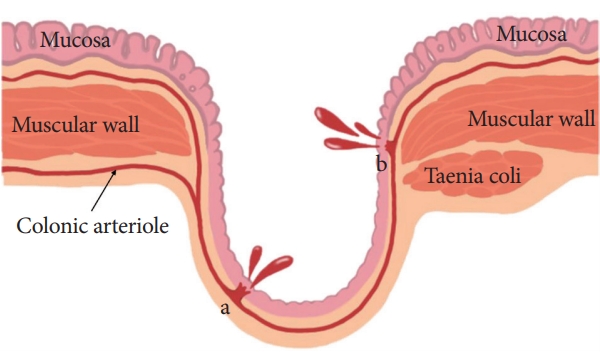
Focused Review Series: Endoscopic Hemostasis: An Overview of Principles and Recent Applications
- Endoscopic Therapy for Acute Diverticular Bleeding
- Masayuki Kato
- Clin Endosc 2019;52(5):419-425. Published online August 20, 2019
- DOI: https://doi.org/10.5946/ce.2019.078
-
Abstract
PDFPubReaderePub
- Diverticular bleeding accounts for approximately 26%–40% of the cases of lower gastrointestinal bleeding. Rupture of the vasa recta at the neck or dome of the diverticula can be the cause of this bleeding. Colonoscopy aids in not only the diagnosis but also the treatment of diverticular bleeding after a steady bowel preparation. Endoscopic hemostasis involves several methods, such as injection/thermal contact therapy, clipping, endoscopic band ligation (EBL), hemostatic powder, and over-the-scope clips. Each endoscopic method can provide a secure initial hemostasis. With regard to the clinical outcomes after an endoscopic treatment, the methods reportedly have no significant differences in the initial hemostasis and early recurring bleeding; however, EBL might prevent the need for transcatheter arterial embolization or surgery. In contrast, the long-term outcomes of the endoscopic treatments, such as a late bleeding and recurrent bleeding at 1 and 2 years, are not well known for diverticular bleeding. With regard to a cure for diverticular bleeding, there should be an improvement in both the endoscopic methods and the multilateral perspectives, such as diet, medicines, interventional approaches, and surgery.
-
Citations
Citations to this article as recorded by- A Review of Colonoscopy in Intestinal Diseases
Seung Hong, Dong Baek
Diagnostics.2023; 13(7): 1262. CrossRef - Short Peptide Nanofiber Biomaterials Ameliorate Local Hemostatic Capacity of Surgical Materials and Intraoperative Hemostatic Applications in Clinics
Zehong Yang, Lihong Chen, Ji Liu, Hua Zhuang, Wei Lin, Changlong Li, Xiaojun Zhao
Advanced Materials.2023;[Epub] CrossRef - A new band ligation device to treat colonic diverticular bleeding
Yunho Jung
Clinical Endoscopy.2022; 55(3): 367. CrossRef - Diagnosis and Treatment of Colonic Diverticular Disease
You Sun Kim
The Korean Journal of Gastroenterology.2022; 79(6): 233. CrossRef - Efficacy of Combination Therapy with Epinephrine Local Injection and Hemostatic Clips on Active Diverticular Bleeding
Seiji Hamada, Akira Teramoto, Ryuta Zukeyama, Shinobu Matsukawa, Tomofumi Fukuhara, Ryo Takaki, Takahiro Utsumi, Masamoto Nakamura, Kasen Kobashikawa, Nobufumi Uchima, Tomokuni Nakayoshi, Fukunori Kinjo
Journal of Clinical Medicine.2022; 11(17): 5195. CrossRef - Diverticular Bleeding: A Clinical Image
Christopher F Brewer, Yayha Al Abed
Cureus.2021;[Epub] CrossRef
- A Review of Colonoscopy in Intestinal Diseases
- 13,292 View
- 503 Download
- 6 Web of Science
- 6 Crossref
Original Article
- Impact of Periampullary Diverticulum on ERCP Performance: A Matched Case-Control Study
- Juan E. Corral, Omar Y. Mousa, Paul T. Kröner, Victoria Gomez, Frank J. Lukens
- Clin Endosc 2019;52(1):65-71. Published online August 21, 2018
- DOI: https://doi.org/10.5946/ce.2018.070

-
Abstract
PDFPubReaderePub
- Background
/Aims: Periampullary diverticulum (PAD) is frequently encountered during endoscopic retrograde cholangiopancreatography (ERCP) and has been associated with stone formation in the bile duct. The effects of PAD on the ERCP procedure have been often debated. We aimed to compare the therapeutic success of ERCP between patients with PAD and matched controls.
Methods
We reviewed all ERCPs with findings of PAD in a national database (n=1,089) and compared them with age- and gendermatched controls in a 1:3 fashion (n=3,267). Demographics, endoscopic findings, visualization of main structures, and therapeutic success rates were compared between groups. Secondary analysis compared PAD cases and controls who had gallstone disease.
Results
The average cohort age was 68.4±14.3 years and 55.1% were male. ERCP success was similar in both groups, and no significant inter-group differences were found in the multivariate analysis. The presence of PAD did not affect the rates of sphincterotomy or visualization of main biliary structures. Secondary analysis showed similar success rates for gallstone removal between patients with PAD and controls.
Conclusions
PAD may not be considered a hinderance to ERCP success. Further research is needed to determine the best approach to cannulate the ampulla and provide endoscopic therapy for different subtypes of PAD. -
Citations
Citations to this article as recorded by- Impact of periampullary diverticulum on biliary cannulation: A retrospective cohort study
Jing Liang Ho, Aruni Seneviratna, Cherng Hann Benjamin Yip
Advances in Digestive Medicine.2023; 10(4): 232. CrossRef - A new classification of periampullary diverticulum: cannulation of papilla on the inner margins of the diverticulum (Type IIa) is more challenging
He-xian Shi, Yong-qiang Ye, Hai-wang Zhao, De-cai Kong, Shan-zhou Huang, Qian Yan, Yu-bin Chen, Ping Zhang, Sheng Chen, Bao-hua Hou, Chuan-zhao Zhang
BMC Gastroenterology.2023;[Epub] CrossRef - Clinical significance of different periampullary diverticulum classifications for endoscopic retrograde cholangiopancreatography cannulation
Ping Yue, Ke-Xiang Zhu, Hai-Ping Wang, Wen-Bo Meng, Jian-Kang Liu, Lei Zhang, Xiao-Liang Zhu, Hui Zhang, Long Miao, Zheng-Feng Wang, Wen-Ce Zhou, Azumi Suzuki, Kiyohito Tanaka, Xun Li
World Journal of Gastroenterology.2020; 26(19): 2402. CrossRef - Clinical significance of different periampullary diverticulum classifications for endoscopic retrograde cholangiopancreatography cannulation
Ping Yue, Ke-Xiang Zhu, Hai-Ping Wang, Wen-Bo Meng, Jian-Kang Liu, Lei Zhang, Xiao-Liang Zhu, Hui Zhang, Long Miao, Zheng-Feng Wang, Wen-Ce Zhou, Azumi Suzuki, Kiyohito Tanaka, Xun Li
World Journal of Gastroenterology.2020; 26(19): 2403. CrossRef - Periampüller divertikül endoskopik retrograd kolanjiyopankreatografide kanülasyon başarısı ve komplikasyon sıklığını etkiler mi?
Bilal TOKA, Salih TOKMAK
Akademik Gastroenteroloji Dergisi.2020; 19(2): 83. CrossRef - Complications increase in which type of duodenal diverticulum? A retrospective cohort study
Murat AKAYDIN, Tamer AKAY, Metin LEBLEBİCİ
Journal of Surgery and Medicine.2020; 4(11): 938. CrossRef - Presence of Periampullary Diverticulum is Not a Hurdle to Successful Endoscopic Retrograde Cholangiopancreatography
Jimin Han
Clinical Endoscopy.2019; 52(1): 7. CrossRef - ERCP Success Rate and Periampullary Diverticula: The Pocket Makes No Difference
Gyanprakash Ketwaroo, Waqar Qureshi
Digestive Diseases and Sciences.2019; 64(5): 1072. CrossRef
- Impact of periampullary diverticulum on biliary cannulation: A retrospective cohort study
- 5,006 View
- 151 Download
- 6 Web of Science
- 8 Crossref
Case Reports
-
Congenital Jejunal Diverticular Bleeding in a Young Adult
- Ji-Yung Lee, Jae-Young Jang, Min-Je Kim, Tae-In Lee, Jung-Wook Kim, Young-Woon Chang
- Clin Endosc 2017;50(5):495-499. Published online June 14, 2017
- DOI: https://doi.org/10.5946/ce.2016.154
-
Abstract
PDFSupplementary MaterialPubReaderePub
- Diverticular bleeding of the small bowel is rare and occurs primarily in adults aged more than 60 years. In younger adults, Meckel’s diverticulum, a true diverticulum that congenitally occurs in the distal ileum, is the most common cause of diverticular bleeding of the small bowel. Unlike Meckel’s diverticula, other kinds of small bowel diverticula are not congenital and their incidence is related to age. Furthermore, congenital true diverticular bleeding of the jejunum in adults is very rare. We report the case of a 24-year-old man with subepithelial tumor-like lesion accompanied with obscure overt gastrointestinal bleeding (OOGIB). This lesion was initially suspected to be a subepithelial tumor based on radiologic tests and capsule endoscopy. He was finally diagnosed with a congenital true diverticulum in the jejunum with the appearance of a Meckel’s diverticulum after surgical resection.
-
Citations
Citations to this article as recorded by- Diverticular hemorrhage of terminal ileum successfully treated by ultra-selective transcatheter arterial embolization using triaxial system: a case report
Yuki Yaginuma, Kenichi Utano, Yuka Utano, Daiki Nemoto, Masato Aizawa, Hajime Matsuida, Noriyuki Isohata, Shungo Endo, Kazutomo Togashi
Clinical Journal of Gastroenterology.2021; 14(2): 517. CrossRef - Small bowel bleeding
Stefania Chetcuti Zammit, Reena Sidhu
Current Opinion in Gastroenterology.2018; 34(3): 165. CrossRef - Diagnosis of Meckel’s Diverticulum Using Colon Capsule Endoscopy for Small Bowel Investigation
Lidia Ciobanu, Oliviu Pascu, Marcel Tanțău
Clinical Endoscopy.2018; 51(4): 395. CrossRef
- Diverticular hemorrhage of terminal ileum successfully treated by ultra-selective transcatheter arterial embolization using triaxial system: a case report
- 6,704 View
- 121 Download
- 4 Web of Science
- 3 Crossref
- Endoscopic Submucosal Dissection of an Inverted Early Gastric Cancer-Forming False Gastric Diverticulum
- Yong-il Lee, Sang-kil Lee
- Clin Endosc 2016;49(1):86-90. Published online January 28, 2016
- DOI: https://doi.org/10.5946/ce.2016.49.1.86
-
Abstract
PDFPubReaderePub
- Endoscopic submucosal dissection (ESD) is a standard treatment for early gastric cancer (EGC) that does not have any risk of lymph node or distant metastases. Here, we report a case of EGC resembling a diverticulum. Diverticular formation makes it difficult for endoscopists to determine the depth of invasion and to subsequently perform ESD. Because the false diverticulum does not have a muscular layer, this lesion can be treated with ESD. Our case was successfully treated with ESD. After ESD, the EGC was confined to the submucosal layer without vertical and lateral margin involvement. This is the first case in which ESD was successfully performed for a case of EGC that coexisted with a false gastric diverticulum. An additional, larger study is needed to determine the efficacy of ESD in various types of EGC, such as a false gastric diverticulum.
-
Citations
Citations to this article as recorded by- A rare case of perforated gastric duplication cyst associated with gastric diverticulum
Joseph M Smith, Jessie A Elliott, Amy E Gillis, Paul F Ridgway
BMJ Case Reports.2021; 14(3): e239971. CrossRef - Upper Gastrointestinal Manifestation of Bezoars and the Etiological Factors: A Literature Review
Samiullah Khan, Kui Jiang, Lan-ping Zhu, Iftikhar-ahmad Khan, Kifayat Ullah, Saima Khan, Xin Chen, Bang-mao Wang
Gastroenterology Research and Practice.2019; 2019: 1. CrossRef - Gastric Diverticulum: A Comprehensive Review
Jamil Shah, Kalpesh Patel, Tagore Sunkara, Charilaos Papafragkakis, Abul Shahidullah
Inflammatory Intestinal Diseases.2018; 3(4): 161. CrossRef
- A rare case of perforated gastric duplication cyst associated with gastric diverticulum
- 7,371 View
- 68 Download
- 4 Web of Science
- 3 Crossref
- Esophageal Cancer in Esophageal Diverticula Associated with Achalasia
- Ah Ran Choi, Nu Ri Chon, Young Hoon Youn, Hyo Chae Paik, Yon Hee Kim, Hyojin Park
- Clin Endosc 2015;48(1):70-73. Published online January 31, 2015
- DOI: https://doi.org/10.5946/ce.2015.48.1.70
-
Abstract
PDFPubReaderePub
The simultaneous occurrence of achalasia and esophageal diverticula is rare. Here, we report the case of a 68-year-old man with multiple esophageal diverticula associated with achalasia who was later diagnosed with early esophageal cancer. He initially presented with dysphagia and dyspepsia, and injection of botulinum toxin to the lower esophageal sphincter relieved his symptoms. Five years later, however, the patient presented with worsening of symptoms, and esophagogastroduodenoscopy (EGD) was performed. The endoscopic findings showed multifocal lugol-voiding lesions identified as moderate dysplasia. We decided to use photodynamic therapy to treat the multifocal dysplastic lesions. At follow-up EGD 2 months after photodynamic therapy, more lugol-voiding lesions representing a squamous cell carcinoma
in situ were found. The patient ultimately underwent surgery for the treatment of recurrent esophageal multifocal neoplasia. After a follow-up period of 3 years, the patient showed a good outcome without symptoms. To manage premalignant lesions such as achalasia with esophageal diverticula, clinicians should be cautious, but have an aggressive approach regarding endoscopic surveillance.-
Citations
Citations to this article as recorded by- Advanced squamous cell carcinoma in an asymptomatic, large, epiphrenic esophageal diverticulum
Tomoaki Yoshida, Satoru Hashimoto, Ken-ichi Mizuno, Hiroshi Ichikawa, Junji Yokoyama, Hajime Umezu, Shuji Terai
Clinical Journal of Gastroenterology.2020; 13(4): 477. CrossRef - Locally Advanced Esophageal Cancer Arising from an Epiphrenic Diverticulum Treated by Curative Esophagectomy Combined with Adjacent Organs Resection
Aina KUNITOMO, Eiji HIGAKI, Tetsuya ABE, Takahiro HOSOI, Seiji ITO, Yasuhiro SHIMIZU
Nihon Rinsho Geka Gakkai Zasshi (Journal of Japan Surgical Association).2019; 80(11): 1999. CrossRef - Imaging in the Evaluation of Endoscopic or Surgical Treatment for Achalasia
Diego Palladino, Andrea Mardighian, Marilina D’Amora, Luca Roberto, Francesco Lassandro, Claudia Rossi, Gianluca Gatta, Mariano Scaglione, Guglielmi Giuseppe
Gastroenterology Research and Practice.2016; 2016: 1. CrossRef
- Advanced squamous cell carcinoma in an asymptomatic, large, epiphrenic esophageal diverticulum
- 6,299 View
- 59 Download
- 4 Web of Science
- 3 Crossref
- A Case of Duodenal Diverticulitis
- Chang Hyeon Seock, M.D., Hae Kyung Kim, M.D., Tae Il Park, M.D., Byung Min John, M.D., Hyeon U Jo, M.D., Jae Seung Kim, M.D., Kee Bum Kim, M.D.* and Byung Soo Na, M.D.*
- Korean J Gastrointest Endosc 2010;41(5):294-297. Published online November 30, 2010
-
Abstract
PDF
- Duodenal diverticulitis is difficult to diagnose because it can mimic other common diseases such as cholecystitis and perforated ulcer. Recently, we experienced a rare case of duodenal diverticulitis that was initially suspected on abdominal computed tomography as focal pancreatitis. Although duodenal diverticulitis has been increasingly recognizable before surgery, with the advent of multi-detector computed tomography, misdiagnosis remains problematic since duodenal diverticulitis is commonly not considered in the differential diagnosis of acute abdominal pain. We have to consider this rare disease entity because delayed diagnosis might be a cause of substantial morbidity and mortality. (Korean J Gastrointest Endosc 2010;41:294-297)
- 2,463 View
- 20 Download
- A Case of Meckel's Diverticulum Diagnosed for Recurrent Hematochezia in Old Age
- Dong Kyun Kim, M.D., Jin Oh Kim, M.D., Hyun Gun Kim, M.D., Tae Hee Lee, M.D., Yoon Ho Jung, M.D., Joo Young Cho, M.D., Joon Seong Lee, M.D. and So Young Jin, M.D.*
- Korean J Gastrointest Endosc 2010;40(5):334-337. Published online May 30, 2010
-
Abstract
PDF
- The small bowel is the most common site of an unknown origin of gastrointestinal bleeding. Meckel's diverticulum is the most common congenital anomaly of the gastrointestinal tract in children. The complications from Meckel's diverticulum such as bleeding decrease with age, and so Meckel's diverticular bleeding is very rare in old age patients. The diagnosis of Meckel's diverticulum may be very difficult and especially in old age patients. Capsule endoscopy and double balloon enteroscopy have recently become useful diagnostic tools for assessing diverticular bleeding and small bowel lesions as these techniques can examine the entire small intestine. We report here on a case of Meckel's diverticulum that was detected by capsule endoscopy and double balloon enteroscopy and this was confirmed by surgery in a 76-year-old man with recurrent hematochezia. (Korean J Gastrointest Endosc 2010;40:334-337)
- 1,932 View
- 10 Download
- Acute Pseudodiverticular Bleeding from a Tuberculous Scar of the Terminal Ileum
- Suh Eun Bae, M.D., Sung-Ae Jung, M.D., Hyun Joo Song, M.D., Min-Jung Kang, M.D., Ji Min Jung, M.D., Seong-Eun Kim, M.D., Ki-Nam Shim, M.D. and Kwon Yoo, M.D.
- Korean J Gastrointest Endosc 2009;38(6):360-363. Published online June 30, 2009
-
Abstract
PDF
- Acquired ileal diverticuli are an uncommon condition and the diagnosis is often difficult when bleeding occurs from this source. Tuberculosis mainly involves the terminal ileum and has associated complications such as obstruction, perforation, stricture and bleeding, but rarely presents with pseudodiverticuli with a fistula. A 42-year-old man presented with massive hematochezia for three days. The patient had a history of pulmonary tuberculosis with complete recovery two times. Emergency sigmoidoscopy, esophagoduodenoscopy and computed tomography of the abdomen could not detect the bleeding focus. The next day, colonoscopy was performed, which demonstrated the opening of pseudodiverticuli at the terminal ileum. There was an exposed vessel in one of the pseudodiverticuli. The patient was treated successfully with epinephrine and ethanol sclerotherapy. A subsequent colonoscopy showed that the exposed vessel was completely healed seven days later. We report a case of acute pseudodiverticular bleeding from a tuberculous scar of the terminal ileum with a review of the relevant literature. (Korean J Gastrointest Endosc 2009;38:360-363)
- 1,621 View
- 4 Download
- A Case of Narrow Opened Priampullary Diverticular Bleeding with Diagnostic Difficulty
- Jin Nam Kim, M.D., Hong Sik Lee, M.D., Jae Hong Ahn, M.D., Seung Young Kim, M.D., Dong Il Kim, M.D., Sang Woo Lee, M.D. and Jae Hyun Choi, M.D.
- Korean J Gastrointest Endosc 2009;38(5):275-378. Published online May 30, 2009
-
Abstract
PDF
- A duodenal diverticulum is most common in the medial aspect of the second portion of the duodenum and rarely causes symptoms. An obstruction, bleeding, perforation, jaundice and pancreatitis are uncommon complications of a duodenal diverticulum. Bleeding from the periampullary diverticulum should be considered in the diagnosis of a patient who presents with upper gastrointestinal bleeding of unknown origin. The second portion of the duodenum is sometimes difficult to observe entirely from the tangent line with the use of a forward-viewing endoscope. The diagnosis and treatment of periampullary diverticular bleeding may be achieved more easily by use of a side-viewing endoscope. We report here a case of narrow opened periampullary diverticular bleeding diagnosed by the use of a side-viewing endoscope with difficulty. (Korean J Gastrointest Endosc 2009; 38:275-278)
- 2,105 View
- 10 Download
- A Case of a Meckel's Diverticular Bleeding Diagnosed by the Use of Double Balloon Enteroscopy
- Woo Young Heo, M.D., Jae Myung Cha, M.D., Sung Won Jung, M.D., Hyun Phil Shin, M.D., Jae Won Choe, M.D., Kang Ro Joo, M.D., Joung Il Lee, M.D. and Jong Beom Park, M.D.*
- Korean J Gastrointest Endosc 2008;37(5):364-368. Published online November 30, 2008
-
Abstract
PDF
- Meckel's diverticulum is one of the most common congenital anomalies, and its incidence is about 2% in the population. Most of the cases are asymptomatic and only 5% of cases are symptomatic with complications, including bleeding, intestinal obstruction, inflammation and perforation. Bleeding from a Meckel's diverticulum is usually painless and is sometimes massive. Recently, the use of double balloon enteroscopy has allowed improved access in patients with obscure gastrointestinal bleeding; however, a case of bleeding from a Meckel's diverticulum treated with double balloon enteroscopy hs rarely been reported. Double balloon enteroscopy can diagnose a Meckel's diverticulum without difficulty as the lesion is usually located at the distal ileum, and the method provides endoscopic hemostasis for the bleeding. Therefore, double balloon enteroscopy might be a useful diagnostic and therapeutic modality for bleeding from a Meckel's diverticulum. We experienced a case of bleeding from a Meckel's diverticulum that was detected and was treated by the use of double-balloon enteroscopy in a 47-year-old man with recurrent episodes of melena. (Korean J Gastrointest Endosc 2008;37:364-368)
- 2,079 View
- 5 Download
- A Case of Esophageal Compression by a Right-sided Aortic Arch and Kommerell's Diverticulum Mimicking an Esophageal Submucosal Tumor
- Young Jig Cho, M.D., Bora Keum, M.D., Youn Ho Kim, M.D., Hwi Kong, M.D., Jin Nam Kim, M.D., Yong Sik Kim, M.D., Yoon Tae Jeen, M.D., Hoon Jai Chun, M.D., Soon Ho Um, M.D., Chang Duck Kim, M.D., Ho Sang Rhu, M.D. and Jin Hai Hyun, M.D.
- Korean J Gastrointest Endosc 2008;36(2):78-82. Published online February 27, 2008
-
Abstract
PDF
- A right-sided aortic arch and Kommerell's diverticulum, remnants of the left dorsal aortic arch in the circulation of the embryo, are uncommon congenital defects of the aorta. They may be asymptomatic in most cases, but symptoms are manifested by compressing mediastinal structures or are related to congenital heart anomalies. If aneurismal dilatation of the diverticulum presents with rupture, it is lethal. We report a case of esophageal compression by a right-sided aortic arch and Kommerell's diverticulum that mimicked an esophageal submucosal tumor in a patient who complained of symptoms during the past ten years of food retention in the upper thorax when a bolus of food was ingested. (Korean J Gastrointest Endosc 2008;36: 78-82)
- 2,060 View
- 14 Download
- Analysis of Risk Factors Associated with the Severity of Biliary Pancreatitis
- Hyo Jeong Oh, M.D., Tae Hyeon Kim, M.D., Chang Soo Choi, M.D., Ji Hye Kweon, M.D., Pyoung Suk Lim, M.D., Sae Ron Shin, M.D.*, Suck Chei Choi, M.D. and Yong-Ho Nah, M.D.
- Korean J Gastrointest Endosc 2007;35(6):385-390. Published online December 30, 2007
-
Abstract
PDF
- Background
/Aims: The aim of the study was to investigate the risk factors for biliary pancreatitis according to severity. Methods: This study retrospectively reviewed 58 patients who underwent endoscopic retrograde cholangiopancreatography for the management of acute biliary pancreatitis between November 2001 and June 2004. The severity of pancreatitis was classified as severe or mild pancreatitis according to the Glasgow scale. Multiple clinical and radiological factors were analyzed for a relationship with the severity of pancreatitis and coexisting biliary pathology. Results: Ten patients (17%) had severe pancreatitis (the SP group) and the remaining 48 patients (83%) had mild pancreatitis (the MP group). The diameter of the common bile duct CBD) and cystic duct, and the number and the size of gallstones were not significantly different between the two groups of patients. The number of patients without a periampullary diverticulum in the SP group (90.0%) was significantly higher than in the MP group (39.6%). Most of the SP patients (90.0%) had CBD stones (<5 mm) or CBD sludge, but the prevalence of CBD stones (<5 mm) or CBD sludge was lower in the MP group (54.2%, p=0.04). The absence of a periampullary diverticulum was identified as a risk factor according to severity by the use of logistic regression analysis (odds ratio=25; p=0.01). Conclusions: The development of severe biliary pancreatitis was influenced by risk factors such as a CBD stone less than 5 mm or sludge and the absence of a periampullary diverticulum. (Korean J Gastrointest Endosc 2007;35:385-390)
- 1,887 View
- 3 Download
- Hemoclipped Dieulafoy's Lesion in Giant Diverticulum in the 3rd Portion of Duodenum
- Mo Se Kim, M.D., Sung Yeun Yang, M.D., Jae Hwan Kim, M.D., Su Kyoung Kwon, M.D., Tae Hee Kim, M.D., Sang Hoon Seol, M.D., Eun Ji Noh, M.D., Doo Gun Chae, M.D.* and Jung Hae Koh, M.D.
- Korean J Gastrointest Endosc 2007;35(6):441-444. Published online December 30, 2007
-
Abstract
PDF
- A duodenal diverticulum is common in the second portion of the duodenum and can occur at any age. An obstruction, bleeding, perforation, diverticulitis are not an uncommon complicationa of duodenal diverticulum. As a rare complication, bleeding in the duodenal diverticulum may be massive, and duodenal diverticulum is resected primarily as a result of the difficulty in determining the site of bleeding. However, there has been a recent increase in endoscopic diagnosis and the treatment of diverticular bleeding. Band ligation increases the risk of duodenal diverticular perforation because of the thin diverticular wall. An endoscopic hemoclip is a preferable method for endoscopic sclerotherapy. We report a 48- year-old man with a giant duodenal diverticulum that was treated with a hemoclip. The duodenal diverticular perforation was treated effectively with supportive care. (Korean J Gastrointest Endosc 2007;35:441-444)
- 2,138 View
- 9 Download
- A Case of a Bleeding Dieulafoy's Lesion in a Duodenal Diverticulum Treated by Endoscopic Hemoclipping
- Nang Hee Kim, M.D., Kyu-Jong Kim, M.D., Seo Ryong Han, M.D., Ji Eun Park, M.D., Ji Hyeon Nam, M.D., Sung Hoon Kim, M.D., Eun Kyung Shin, M.D., Do Hyun Kim, M.D., Jun Young Song, M.D., Sung Eun Kim, M.D., Won Moon, M.D., Moo In Park, M.D. and Seun Ja Park,
- Korean J Gastrointest Endosc 2007;35(4):258-261. Published online October 30, 2007
-
Abstract
PDF
- A duodenal diverticulum is common and usually originates in the second portion of the duodenum. The majority of diverticula are asymptomatic; however, they may sometimes present with symptoms such as obstruction, hemorrhage, perforation, jaundice and pancreatitis. Active bleeding from a duodenal diverticulum is rare, and moreover, Dieulafoy's lesion as a cause is quite rare with very few cases reported so far. The use of endoscopic methods instead of surgery in achieving hemostasis has been on the increase with the widespread use and improvement in endoscope instrumentation and accessories. Of these methods, the use of endoscopic hemoclipping for Dieulafoy's lesion is considered more effective and safe than the use of other methods, such as injection and thermal methods. We report here a case of a bleeding Dieulafoy's lesion in a duodenal diverticulum treated by endoscopic hemoclipping. (Korean J Gastrointest Endosc 2007;35:258-261)
- 2,028 View
- 5 Download
- Analysis of Endoscopic Sphincterotomy in Patients with Periampullary Diverticulum: according to the Location of the Diverticulum
- Hyun Ah Yoon, M.D., Myung Hwan Roh, M.D., Yang Hyun Baek, M.D., Yung Hoon Kim, M.D., Hyun Seung Yoo, M.D., Sung Wook Lee, M.D., Jin Seok Jang, M.D., Jong Hun Lee, M.D., Sang Yung Han, M.D. and Seok Reyol Choi, M.D.
- Korean J Gastrointest Endosc 2007;35(3):152-158. Published online September 30, 2007
-
Abstract
PDF
- Background
/Aims: Endoscopic sphincterotomy (EST) involves more complications and medical problems when a periampullary diverticulum (PD) is present. The data about EST for treating a small population of PD patients is controversial and any recent data is rare. The aim of this study is to evaluate the results of performing EST for a large population of PD patients. Methods: We retrospectively enrolled 178 patients with PD and 178 patients without PD and these patients underwent EST for removal of common bile duct (CBD) stones during the years 2003~2005 at Dong-A University Hospital. We classified PD patients, according to the location of the ampulla and diverticulum, into 3 groups and we considered removal of the CBD stones as success. Results: The success rates of EST in the two groups were similar: 91.0% in the PD group and 98.8% in the control group (p=0.0341). Failures were more frequently observed when the papilla was located inside of the diverticulum than for the other locations (p=0.0341). The complications cholangitis and pancreatitis after EST were similar for the two groups, but bleeding was more frequently observed in the PD group (p=0.0067). Conclusions: More skill for performing EST is needed to prevent bleeding in PD patients and it is more difficult to remove CBD stones when the papilla was located inside of the diverticulum. (Korean J Gastrointest Endosc 2007;35:152-158)
- 1,926 View
- 5 Download
- The Prevalence and Clinical Features of Colonic Diverticulosis Diagnosed with Colonscopy
- Chang Soo Choi, M.D., Eun Young Cho, M.D., Ji Hye Kweon, M.D., Pyoung Suk Lim, M.D., Hye Jung No, M.D., Ki Hoon Kim, M.D., Jeong Geun Lee, M.D., Geom Seog Seo, M.D., Tae Hyeon Kim, M.D. and Suck Chei Choi, M.D.
- Korean J Gastrointest Endosc 2007;35(3):146-151. Published online September 30, 2007
-
Abstract
PDF
- Background
/Aims: Recently, due to the adoption of a Western lifestyle, the incidence of colonic diverticulosis is increasing in the Korean population. The purpose of this study is to review the prevalence and clinical characteristics of colonic diverticulosis as diagnosed by a colonoscopic examination. Methods: We retrospectely analyzed the medical records of 3,352 patients that had undergone a colonoscopy from 1 January 2002 to 31 July 31 2005. We recorded the extent (1, 2∼5, 6>) and the location of a diverticulum. We also reviewed the medical records of the patients that had symptoms and other clinical features. Results: A total of 2,831 patients were selected. The overall prevalence of colonic diverticulosis was 10.1%; the mean patient age was 53±11 years and the ratio of males to females was 2.3:1. A diverticulum occurred more frequently in the right side colon (79.0%) than the left side colon (13.9%). The most common symptom was abdominal pain (63.5%). Complications were diverticulitis (2.0%) and bleeding (0.3%). The prevalence of a colon polyp was 39.1% and the prevalence of a fatty liver was 26.4% in the diverticular patients. Conclusions: The prevalence of colonic diverticulosis is increasing in the Korean population and a diverticulum occurred more frequently in the right side colon. (Korean J Gastrointest Endosc 2007; 35:146-151)
- 2,348 View
- 26 Download
- Massive Upper Gastrointestinal Bleeding from a Traction Type of Diverticulum in the Midesophagus
- Chang Soo Jang, M.D., Kwang An Kwon, M.D., Soo Jin Choi, M.D.*, Yeon Suk Kim, M.D., Yang Suh Ku, M.D., Kee Sup Song, M.D., Uk Sun Chang, M.D., Sang Kyun Yu, M.D., Dong Kyun Park, M.D., Yu Kyung Kim, M.D. and Ju Hyun Kim, M.D.
- Korean J Gastrointest Endosc 2007;34(4):200-204. Published online April 30, 2007
-
Abstract
PDF
- The common sites of esophageal diverticula are the pharyngoesophageal junction, midesophagus and epiphrenic. The pathophysiological mechanisms of acquired esophageal diverticula are traction and pulsion forces. Traction diverticula of the midesophagus are usually asymptomatic, and found incidentally on an esophagogastroduodenoscopy or barium contrast esophagogram. Midesophageal traction diverticula are caused by inflammatory processes between the external wall of the esophagus and the adjacent structure. Pneumonia, bronchoesophageal fistula and gastrointestinal bleeding can occur due to an extension of inflammatory process into the lung or blood vessels. There are a few reports of midesophageal diverticular bleeding. We present a case of massive upper gastrointestinal bleeding from a traction diverticulum of the midesophagus that was successfully managed by endoscopic treatment. (Korean J Gastrointest Endosc 2007;34:200204)
- 1,937 View
- 6 Download
- Gastric Diverticulum in an Infant
- Joon Sung Kim, M.D. and Sang Kyu Park, M.D.
- Korean J Gastrointest Endosc 2007;34(2):99-102. Published online March 2, 2007
-
Abstract
PDF
- Gastric diverticulum is an uncommon form of diverticular disease that can occur at all ages but is generally encountered between the ages of 20 to 60 years. Only 4% of gastric diverticula occur in patients younger than 20 years old. Apart from Meckel's diverticulum, gastrointestinal tract diverticula are extremely rare, particularly during infancy and childhood with the stomach being the least common site of occurrence. We report a case of gastric diverticulum in a 40-day-old infant who presented with frequent vomiting and a failure to thrive. An upper gastrointestinal contrast study revealed a diverticulum on the posterior wall of the stomach located approximately 2 cm below the esophagogastric junction. The same lesion was also identified by esophagogastroduodenoscopy. Congenital gastric divertilulum should be considered in a differential diagnosis of vomiting and/or a failure to thrive in infancy.
- 1,802 View
- 12 Download
- A Case of Zenker's Diverticulum Treated by Argon Plasma Coagulation
- Jin Nam Kim, M.D., Jong-Jae Park, M.D., Seung Young Kim, M.D., Jae Youn Park, M.D., Moon Kyoung Joo, M.D., Jin Soo Chang, M.D., Do Won Choi, M.D., Seong Nam Oh, M.D., Woo Sik Han, M.D., Youn Ho Kim, M.D., Jae Seon Kim, M.D. and Young-Tae Bak, M.D.
- Korean J Gastrointest Endosc 2006;33(3):159-162. Published online September 30, 2006
-
Abstract
PDF
- Zenker's diverticulum occurs mainly in elderly patients with typical symptoms including dysphagia, regurgitation, chronic cough, aspiration, and weight loss. A diagnosis is easily established on upper endoscopy or barium studies. The treatment is surgery or endoscopic cricopharyngeal myotomy. Endoscopic procedures include staple assisted diverticulostomy, CO2 laser, transparent oblique-endhood attached endoscopic diverticulostomy, and argon plasma coagulation. Minimally invasive endoscopic treatments are associated with a shorter operating time, shorter postoperative hospital stay, quicker resumption of oral intake, and fewer overall complications. Argon plasma coagulation can be performed in any regular endoscopy unit and is less invasive, economical, faster, and well-tolerated. In particular, older patients in a poor general condition, at high surgical risk or with contraindications to general anesthesia can be treated with argon plasma coagulation. (Korean J Gastrointest Endosc 2006;33:159162)
- 1,921 View
- 10 Download
- A Case of Bleeding Meckel's Diverticulm Diagnosed by Wireless Capsule Endoscopy
- Hyun Joo Song, M.D., Ki-Nam Shim, M.D., Kum Hei Ryu, M.D., Hye Jung Yeom, M.D., Tae Hun Kim, M.D., Sung-Ae Jung, M.D., Kwon Yoo, M.D. and Hea-Soo Koo, M.D.*
- Korean J Gastrointest Endosc 2006;32(6):387-391. Published online June 30, 2006
-
Abstract
PDF
- Meckel's diverticulum is a remnant of the vitelline duct located in the distal ileum, and it is the most common cause of small bowel bleeding in patients who are under the age of 25 years. The ectopic gastric mucosa in Meckel's diverticulum causes ulceration and acute gastrointestinal bleeding. Capsule endoscopy is now a valuable tool for diagnosing obscure gastrointestinal bleeding. However, the identification of a Meckel's diverticulum by wireless capsule endoscopy has rarely been reported on. An 18-year-old man was admitted for recurrent melena and anemia. He underwent a small bowel series that showed a jejunal diverticulum, and capsule endoscopy then revealed a jejunal diverticulum with multiple ulcerations. After 2 months, he had fresh hematochezia and so he underwent small bowel segemental resection that included the jejunal diverticulum. The operation revealed Meckels' diverticulum at 180 cm distant from the ileocecal valve at the mesenteric side. We report here on a case of bleeding Meckel's diverticulum that was diagnosed by wireless capsule endoscopy, and we include a review of the relevant literature. (Korean J Gastrointest Endosc 2006;32:387391)
- 2,368 View
- 11 Download
- Duodenal Perforation due to Hemoclipping for the Dieulafoy's Lesion in a Duodenal Diverticulum
- Hyeuk Park, M.D., Kwang Hyun Ko, M.D., Jeong Ki Kim, M.D., Hong Youp Choi, M.D., Sung Pyo Hong, M.D., Sung Kyu Hwang, M.D., Pil Won Park, M.D. and Kyu Sung Rim, M.D.
- Korean J Gastrointest Endosc 2005;30(3):160-163. Published online March 31, 2005
-
Abstract
PDF
- Duodenal diverticulum usually originates in the second portion of the duodenum and occasionally causes duodenal obstruction, hemorrhage, perforation and diverticulitis. A bleeding from Dieulafoy's lesion in a duodenal diverticulum is rare. It is not easily dignosed and treated by forward viewing endoscopy. Recently, a case was reported describing the hemorrhage from the Dieulafoy's lesion in a duodenal diverticulum which was treated by hemoclip with forward viewing endoscopy. Hemoclip application is considered to be the most appropriate endoscopic treatment, because sclerotherapy, electrocoagulation or band ligation for Dieulafoy's lesion in the duodenal diverticulum may increase risk of duodenal perforation. We report a case of duodenal perforation due to hemoclip application for the treatment of Dieulafoy's lesion in a duodenal diverticulum. (Korean J Gastrointest Endosc 2005;30:160163)
- 1,803 View
- 9 Download
- Massive Bleeding from the Dieulafoy Lesion at the Brim of Large Periampullary Diverticulum
- Wan Sik Lee, Jae Hong Park, Jeung Ho Park, Soo Jung Lee, Chang Hwan Park, Young Eun Joo, Hyun Soo Kim, Sung Kyu Choi, Jong Sun Rew, Sei Jong Kim
- Korean J Gastrointest Endosc 2003;27(5):463-463. Published online November 20, 2003
- 1,535 View
- 0 Download
 First
First Prev
Prev


