

Search
- Page Path
- HOME > Search
Systematic Review and Meta-analysis
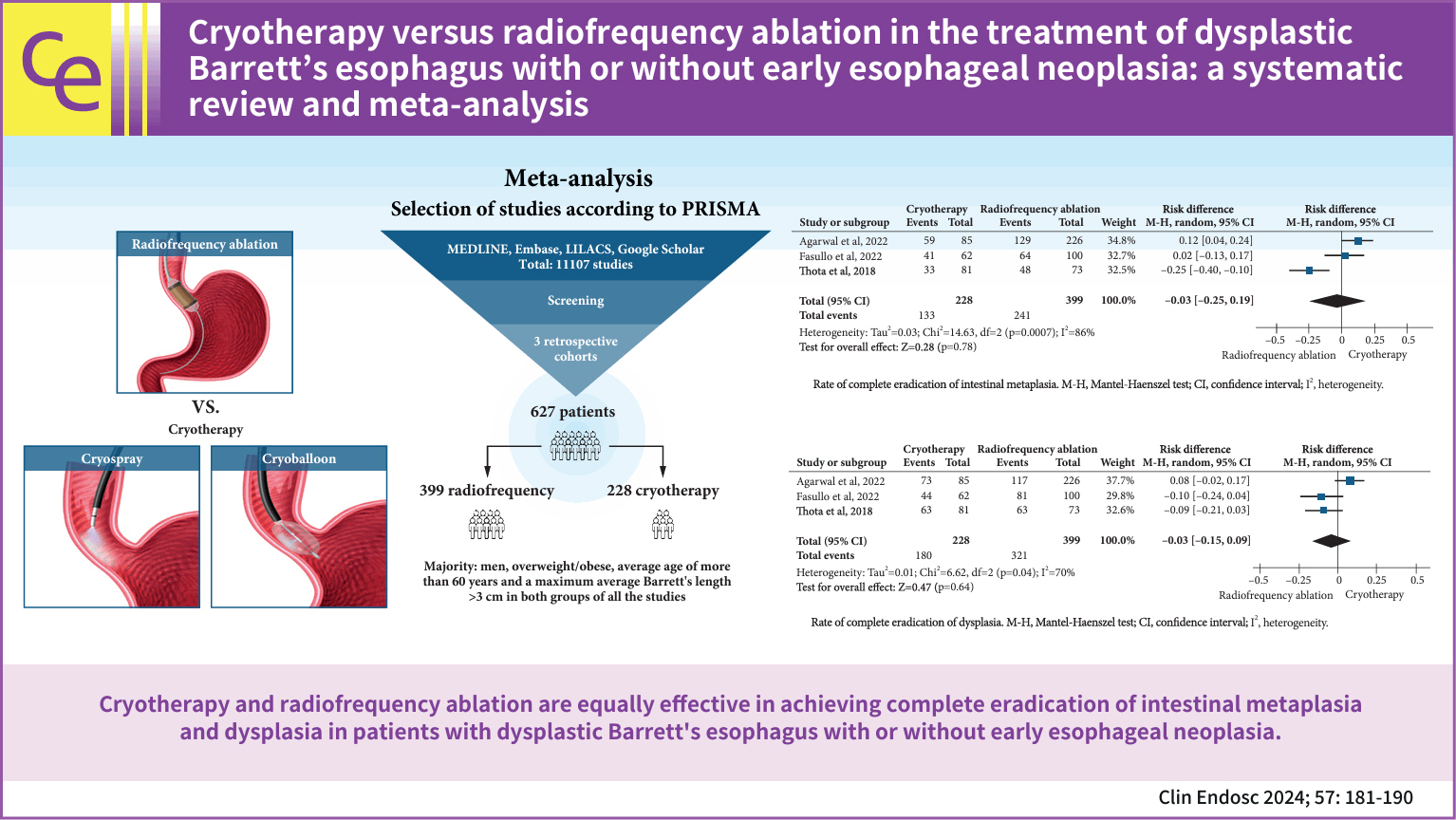
- Cryotherapy versus radiofrequency ablation in the treatment of dysplastic Barrett’s esophagus with or without early esophageal neoplasia: a systematic review and meta-analysis
- Igor Logetto Caetité Gomes, Diogo Turiani Hourneaux de Moura, Igor Braga Ribeiro, Sérgio Barbosa Marques, Alexandre de Sousa Carlos, Beanie Conceição Medeiros Nunes, Bruno Salomão Hirsch, Guilherme Henrique Peixoto de Oliveira, Roberto Paolo Trasolini, Wanderley Marques Bernardo, Eduardo Guimarães Hourneaux de Moura
- Clin Endosc 2024;57(2):181-190. Published online January 17, 2024
- DOI: https://doi.org/10.5946/ce.2023.065
-
 Graphical Abstract
 Abstract
 PDF
PDF Supplementary Material
 PubReader
 ePub
ePub - Background
/Aims: Radiofrequency ablation (RFA) is the first-line therapy for dysplastic Barrett’s esophagus (BE). Therefore, cryotherapy has emerged as an alternative treatment option. This study aimed to compare the efficacies of these two techniques based on the rates of complete eradication of intestinal metaplasia (CE-IM) and dysplasia (CE-D). Adverse events and recurrence have also been reported.
Methods
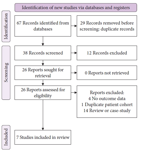
An electronic search was conducted using the Medline (PubMed), Embase, LILACS, and Google Scholar databases until December 2022. Studies were included comparing cryotherapy and RFA for treating dysplastic BE with or without early esophageal neoplasia. This study was performed in accordance with the Preferred Reporting Items for Systematic Reviews and Meta-Analyses guidelines.
Results
Three retrospective cohort studies involving 627 patients were included. Of these, 399 patients underwent RFA, and 228 were treated with cryotherapy. There was no difference in CE-IM (risk difference [RD], –0.03; 95% confidence interval [CI], –0.25 to 0.19; p=0.78; I2=86%) as well as in CE-D (RD, –0.03; 95% CI, –0.15 to 0.09; p=0.64; I2=70%) between the groups. The absolute number of adverse events was low, and there was no difference in the recurrence rate.
Conclusions
Cryotherapy and RFA were equally effective in treating dysplastic BE, with or without early esophageal neoplasia.
- 3,030 View
- 205 Download

Reviews
- Advanced endoscopic imaging for detection of Barrett’s esophagus
- Netanel Zilberstein, Michelle Godbee, Neal A. Mehta, Irving Waxman
- Clin Endosc 2024;57(1):1-10. Published online January 5, 2024
- DOI: https://doi.org/10.5946/ce.2023.031
-
Abstract
PDFPubReaderePub
- Barrett’s esophagus (BE) is the precursor to esophageal adenocarcinoma (EAC), and is caused by chronic gastroesophageal reflux. BE can progress over time from metaplasia to dysplasia, and eventually to EAC. EAC is associated with a poor prognosis, often due to advanced disease at the time of diagnosis. However, if BE is diagnosed early, pharmacologic and endoscopic treatments can prevent progression to EAC. The current standard of care for BE surveillance utilizes the Seattle protocol. Unfortunately, a sizable proportion of early EAC and BE-related high-grade dysplasia (HGD) are missed due to poor adherence to the Seattle protocol and sampling errors. New modalities using artificial intelligence (AI) have been proposed to improve the detection of early EAC and BE-related HGD. This review will focus on AI technology and its application to various endoscopic modalities such as high-definition white light endoscopy, narrow-band imaging, and volumetric laser endomicroscopy.
-
Citations
Citations to this article as recorded by
- Advancements in Barrett's esophagus detection: The role of artificial intelligence and its implications
Sara Massironi
World Journal of Gastroenterology.2024; 30(11): 1494. CrossRef
- Advancements in Barrett's esophagus detection: The role of artificial intelligence and its implications
- 2,823 View
- 208 Download
- 1 Web of Science
- 1 Crossref
- Role of linked color imaging for upper gastrointestinal disease: present and future
- Sang Pyo Lee
- Clin Endosc 2023;56(5):546-552. Published online June 9, 2023
- DOI: https://doi.org/10.5946/ce.2023.015
-
Abstract
PDFPubReaderePub
- Techniques for upper gastrointestinal endoscopy are advancing to facilitate lesion detection and improve prognosis. However, most early tumors in the upper gastrointestinal tract exhibit subtle color changes or morphological features that are difficult to detect using white light imaging. Linked color imaging (LCI) has been developed to overcome these shortcomings; it expands or reduces color information to clarify color differences, thereby facilitating the detection and observation of lesions. This article summarizes the characteristics of LCI and advances in LCI-related research in the upper gastrointestinal tract field.
-
Citations
Citations to this article as recorded by- Upper gastrointestinal signs and symptoms: assessment, management and referral pathways
Hasan Alsararatee
Gastrointestinal Nursing.2024; 22(4): 192. CrossRef - Endoscopic submucosal dissection for early gastric cancer: It is time to consider the quality of its outcomes
Gwang Ha Kim
World Journal of Gastroenterology.2023; 29(43): 5800. CrossRef
- Upper gastrointestinal signs and symptoms: assessment, management and referral pathways
- 2,237 View
- 195 Download
- 1 Web of Science
- 2 Crossref
Original Articles
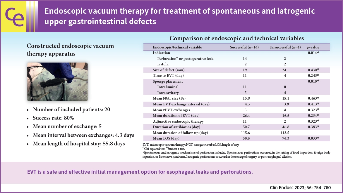
- Endoscopic vacuum therapy for treatment of spontaneous and iatrogenic upper gastrointestinal defects
- Kavea Panneerselvam, Jake S. Jacob, Ronald E. Samuel, Andy Tau, Gyanprakash A. Ketwaroo, Wasif M. Abidi, Robert J. Sealock
- Clin Endosc 2023;56(6):754-760. Published online May 9, 2023
- DOI: https://doi.org/10.5946/ce.2022.177
-
Graphical Abstract
Abstract
PDFPubReaderePub
- Background
/Aims: Endoscopic vacuum therapy (EVT) can heal a variety of defects within the gastrointestinal (GI) tract via applying negative pressure, which reduces the defect size, aspirates the infected fluid, and promotes granulation tissue. Here we present our experience with EVT as it relates to both spontaneous and iatrogenic upper GI tract perforations, leaks, and fistulas.
Methods
This retrospective study was conducted at four large hospital centers. All patients who underwent EVT between June 2018 and March 2021 were included. Data on multiple variables were collected, including demographics, defect size and location, number and intervals of EVT exchanges, technical success, and hospital length of stay. Student t-test and the chi-squared test were used to analyze the data.
Results
Twenty patients underwent EVT. The most common defect cause was spontaneous esophageal perforation (50%). The most common defect location was the distal esophagus (55%). The success rate was 80%. Seven patients were treated with EVT as the primary closure method. The mean number of exchanges was five with a mean interval of 4.3 days between exchanges. The mean length of hospital stay was 55.8 days.
Conclusions
EVT is a safe and effective initial management option for esophageal leaks and perforations. -
Citations
Citations to this article as recorded by- Endoscopic vacuum therapy: management of upper gastrointestinal anastomotic leaks and esophageal perforations
María de Armas Conde, Carmen Díaz-López , Vanessa Concepción-Martín, María Del Pilar Borque-Barrera
Revista Española de Enfermedades Digestivas.2024;[Epub] CrossRef - Management of fistulas in the upper gastrointestinal tract
Maria Valeria Matteo, Maria Mihaela Birligea, Vincenzo Bove, Valerio Pontecorvi, Martina De Siena, Loredana Gualtieri, Federico Barbaro, Cristiano Spada, Ivo Boškoski
Best Practice & Research Clinical Gastroenterology.2024; : 101929. CrossRef - Endoscopic Vacuum Therapy of Upper Gastrointestinal Anastomotic Leaks: How to Deal with the Challenges (with Video)
Laurent Monino, Tom G. Moreels
Life.2023; 13(6): 1412. CrossRef
- Endoscopic vacuum therapy: management of upper gastrointestinal anastomotic leaks and esophageal perforations
- 2,063 View
- 132 Download
- 1 Web of Science
- 3 Crossref
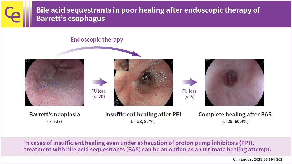
- Bile acid sequestrants in poor healing after endoscopic therapy of Barrett’s esophagus
- Lukas Welsch, Andrea May, Tobias Blasberg, Jens Wetzka, Elisa Müller, Myriam Heilani, Mireen Friedrich-Rust, Mate Knabe
- Clin Endosc 2023;56(2):194-202. Published online March 9, 2023
- DOI: https://doi.org/10.5946/ce.2022.121
-
Graphical Abstract
Abstract
PDFPubReaderePub
- Background
/Aims: Endoscopic therapy for neoplastic Barrett’s esophagus (BE) has become the standard of care over the past two decades. In clinical practice, we regularly encounter patients who fail to achieve complete squamous epithelialization of the esophagus. Although the therapeutic strategies in the individual stages of BE, dysplasia, and esophageal adenocarcinoma are well studied and largely standardized, the problem of inadequate healing after endoscopic therapy is only marginally considered. This study aimed to shed light on the variables influencing inadequate wound healing after endoscopic therapy and the effect of bile acid sequestrants (BAS) on healing.
Methods
Retrospective analysis of endoscopically treated neoplastic BE in a single referral center.
Results
In 12.1% out of 627 patients, insufficient healing was present 8 to 12 weeks after previous endoscopic therapy. The average follow-up duration was 38.8±18.4 months. Complete healing was achieved in 13 patients already after intensifying proton pump inhibitor therapy. Out of 48 patients under BAS, 29 patients (60.4%) showed complete healing. An additional eight patients (16.7%) improved, but only partial healing was achieved. Eleven (22.9%) patients showed no response to BAS augmented therapy.
Conclusions
In cases of insufficient healing even under exhaustion of proton pump inhibitors, treatment with BAS can be an option as an ultimate healing attempt. -
Citations
Citations to this article as recorded by
- 2,516 View
- 160 Download
- 1 Web of Science
- 2 Crossref
Systematic Review and Meta-Analysis
- Hybrid argon plasma coagulation in Barrett’s esophagus: a systematic review and meta-analysis
- Sagar N. Shah, Nabil El Hage Chehade, Amirali Tavangar, Alyssa Choi, Marc Monachese, Kenneth J. Chang, Jason B. Samarasena
- Clin Endosc 2023;56(1):38-49. Published online January 30, 2023
- DOI: https://doi.org/10.5946/ce.2022.179
-
Abstract
PDFSupplementary MaterialPubReaderePub
- Background
/Aims: Patients with Barrett’s esophagus are at increased risk of developing esophageal adenocarcinoma. Endoscopic therapies aim to eradicate dysplastic and metaplastic tissues. Hybrid argon plasma coagulation (hybrid-APC) utilizes submucosal fluid injection to create a protective cushion prior to ablation that shields the submucosa from injury. We performed a pooled meta-analysis to evaluate the safety and efficacy of hybrid-APC.
Methods
We conducted a systematic search of major electronic databases in April 2022. Studies that included patients with dysplastic and non-dysplastic Barrett’s esophagus undergoing treatment with hybrid-APC were eligible for inclusion. Outcome measures included complete remission of intestinal metaplasia (CR-IM), stricture formation, serious adverse events, and number of sessions necessary to achieve CR-IM.
Results
Overall pooled CR-IM rate for patients undergoing hybrid-APC was 90.8% (95% confidence interval [CI], 0.872–0.939; I2=0%). Pooled stricture rate was 2.0% (95% CI, 0.005–0.042; I2=0%). Overall serious adverse event rate was 2.7% (95% CI, 0.007–0.055; I2=0%).
Conclusions
Results of the current meta-analysis suggest that hybrid-APC is associated with high rates of CR-IM and a favorable safety profile. Interpretation of these results is limited by the inclusion of retrospective cohort and case series data. Randomized controlled trials that standardize treatment and outcome evaluation protocols are necessary to understand how this treatment option is comparable to the current standards of care. -
Citations
Citations to this article as recorded by- Application of electrosurgery in gastrointestinal endoscopy
Progress in Medical Devices.2024;[Epub] CrossRef - Hybrid Argon Plasma Coagulation for Barrett’s Esophagus and for Colonic Mucosal Resection—A Systematic Review and Meta-Analysis
Maria Manuela Estevinho, Rolando Pinho, João Carlos Silva, João Correia, Pedro Mesquita, Teresa Freitas
Biomedicines.2023; 11(4): 1139. CrossRef - Hybrid-APC treatment for gastric vascular ectasia of
atypical location after failed radiofrequency ablation
José Manuel Palma García, Raúl Honrubia López, Cristina Fernández de Castro, Carmen Comas Redondo
Revista Española de Enfermedades Digestivas.2023;[Epub] CrossRef - Thermal ablative therapies in the gastrointestinal tract
Hendrik Manner
Current Opinion in Gastroenterology.2023; 39(5): 370. CrossRef - Endoscopic Management of Dysplastic Barrett’s Oesophagus and Early Oesophageal Adenocarcinoma
Leonardo Henry Eusebi, Andrea Telese, Chiara Castellana, Rengin Melis Engin, Benjamin Norton, Apostolis Papaefthymiou, Rocco Maurizio Zagari, Rehan Haidry
Cancers.2023; 15(19): 4776. CrossRef - Critical Decision Making: Technical Aspects of Esophageal Ablation
Felice Schnoll-Sussman
Foregut: The Journal of the American Foregut Society.2023; 3(3): 314. CrossRef
- Application of electrosurgery in gastrointestinal endoscopy
- 2,724 View
- 154 Download
- 5 Web of Science
- 6 Crossref
Review
- Role of artificial intelligence in diagnosing Barrett’s esophagus-related neoplasia
- Michael Meinikheim, Helmut Messmann, Alanna Ebigbo
- Clin Endosc 2023;56(1):14-22. Published online January 17, 2023
- DOI: https://doi.org/10.5946/ce.2022.247
-
Abstract
PDFPubReaderePub
- Barrett’s esophagus is associated with an increased risk of adenocarcinoma. Thorough screening during endoscopic surveillance is crucial to improve patient prognosis. Detecting and characterizing dysplastic or neoplastic Barrett’s esophagus during routine endoscopy are challenging, even for expert endoscopists. Artificial intelligence-based clinical decision support systems have been developed to provide additional assistance to physicians performing diagnostic and therapeutic gastrointestinal endoscopy. In this article, we review the current role of artificial intelligence in the management of Barrett’s esophagus and elaborate on potential artificial intelligence in the future.
-
Citations
Citations to this article as recorded by- Endoskopische Therapie von Barrett-Neoplasien und Magenfrühkarzinomen
Florian Berreth, Jan Peveling-Oberhag, Jörg G. Albert
best practice onkologie.2024; 19(1-2): 28. CrossRef - The Role of Screening and Early Detection in Upper Gastrointestinal Cancers
Jin Woo Yoo, Monika Laszkowska, Robin B. Mendelsohn
Hematology/Oncology Clinics of North America.2024; 38(3): 693. CrossRef - Artificial intelligence in gastroenterology: where are we and where are we going?
Laurence B Lovat
Gastrointestinal Nursing.2024; 22(Sup3): S6. CrossRef - As how artificial intelligence is revolutionizing endoscopy
Jean-Francois Rey
Clinical Endoscopy.2024; 57(3): 302. CrossRef - Screening and Diagnostic Advances of Artificial Intelligence in Endoscopy
Muhammed Yaman Swied, Mulham Alom, Obada Daaboul, Abdul Swied
Innovations in Digital Health, Diagnostics, and Biomarkers.2024; 4(2024): 31. CrossRef - Endoskopische Therapie von Barrett-Neoplasien und Magenfrühkarzinomen
Florian Berreth, Jan Peveling-Oberhag, Jörg G. Albert
Die Gastroenterologie.2023; 18(3): 186. CrossRef
- Endoskopische Therapie von Barrett-Neoplasien und Magenfrühkarzinomen
- 2,436 View
- 247 Download
- 2 Web of Science
- 6 Crossref
Original Article
- Epidemiology of early esophageal adenocarcinoma
- Thuy-Van P. Hang, Zachary Spiritos, Anthony M. Gamboa, Zhengjia Chen, Seth Force, Vaishali Patel, Saurabh Chawla, Steven Keilin, Nabil F. Saba, Bassel El-Rayes, Qiang Cai, Field F. Willingham
- Clin Endosc 2022;55(3):372-380. Published online February 11, 2022
- DOI: https://doi.org/10.5946/ce.2021.152

-
Abstract
PDFPubReaderePub
- Background
/Aims: Endoscopic resection has become the preferred treatment approach for select early esophageal adenocarcinoma (EAC); however, the epidemiology of early stage disease has not been well defined.
Methods
Surveillance Epidemiology and End Results (SEER) data were analyzed to determine age-adjusted incidence rates among major epithelial carcinomas, including EAC, from 1973 to 2017. The percent change in incidence over time was compared according to tumor subtype. Early T-stage, node-negative EAC without metastasis was examined from 2004 to 2017 when precise T-stage data were available.
Results
The percent change in annual incidence from 1973 to 2017 was 767% for EAC. Joinpoint analysis showed that the average annual percent change in EAC from 1973 to 2017 was 5.11% (95% confidence interval, 4.66%–5.56%). The annual percent change appeared to plateau between 2004 and 2017; however, early EAC decreased from 2010 to 2017, with an annual percent change of -5.78%.
Conclusions
There has been a 7-fold increase in the incidence of EAC, which was significantly greater than that of the other major epithelial malignancies examined. More recently, the incidence of early EAC has been decreasing. Approximately one in five patients has node negative, potentially resectable early stage disease. -
Citations
Citations to this article as recorded by- Concise Commentary: It’s All Downhill from Here—How Diagnostic and Therapeutic Advances May Decrease the Incidence Rates of Gastroesophageal Junction and Esophageal Adenocarcinoma
Anthony Gamboa, Rishi Naik
Digestive Diseases and Sciences.2024; 69(1): 254. CrossRef - Descriptive Epidemiology of Early-Onset Gastrointestinal Cancers in Iran, 2014-2018
Mohammad Sadra Gholami Chahkand, Fatemeh Esmaeilpour Moallem, Fatemeh Ghasemi-Kebria, Reza Malekzadeh, Gholamreza Roshandel, Mohammad Taher
Middle East Journal of Digestive Diseases.2024; 16(1): 28. CrossRef - Epidemiologie der Adenokarzinome des Ösophagus und des ösophagogastralen Übergangs
Sabine Luttmann, Andrea Eberle, Joachim Hübner
Die Onkologie.2023; 29(6): 470. CrossRef - Evaluation of Esophageal Dysphagia in Elderly Patients
Khanh Hoang Nicholas Le, Eric E. Low, Rena Yadlapati
Current Gastroenterology Reports.2023; 25(7): 146. CrossRef - Histology Shift in Esophageal Cancer Between Biopsies and Resections After Neoadjuvant Therapy: A Pilot Study
Tieying Hou, Zhaohai Yang, Qingzhao Zhang, Xuchen Zhang, Xiaoyan Liao, Jingmei Lin
International Journal of Surgical Pathology.2023;[Epub] CrossRef - Molecular Biology and Clinical Management of Esophageal Adenocarcinoma
Shulin Li, Sanne Johanna Maria Hoefnagel, Kausilia Krishnawatie Krishnadath
Cancers.2023; 15(22): 5410. CrossRef - Progress in Clinical Management of Esophago-Jejunal Anastomotic Fistula with Total Gastrectomy for Adenocarcinoma of the Esophagogastric Junction
天伟 赖
Advances in Clinical Medicine.2023; 13(11): 17210. CrossRef - Cranberry Proanthocyanidins Mitigate Reflux-Induced Transporter Dysregulation in an Esophageal Adenocarcinoma Model
Yun Zhang, Katherine M. Weh, Bridget A. Tripp, Jennifer L. Clarke, Connor L. Howard, Shruthi Sunilkumar, Amy B. Howell, Laura A. Kresty
Pharmaceuticals.2023; 16(12): 1697. CrossRef - Lessons learned in clinical epidemiology of esophageal adenocarcinoma
Hye Kyung Jung
Clinical Endoscopy.2022; 55(3): 365. CrossRef
- Concise Commentary: It’s All Downhill from Here—How Diagnostic and Therapeutic Advances May Decrease the Incidence Rates of Gastroesophageal Junction and Esophageal Adenocarcinoma
- 4,692 View
- 266 Download
- 5 Web of Science
- 9 Crossref
Case Report
- Less invasive transoral resection of esophageal fibrovascular polyps: case reports
- Janusz Włodarczyk, Tomasz Smęder
- Clin Endosc 2022;55(5):683-687. Published online December 6, 2021
- DOI: https://doi.org/10.5946/ce.2021.144
-
Abstract
PDFPubReaderePub
- We report five patients treated for esophageal fibrovascular polyps using a minimally invasive technique. Esophageal fibrovascular polyps are benign pedunculated submucosal tumors of considerable size. The treated polyps size ranged from 1.5 to 13 cm. The polyps were removed by relocation to the oral cavity under endoscopic control. No perioperative complications occurred after the treatment. The follow-up of patients after surgery was 9–89 months, with no evidence of polyp recurrence. Thus, the described treatment is safe but requires experience with endoscopy as well as esophageal surgery.
- 3,010 View
- 169 Download
Focused Review Series: Endoscopic Management of Postoperative Gastrointestinal Complication: What’s New?
- Current Status of Endoscopic Vacuum Therapy in the Management of Esophageal Perforations and Post-Operative Leaks
- Imogen Livingstone, Lily Pollock, Bruno Sgromo, Sotiris Mastoridis
- Clin Endosc 2021;54(6):787-797. Published online November 16, 2021
- DOI: https://doi.org/10.5946/ce.2021.240
-
Abstract
PDFPubReaderePub
- Esophageal wall defects, including perforations and postoperative leaks, are associated with high morbidity and mortality and pose a significant management challenge. In light of the high morbidity of surgical management or revision, in recent years, endoscopic vacuum therapy (EVT) has emerged as a novel alternative treatment strategy. EVT involves transoral endoscopic placement of a polyurethane sponge connected to an externalized nasogastric tube to provide continuous negative pressure with the intention of promoting defect healing, facilitating cavity drainage, and ameliorating sepsis. In the last decade, EVT has become increasingly adopted in the management of a diverse spectrum of esophageal defects. Its popularity has been attributed in part to the growing body of evidence suggesting superior outcomes and defect closure rates in excess of 80%. This growing body of evidence, coupled with the ongoing evolution of the technology and techniques of deployment, suggests that the utilization of EVT has become increasingly widespread. Here, we aimed to review the current status of the field, addressing the mechanism of action, indications, technique methodology, efficacy, safety, and practical considerations of EVT implementation. We also sought to highlight future directions for the use of EVT in esophageal wall defects.
-
Citations
Citations to this article as recorded by- Multi-modality management of defects in the gastrointestinal tract: Where the endoscope meets the scalpel: Endoscopic vacuum therapy in the upper gastrointestinal tract
Lisanne M.D. Pattynama, Wietse J. Eshuis, Stefan Seewald, Roos E. Pouw
Best Practice & Research Clinical Gastroenterology.2024; : 101901. CrossRef - Management of esophageal anastomotic leaks, a systematic review and network meta-analysis
William Murray, Mathew G Davey, William Robb, Noel E Donlon
Diseases of the Esophagus.2024;[Epub] CrossRef - Management of an Aortoesophageal Fistula With Esophageal Endoluminal Wound Vacuum Therapy
Antoine Nehme, Samuel Brown, Salman Zaheer, Alexander Leung
Annals of Thoracic Surgery Short Reports.2024;[Epub] CrossRef - Treatment of Esophageal-Pleural Fistula After Diverticulectomy Using Transluminal Vacuum Therapy in a Patient with HIV Infection
M. A. Panasyuk, G. Yu. Aldaranov, V. N. Makhutov, E. G. Grigoriev
Russian Sklifosovsky Journal "Emergency Medical Care".2024; 13(1): 156. CrossRef - Homemade endoscopic vacuum therapy device for the management of transmural gastrointestinal defects
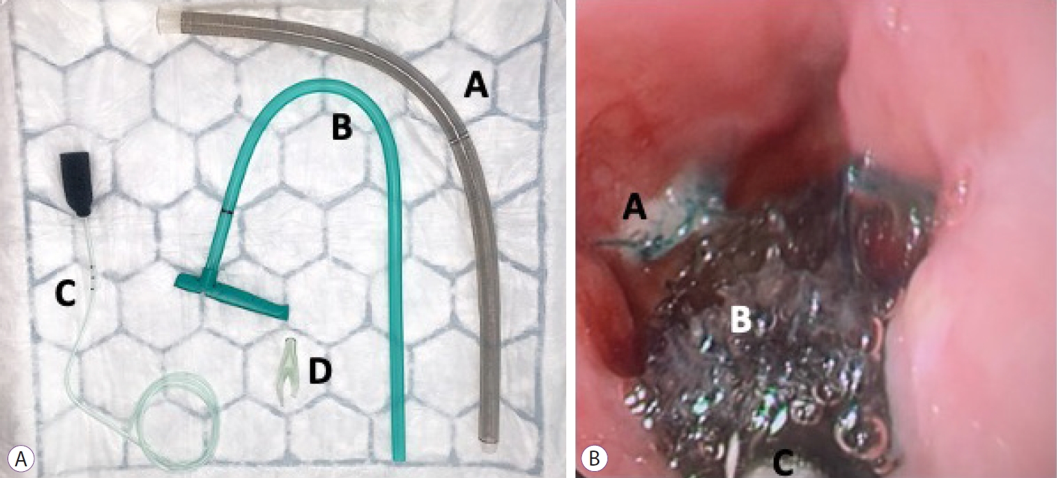
Diogo Turiani Hourneaux de Moura, Bruno Salomão Hirsch, Thomas R. McCarty, Marcos Eduardo Lera dos Santos, Hugo Gonçalo Guedes, Guilherme Francisco Gomes, Flaubert Sena de Medeiros, Eduardo Guimarães Hourneaux de Moura
Digestive Endoscopy.2023; 35(6): 745. CrossRef - Choosing the best endoscopic approach for post-bariatric surgical leaks and fistulas: Basic principles and recommendations
Victor Lira de Oliveira, Alexandre Moraes Bestetti, Roberto Paolo Trasolini, Eduardo Guimarães Hourneaux de Moura, Diogo Turiani Hourneaux de Moura
World Journal of Gastroenterology.2023; 29(7): 1173. CrossRef - Vacuum-Assisted Closure Treats Refractory Esophageal Leak in a Pediatric Patient
Evan K Lin, Felicia Lee, Jasmin Cao, Christian Saliba, Vivian Lu, Raymond I Okeke, Justin Sobrino, Christopher Blewett
Cureus.2023;[Epub] CrossRef - Esophageal Perforation
Kelly Fairbairn, Stephanie G. Worrell
Thoracic Surgery Clinics.2023; 33(2): 117. CrossRef - Endoscopic Treatment of Non-malignant Esophageal Perforation: Time to Go Vacuum?
Diogo Turiani Hourneaux de Moura, Bruno Salomão Hirsch, Heli Clóvis de Medeiros Neto, Victor Lira de Oliveira, Alexandre Moraes Bestetti, Bruna Furia Buzetti Hourneaux de Moura, Mouen A. Khashab, Eduardo Guimarães Hourneaux de Moura
Current Treatment Options in Gastroenterology.2023; 21(2): 95. CrossRef - Prophylactic endoluminal vacuum therapy after major gastrointestinal surgery: a systematic review
Olga Adamenko, Carlo Ferrari, Stefan Seewald, Jan Schmidt
Updates in Surgery.2022; 74(4): 1177. CrossRef - Endoscopic Management of Esophageal Cancer
Christopher Paiji, Alireza Sedarat
Cancers.2022; 14(15): 3583. CrossRef
- Multi-modality management of defects in the gastrointestinal tract: Where the endoscope meets the scalpel: Endoscopic vacuum therapy in the upper gastrointestinal tract
- 4,578 View
- 240 Download
- 7 Web of Science
- 11 Crossref
Original Article
- Endoscopic Management of Anastomotic Leakage after Esophageal Surgery: Ten Year Analysis in a Tertiary University Center
- Nader El-Sourani, Sorin Miftode, Maximilian Bockhorn, Alexander Arlt, Christian Meinhardt
- Clin Endosc 2022;55(1):58-66. Published online October 14, 2021
- DOI: https://doi.org/10.5946/ce.2021.099

-
Abstract
PDFPubReaderePub
- Background
/Aims: Anastomotic leakage after esophageal surgery remains a feared complication. During the last decade, management of this complication changed from surgical revision to a more conservative and endoscopic approach. However, the treatment remains controversial as the indications for conservative, endoscopic, and surgical approaches remain non-standardized.
Methods
Between 2010 and 2020, all patients who underwent Ivor Lewis esophagectomy for underlying malignancy were included in this study. The data of 28 patients diagnosed with anastomotic leak were further analyzed.
Results
Among 141 patients who underwent resection, 28 (19.9%) developed an anastomotic leak, eight (28.6%) of whom died. Thirteen patients were treated with endoluminal vacuum therapy (EVT), seven patients with self-expanding metal stents (SEMS) four patients with primary surgery, one patient with a hemoclip, and three patients were treated conservatively. EVT achieved closure in 92.3% of the patients with a large defect and no EVT-related complications. SEMS therapy was successful in clinically stable patients with small defect sizes.
Conclusions
EVT can be successfully applied in the treatment of anastomotic leakage in critically ill patients, while SEMS should be limited to clinically stable patients with a small defect size. Surgery is only warranted in patients with sepsis with graft necrosis. -
Citations
Citations to this article as recorded by- Placing vacuum sponges in esophageal anastomotic leaks — how we do it
Florian Hentschel, Götz Mollenhauer, Björn Siemssen, Christoph Paasch, René Mantke, Stefan Lüth
Langenbeck's Archives of Surgery.2024;[Epub] CrossRef - Management of esophageal anastomotic leaks, a systematic review and network meta-analysis
William Murray, Mathew G Davey, William Robb, Noel E Donlon
Diseases of the Esophagus.2024;[Epub] CrossRef - Multicenter study on the incidence and treatment of mediastinal leaks after esophagectomy (MuMeLe 2)
Filippo Ascari, Stefano De Pascale, Riccardo Rosati, Simone Giacopuzzi, Francesco Puccetti, Jacopo Weindelmayer, Sofia Cusin, Barbara Leone, Uberto Fumagalli Romario
Journal of Gastrointestinal Surgery.2024;[Epub] CrossRef - Endoscopic vacuum therapy for anastomotic leakage after esophagectomy: a retrospective analysis at a tertiary university center
Nader El-Sourani, Sorin Miftode, Maximilian Bockhorn
Surgery Open Science.2023; 11: 69. CrossRef - RETRACTED ARTICLE: Changes in diagnosis and management of anastomotic leakage after esophagectomy for underlying malignancy reduce postoperative mortality and improve patient outcome
Nader El-Sourani, Sorin Miftode, Achim Troja, Fadl Alfarawan, Maximilian Bockhorn
European Surgery.2023; 55(2): 77. CrossRef - Endoscopic Vacuum Therapy (EVT) versus Self-Expandable Metal Stent (SEMS) for Anastomotic Leaks after Upper Gastrointestinal Surgery: Systematic Review and Meta-Analysis
Francesco Vito Mandarino, Alberto Barchi, Ferdinando D’Amico, Lorella Fanti, Francesco Azzolini, Edi Viale, Dario Esposito, Riccardo Rosati, Gionata Fiorino, Willem Adrianus Bemelman, Ugo Elmore, Lavinia Barbieri, Francesco Puccetti, Sabrina Gloria Giulia
Life.2023; 13(2): 287. CrossRef - Endoscopic vacuum therapy significantly improves clinical outcomes of anastomotic leakages after 2-stage, 3-stage, and transhiatal esophagectomies
Jonas Maier, A. Kandulski, N. E. Donlon, J. M. Werner, A. Mehrl, M. Müller, A. Doenecke, H. J. Schlitt, M. Hornung, A. R. R. Weiss
Langenbeck's Archives of Surgery.2023;[Epub] CrossRef - The Use of Esophageal Stents in the Management of Postoperative Fistulas—Current Status, Clinical Outcomes and Perspectives—Review
Cristian Gelu Rosianu, Petre Hoara, Florin Achim, Rodica Birla, Alexandra Bolocan, Ahmed Mohssen, Narcis Copca, Silviu Constantinoiu
Life.2023; 13(4): 966. CrossRef - Don't be afraid of black holes: Vacuum sponge and vacuum stent treatment of leaks in the upper GI tract—a case series and mini-review
Christian Schäfer
Frontiers in Surgery.2023;[Epub] CrossRef - Treatment and Prevention of Postoperative Leakage after Gastrectomy for Gastric Cancer
Sang-Ho Jeong, Jin-Kwon Lee, Kyung Won Seo, Jae-Seok Min
Journal of Clinical Medicine.2023; 12(12): 3880. CrossRef - Endoscopic Vacuum Therapy of Upper Gastrointestinal Anastomotic Leaks: How to Deal with the Challenges (with Video)
Laurent Monino, Tom G. Moreels
Life.2023; 13(6): 1412. CrossRef - Endoscopic vacuum therapy versus self-expandable metal stent for treatment of anastomotic leaks < 30 mm following oncologic Ivor-Lewis esophagectomy: a matched case–control study
Francesco Vito Mandarino, Alberto Barchi, Lorenzo Leone, Lorella Fanti, Francesco Azzolini, Edi Viale, Dario Esposito, Noemi Salmeri, Francesco Puccetti, Lavinia Barbieri, Andrea Cossu, Elio Treppiedi, Ugo Elmore, Riccardo Rosati, Silvio Danese
Surgical Endoscopy.2023; 37(9): 7039. CrossRef - The Optimal Treatment Strategy for Postoperative Anastomotic Leakage After Esophagectomy: a Comparative Analysis Between Endoscopic Vacuum Therapy and Conventional Treatment
Joonseok Lee, Jae Hyun Jeon, Seung Hwan Yoon, Beatrice Chia-Hui Shih, Woohyun Jung, Yoohwa Hwang, Sukki Cho, Kwhanmien Kim, Sanghoon Jheon
Journal of Gastrointestinal Surgery.2023; 27(12): 2899. CrossRef - Endoscopic Endoluminal Vacuum Therapy or Self-Expandable Metallic Stent: Treatment Option in Anastomotic Leakage after Esophageal Surgery
Chul-Hyun Lim
Clinical Endoscopy.2022; 55(1): 41. CrossRef - Treating an Intractable Jejunocutaneous Fistula by Endoscopic Metallic Stent Placement: A Case Report of Successful Palliative Endoscopic Treatment in a Case Demonstrating Peritoneal Dissemination with Terminal Stage Gastric Cancer
Hironori Tanaka, Kazuhiro Ota, Noriaki Sugawara, Taro Iwatsubo, Shimpei Kawaguchi, Yosuke Mori, Noriyuki Nakajima, Akitoshi Hakoda, Yuichi Kojima, Yoshihiro Inoue, Toshihisa Takeuchi, Kazuhide Higuchi
Internal Medicine.2022; 61(22): 3343. CrossRef - Management of intra-thoracic anastomotic leakages after esophagectomy: updated systematic review and meta-analysis of endoscopic vacuum therapy versus stenting
Pasquale Scognamiglio, Matthias Reeh, Nathaniel Melling, Marcus Kantowski, Ann-Kathrin Eichelmann, Seung-Hun Chon, Nader El-Sourani, Gerhard Schön, Alexandra Höller, Jakob R. Izbicki, Michael Tachezy
BMC Surgery.2022;[Epub] CrossRef - Risk Factors and Effect of Intrathoracic Anastomotic Leakage after Esophagectomy for Underlying Malignancy—A Ten-Year Analysis at a Tertiary University Centre
Nader El-Sourani, Sorin Miftode, Fadl Alfarawan, Achim Troja, Maximilian Bockhorn
Clinics and Practice.2022; 12(5): 782. CrossRef
- Placing vacuum sponges in esophageal anastomotic leaks — how we do it
- 4,900 View
- 252 Download
- 17 Web of Science
- 17 Crossref
Focused Review Series: Cutting Edge of Advanced Therapeutic Endoscopy
-
Role of Peroral Endoscopic Myotomy (POEM) in the Management of Esophageal Diverticula

- Bogdan P. Miutescu, Sarah Khan, Shruti Mony, Mouen A. Khashab
- Clin Endosc 2020;53(6):646-651. Published online November 26, 2020
- DOI: https://doi.org/10.5946/ce.2020.262
-
Abstract
PDFSupplementary MaterialPubReaderePub
- Esophageal diverticula are uncommon; however, when present, they can cause symptoms of dysphagia, regurgitation, and chest pain. Based on location and pathophysiological characteristics, they are classified as pulsion- and traction-type diverticula. In the past, the open surgical approach was the only treatment available; however, in the past few decades, transoral incisionless approaches in the form of rigid and flexible endoscopy have gained popularity. Diverticular peroral endoscopic myotomy has emerged as an alternative treatment option. In this paper, we reviewed the role of peroral endoscopic myotomy as a treatment option for different types of esophageal diverticula. Although a safe and effective procedure, this novel submucosal tunneling technique for the treatment of esophageal diverticula requires further validation, and head-to-head comparisons between the different approaches for the treatment of esophageal diverticula are warranted.
-
Citations
Citations to this article as recorded by- Efficacy and safety of peroral endoscopic myotomy for esophageal diverticula
Elise M. Wessels, Jeroen M. Schuitenmaker, Barbara A.J. Bastiaansen, Paul Fockens, Gwen M.C. Masclee, Albert J. Bredenoord
Endoscopy International Open.2023; 11(05): E546. CrossRef - Peroral endoscopic myotomy (POEM) for esophageal diverticula
Jayanta SAMANTA, Zaheer NABI, Jahnvi DHAR, Harshal S. MANDAVDHARE
Minerva Gastroenterology.2023;[Epub] CrossRef - Multimodal Endoscopic Management of Esophageal Perforations as a Complication of Peroral Endoscopic Myotomy for a Zenker's Diverticulum
Erlison Mauricio Daza Castro, Carlos Fernando Fuentes, Andrea Carolina Córdoba Guzmán, Diego Aponte, José Nicolás Rocha, Carlos González, Luis Carlos Sabbagh
ACG Case Reports Journal.2023; 10(6): e01059. CrossRef - A rare case of bilateral Killian-Jamieson diverticula treated endoscopically
Catarina Félix, Pedro Barreiro, José Rodrigues, Rui Mendo, Catarina O’Neill, Cristina Chagas
Endoscopy.2022; 54(06): E283. CrossRef - Peroral endoscopic myotomy, septotomy, and restoration of esophageal lumen with over-the-scope clips: closing the circle of esophageal diverticula management
Eduardo Albéniz, Fermín Estremera-Arévalo, Marta Gómez Alonso, Pedro J. Rosón, Francisco J. Gallego Rojo, Juan Vila, Sheyla Montori
Endoscopy.2022; 54(11): E666. CrossRef - Successful D-POEM after failed surgical myotomy and diverticulectomy
Andrew Ross Leopold, Raymond E. Kim
VideoGIE.2022; 7(6): 211. CrossRef - Peroral Endoscopic Myotomy for the Treatment of Esophageal Diverticula
Antonio Facciorusso, Daryl Ramai, Yervant Ichkhanian, Rena Yadlapati, Vito Annese, Sachin Wani, Mouen A. Khashab
Journal of Clinical Gastroenterology.2022; 56(10): 853. CrossRef
- Efficacy and safety of peroral endoscopic myotomy for esophageal diverticula
- 6,115 View
- 197 Download
- 8 Web of Science
- 7 Crossref
Case Reports
-
Hybrid Peroral Endoscopic Myotomy for Achalasia with Prior Treatment Failure
- In Kyung Yoo, Abdullah OzgurYeniova, Joo Young Cho
- Clin Endosc 2021;54(1):127-130. Published online April 2, 2020
- DOI: https://doi.org/10.5946/ce.2020.013

-
Abstract
PDFSupplementary MaterialPubReaderePub
- Achalasia is a neurodegenerative motility disorder caused by enteric neuron damage in the lower esophageal sphincter. Peroral endoscopic myotomy (POEM) is a standard treatment method for achalasia. Previous treatment modalities may affect the outcome of POEM as they cause submucosal fibrosis. We report a new technique called “hybrid POEM” for the treatment of patients with achalasia who had been previously treated with pneumatic balloon dilatation. We performed two techniques of POEM simultaneously, the standard POEM for the upper part of the submucosal tunnel and open POEM for the stenotic part of the esophagogastric junction. We dissected the mucosa and submucosa, and performed myotomy simultaneously. We overcame submucosal fibrosis of the esophagogastric junction, which was caused by the previous hybrid POEM treatment. The risks of mucosal incision and technical challenge of submucosal tunneling for the fibrotic area may be reduced by hybrid POEM.
- 4,061 View
- 133 Download
- Submucosal Tunneling Muscle Biopsy for Esophageal Motility Disorders: A Case Report
- Aleksandr A. Smirnov, Maya M. Kiriltseva, Aleksandr N. Burakov, Maksim V. Maksimov, Anna V. Botina, Marina M. Saadulaeva, Nadezda V. Konkina
- Clin Endosc 2020;53(3):370-373. Published online August 20, 2019
- DOI: https://doi.org/10.5946/ce.2019.109
-
Abstract
PDFPubReaderePub
- Submucosal tunneling endoscopic technique can be useful in obtaining esophageal muscle specimens in patients with esophageal motility disorders. Here, we describe the case of a patient with systemic sclerosis. Histological verification of the esophageal involvement in the pathological process was required for the treatment. There were no intra- and post- operational complications.
-
Citations
Citations to this article as recorded by- Updates in the Field of Submucosal Endoscopy
Tadateru Maehata, Yoshinori Sato, Yusuke Nakamoto, Masaki Kato, Akiyo Kawashima, Hirofumi Kiyokawa, Hiroshi Yasuda, Hiroyuki Yamamoto, Keisuke Tateishi
Life.2022; 13(1): 104. CrossRef
- Updates in the Field of Submucosal Endoscopy
- 4,319 View
- 87 Download
- 1 Web of Science
- 1 Crossref
- Buried Barrett’s Esophagus with High-Grade Dysplasia after Radiofrequency Ablation
- Joana Castela, Miguel Serrano, Susana Mão de Ferro, Daniela Vinha Pereira, Paula Chaves, António Dias Pereira
- Clin Endosc 2019;52(3):269-272. Published online October 5, 2018
- DOI: https://doi.org/10.5946/ce.2018.124
-
Abstract
PDFPubReaderePub
- Radiofrequency ablation therapy is an effective endoscopic option for the eradication of Barrett’s esophagus that appears to reduce the risk of esophageal cancer. A concern associated with this technique is the development of subsquamous/buried intestinal metaplasia, whose clinical relevance and malignant potential have not yet been fully elucidated. Fewer than 20 cases of subsquamous neoplasia after the successful radiofrequency ablation of Barrett’s esophagus have been reported to date. Here, we describe a new case of subsquamous neoplasia (high-grade dysplasia) following radiofrequency ablation that was managed with endoscopic resection. Our experience suggests that a meticulous endoscopic inspection prior to and after radiofrequency ablation is fundamental to reduce the risk of buried neoplasia development.
-
Citations
Citations to this article as recorded by- Allaying uncertainty in diagnosing buried Barrett's esophagus
Ryan Demkowicz, Prashanthi N. Thota, Tanmayee Benjamin, Rocio Lopez, Haiyan Lu, Deepa T. Patil, Erinn Downs-Kelly, Jennifer A. Jeung, Keith K. Lai, James Lapinski, Erica C. Savage, John R. Goldblum, Ilyssa O. Gordon
Annals of Diagnostic Pathology.2021; 51: 151672. CrossRef - Endoscopic features of buried Barrett’s mucosa
Linda S. Yang, Bronte A. Holt, Richard Williams, Richard Norris, Edward Tsoi, Georgina Cameron, Paul Desmond, Andrew C.F. Taylor
Gastrointestinal Endoscopy.2021; 94(1): 14. CrossRef - Post-ablation buried neoplasia in Barrett’s esophagus
Prabhat Kumar, Ilyssa O. Gordon, Prashanthi N. Thota
Scandinavian Journal of Gastroenterology.2021; 56(5): 624. CrossRef - Role of optical coherence tomography in Barrett’s esophagus
Nikhil Gupta, Raghav Yelamanchi, Himanshu Agrawal, Nitin Agarwal
Artificial Intelligence in Gastrointestinal Endoscopy.2021; 2(4): 149. CrossRef - Indications, contraindications and limitations of endoscopic therapy for Barrett’s esophagus and early esophageal adenocarcinoma
Carol Rouphael, Mythri Anil Kumar, Madhusudhan R. Sanaka, Prashanthi N. Thota
Therapeutic Advances in Gastroenterology.2020; 13: 175628482092420. CrossRef - Risk Factors for Self-Expandable Metal Stent Complications in the Treatment of Esophageal Cancer: A Scoping Review
Connor K. Wilson, Sara R. Frankowski, Susan C. Steelman, Issam Makhoul
SN Comprehensive Clinical Medicine.2020; 2(8): 1163. CrossRef - Multifocal Cryoballoon Ablation for Eradication of Barrett's Esophagus-Related Neoplasia: A Prospective Multicenter Clinical Trial
Marcia Irene Canto, Arvind J. Trindade, Julian Abrams, Michael Rosenblum, John Dumot, Amitabh Chak, Prasad Iyer, David Diehl, Harshit S. Khara, F. Scott Corbett, Matthew McKinley, Eun Ji Shin, Irving Waxman, Anthony Infantolino, Christina Tofani, Jason Sa
American Journal of Gastroenterology.2020; 115(11): 1879. CrossRef - Inflammatory bowel disease- and Barrett’s esophagus-associated neoplasia: the old, the new, and the persistent struggles
Dipti M Karamchandani, Qin Zhang, Xiao-Yan Liao, Jing-Hong Xu, Xiu-Li Liu
Gastroenterology Report.2019; 7(6): 379. CrossRef
- Allaying uncertainty in diagnosing buried Barrett's esophagus
- 6,691 View
- 161 Download
- 6 Web of Science
- 8 Crossref
Focused Review Series: Updates on Capsule Endoscopy from Esophagus to Colon
- Current and Future Use of Esophageal Capsule Endoscopy
- Junseok Park, Young Kwan Cho, Ji Hyun Kim
- Clin Endosc 2018;51(4):317-322. Published online July 31, 2018
- DOI: https://doi.org/10.5946/ce.2018.101
-
Abstract
PDFPubReaderePub
- Capsule endoscopy can be a diagnostic option for patients with esophageal diseases who cannot tolerate esophagogastroduodenoscopy.Functional modifications of the capsule allow for thorough examination of the esophagus. Esophageal capsule endoscopy has so farfailed to show sufficient performance to justify the replacement of traditional endoscopy for the diagnosis of esophageal diseasesbecause the esophagus has a short transit time and common pathologies appear near the esophagogastric junction. However,technological improvements are being introduced to overcome the limitations of capsule endoscopy, which is expected to become agood alternative to conventional endoscopy.
-
Citations
Citations to this article as recorded by- Barrett’s Esophagus: Who and How Do We Screen?
Bibek Saha, Anjul Verma, Prasad G. Iyer
Current Treatment Options in Gastroenterology.2024; 22(2): 23. CrossRef - Detachable string magnetically controlled capsule endoscopy for the noninvasive diagnosis of esophageal diseases: A prospective, blind clinical study
Yan-Ling Yang, Huang-Wen Qin, Zhao-Yu Chen, Hui-Ning Fan, Yi Yu, Wei Da, Jin-Shui Zhu, Jing Zhang
World Journal of Gastroenterology.2024; 30(9): 1121. CrossRef - Cascade-EC Network: Recognition of Gastrointestinal Multiple Lesions Based on EfficientNet and CA_stm_Retinanet
Xudong Guo, Lei Xu, Shengnan Li, Meidong Xu, Yuan Chu, Qinfen Jiang
Journal of Imaging Informatics in Medicine.2024;[Epub] CrossRef - The evolving role of small-bowel capsule endoscopy
Silvia Pecere, Michele Francesco Chiappetta, Livio Enrico Del Vecchio, Edward Despott, Xavier Dray, Anastasios Koulaouzidis, Lorenzo Fuccio, Alberto Murino, Emanuele Rondonotti, Manon Spaander, Cristiano Spada
Best Practice & Research Clinical Gastroenterology.2023; 64-65: 101857. CrossRef - The Future of Minimally Invasive Capsule Panendoscopy: Robotic Precision, Wireless Imaging and AI-Driven Insights
Miguel Mascarenhas, Miguel Martins, João Afonso, Tiago Ribeiro, Pedro Cardoso, Francisco Mendes, Patrícia Andrade, Helder Cardoso, João Ferreira, Guilherme Macedo
Cancers.2023; 15(24): 5861. CrossRef - Design and implementation of a highly integrated dual hemisphere capsule robot
Yongshun Zhang, Xu Liu, Guanxi Liu, Xuan Ji, Huiyuan Yang, Zhenhu Liu
Biomedical Microdevices.2022;[Epub] CrossRef - Capsule endoscopy - a non-invasive modality to investigate the GI tract: out with the old and in with the new?
Priya Oka, Mark McAlindon, Reena Sidhu
Expert Review of Gastroenterology & Hepatology.2022; 16(7): 591. CrossRef - Proton pump inhibitor in the prevention of upper gastrointestinal mucosal injury associated with dual antiplatelet therapy after coronary artery bypass grafting (DACAB-GI-2): study protocol for a randomized controlled trial
Yunpeng Zhu, Xiaojin Wang, Yi Yang, Lei Liu, Qiang Zhao, Lifen Yu
Trials.2022;[Epub] CrossRef - Expanding beyond endoscopy: A review of non-invasive modalities in Barrett’s esophagus screening and surveillance
Dariush Shahsavari, Praneeth Kudaravalli, John Erikson L Yap, Kenneth J Vega
World Journal of Gastroenterology.2022; 28(32): 4516. CrossRef - Dynamic tracking effect of a magnetic navigated dual hemisphere capsule robot
Yongshun Zhang, Xu Liu, Zhenhu Liu, Zihao Zhao, Hai Dong, Dianlong Wang
Robotica.2022; 40(12): 4586. CrossRef - Artificial intelligence and deep learning for small bowel capsule endoscopy
Roberto Trasolini, Michael F. Byrne
Digestive Endoscopy.2021; 33(2): 290. CrossRef - Esophageal Cancer: An Updated Review
Michael DiSiena, Alexander Perelman, John Birk, Houman Rezaizadeh
Southern Medical Journal.2021; 114(3): 161. CrossRef - Detachable string magnetically controlled capsule endoscopy for complete observation of the upper gastrointestinal tract
Hui Xiu, Yanyan Lu, Xishuang Liu, Fuguo Liu, Lingyu Zhang, Chengye Zhao, Xueguo Sun
European Journal of Gastroenterology & Hepatology.2021; 33(4): 508. CrossRef - Novel Clinical Applications and Technical Developments in Video Capsule Endoscopy
Shahrad Hakimian, Mark Hanscom, David R. Cave
Gastrointestinal Endoscopy Clinics of North America.2021; 31(2): 399. CrossRef - Next-generation ingestible devices: sensing, locomotion and navigation
Fahad N Alsunaydih, Mehmet R Yuce
Physiological Measurement.2021; 42(4): 04TR01. CrossRef - An intelligent compression system for wireless capsule endoscopy images
Dallel Bouyaya, Said Benierbah, Mohammed Khamadja
Biomedical Signal Processing and Control.2021; 70: 102929. CrossRef - Examination of Entire Gastrointestinal Tract: A Perspective of Mouth to Anus (M2A) Capsule Endoscopy
Ji Hyung Nam, Kwang Hoon Lee, Yun Jeong Lim
Diagnostics.2021; 11(8): 1367. CrossRef - Lavage, Simethicone, and Prokinetics—What to Swallow with a Video Capsule
Martin Keuchel, Niehls Kurniawan, Marc Bota, Peter Baltes
Diagnostics.2021; 11(9): 1711. CrossRef - Innovations in Screening Tools for Barrett’s Esophagus and Esophageal Adenocarcinoma
Matthew G. Bell, Prasad G. Iyer
Current Gastroenterology Reports.2021;[Epub] CrossRef - Applicability of colon capsule endoscopy as pan-endoscopy: From bowel preparation, transit, and rating times to completion rate and patient acceptance
Fanny E.R. Vuik, Sarah Moen, Stella A.V. Nieuwenburg, Eline H. Schreuders, Ernst J. Kuipers, Manon C.W. Spaander
Endoscopy International Open.2021; 09(12): E1852. CrossRef - Development and Application of Magnetically Controlled Capsule Endoscopy in Detecting Gastric Lesions
Yaoping Zhang, Yanning Zhang, Xiaojun Huang, Amosy M'Koma
Gastroenterology Research and Practice.2021; 2021: 1. CrossRef - Development and validation of a risk prediction model to diagnose Barrett's oesophagus (MARK-BE): a case-control machine learning approach
Avi Rosenfeld, David G Graham, Sarah Jevons, Jose Ariza, Daryl Hagan, Ash Wilson, Samuel J Lovat, Sarmed S Sami, Omer F Ahmad, Marco Novelli, Manuel Rodriguez Justo, Alison Winstanley, Eliyahu M Heifetz, Mordehy Ben-Zecharia, Uria Noiman, Rebecca C Fitzge
The Lancet Digital Health.2020; 2(1): e37. CrossRef - Better view by detachable string magnetically controlled capsule endoscopy for esophageal observation: a retrospective comparative study
J Song, T Bai, L Zhang, X-L Xiang, X-P Xie, X-H Hou
Diseases of the Esophagus.2020;[Epub] CrossRef - Recent advances in understanding and preventing oesophageal cancer
James Franklin, Janusz Jankowski
F1000Research.2020; 9: 276. CrossRef - USB capsule endoscope for retrograde imaging of the esophagus
Ivan Martincek, Peter Banovcin, Matej Goraus, Martin Duricek
Journal of Biomedical Optics.2020;[Epub] CrossRef - Follow-up on: optimizing lesion detection in small bowel capsule endoscopy and beyond: from present problems to future solutions
Michael Vasilakakis, Anastasios Koulaouzidis, Diana E Yung, John N Plevris, Ervin Toth, Dimitris K Iakovidis
Expert Review of Gastroenterology & Hepatology.2019; 13(2): 129. CrossRef - Gastrointestinal diagnosis using non-white light imaging capsule endoscopy
Gerard Cummins, Benjamin F. Cox, Gastone Ciuti, Thineskrishna Anbarasan, Marc P. Y. Desmulliez, Sandy Cochran, Robert Steele, John N. Plevris, Anastasios Koulaouzidis
Nature Reviews Gastroenterology & Hepatology.2019; 16(7): 429. CrossRef - Reflux esophagitis, functional and non-functional
Serhat Bor
Best Practice & Research Clinical Gastroenterology.2019; 40-41: 101649. CrossRef
- Barrett’s Esophagus: Who and How Do We Screen?
- 6,125 View
- 133 Download
- 28 Web of Science
- 28 Crossref
Review
- Quality Indicators in Barrett’s Esophagus: Time to Change the Status Quo
- Samuel Han, Sachin Wani
- Clin Endosc 2018;51(4):344-351. Published online July 31, 2018
- DOI: https://doi.org/10.5946/ce.2018.099
-
Abstract
PDFPubReaderePub
- The push for high quality care in all fields of medicine highlights the importance of establishing and adhering to quality indicators.In response, several gastrointestinal societies have established quality indicators specific to Barrett’s esophagus, which serve to createthresholds for performance while standardizing practice and guiding value-based care. Recent studies, however, have consistentlydemonstrated the lack of adherence to these quality indicators, particularly in surveillance (appropriate utilization of endoscopy andobtaining biopsies using the Seattle protocol) and endoscopic eradication therapy practices. These findings suggest that innovativeinterventions are needed to address these shortcomings in order to deliver high quality care to patients with Barrett’s esophagus.
-
Citations
Citations to this article as recorded by- Measuring and improving quality in esophageal care and swallowing disorders
Alexander T Reddy, Joshua P Lee, David A Leiman
Diseases of the Esophagus.2024;[Epub] CrossRef - Impact of Residing in Below Median Household Income Districts on Outcomes in Patients with Advanced Barrett’s Esophagus
Suqing Li, Yusuke Fujiyoshi, Sechiv Jugnundan, Gary May, Norman Marcon, Jeffrey Mosko, Christopher Teshima
Journal of the Canadian Association of Gastroenterology.2023; 6(4): 137. CrossRef - Clinical variation in surveillance and management of Barrett’s esophagus: A cross-sectional study of gastroenterologists and gastrointestinal surgeons
Jamielyn DC Cruz, David Paculdo, Divya Ganesan, Meredith Baker, Rebecca J Critchley-Thorne, Nicholas J Shaheen, Sachin Wani, John W Peabody
Medicine.2022; 101(51): e32187. CrossRef - Risk Factors for Self-Expandable Metal Stent Complications in the Treatment of Esophageal Cancer: A Scoping Review
Connor K. Wilson, Sara R. Frankowski, Susan C. Steelman, Issam Makhoul
SN Comprehensive Clinical Medicine.2020; 2(8): 1163. CrossRef
- Measuring and improving quality in esophageal care and swallowing disorders
- 5,789 View
- 102 Download
- 3 Web of Science
- 4 Crossref
Case Report
- Magnifying Endoscopy for Esophageal Ectopic Sebaceous Glands
- Mu Song Jeon, Gwang Ha Kim, Dong Young Jeong, Byeong Kyu Park, Moon Won Lee, So-Jeong Lee, Do Youn Park
- Clin Endosc 2018;51(5):495-497. Published online February 26, 2018
- DOI: https://doi.org/10.5946/ce.2017.187
-
Abstract
PDFPubReaderePub
- Ectopic sebaceous glands are found very rarely in the esophagus; heretofore, several cases have been reported. The sebaceous gland is originally a source of an endodermal origin; however, there have been controversies regarding whether the origin of the esophageal ectopic sebaceous gland is ectodermal or endodermal. Ectopic sebaceous glands of the esophagus usually do not cause symptoms; thus, they are often found incidentally on endoscopy for routine health screening. Endoscopic findings are characterized by single or multiple yellow patches or nodular lesions of various sizes, sometimes with small central openings. We report two cases of esophageal ectopic sebaceous glands found incidentally during endoscopy with magnifying endoscopic findings. The lesions were in the mid-esophagus and lower esophagus, respectively, and both endoscopic findings were similar as multiple yellowish patches or plaques. Magnifying endoscopy revealed the openings of the excretory ducts surrounded by circular microvessels in both cases.
-
Citations
Citations to this article as recorded by- Multiple heterotopic sebaceous glands in the oesophagus: A case report and literature review
Yuan Fang, Zhi Wang, Yong Qiang Yang, Bei Wen Song, Wen Bin Gou
Tropical Doctor.2024; 54(1): 49. CrossRef - Clinicopathologic Characteristics of Esophageal Ectopic Sebaceous Glands: Chronological Changes and Immunohistochemical Analysis
Hirotsugu Hashimoto, Hajime Horiuchi, Sakiko Miura, Shunya Takayanagi, Toshiaki Gunji, Teppei Morikawa
International Journal of Surgical Pathology.2021; 29(4): 378. CrossRef - The clinical and endoscopic features of esophageal ectopic sebaceous glands
Hui‐Fen Chen, Hsi‐Chang Lee, Min‐Kai Liao, Ting‐An Chang, Chih‐Lin Lin, Li‐Ying Liao, Kuan‐Yang Chen
Advances in Digestive Medicine.2020; 7(4): 179. CrossRef - Case Report of a Proposed, Novel, Endoscopic “Whitehead Pimple” Sign of Ectopic Esophageal Sebaceous Glands Based on Their Mimicking the Dermatologic and Histopathologic Characteristics of Cutaneous Whitehead Pimples/Closed Comedones
Amy Le, Mitual Amin, Mitchell S. Cappell
Digestive Diseases and Sciences.2019; 64(7): 2049. CrossRef - Ectopic Sebaceous Gland in Esophagus Presenting as Subepithelial Tumor
Dong Han Yeom, Han Seung Ryu
Chonnam Medical Journal.2019; 55(3): 168. CrossRef
- Multiple heterotopic sebaceous glands in the oesophagus: A case report and literature review
- 6,705 View
- 129 Download
- 4 Web of Science
- 5 Crossref
Review
- Oroesophageal Fish Bone Foreign Body
- Heung Up Kim
- Clin Endosc 2016;49(4):318-326. Published online July 26, 2016
- DOI: https://doi.org/10.5946/ce.2016.087
-
Abstract
PDFPubReaderePub
- Fish bone foreign body (FFB) is the most frequent food-associated foreign body (FB) in adults, especially in Asia, versus meat in Western countries. The esophageal sphincter is the most common lodging site. Esophageal FB disease tends to occur more frequently in men than in women. The first diagnostic method is laryngoscopic examination. Because simple radiography of the neck has low sensitivity, if perforation or severe complications requiring surgery are expected, computed tomography should be used. The risk factors associated with poor prognosis are long time lapse after FB involvement, bone type, and longer FB (>3 cm). Bleeding and perforation are more common in FFB disease than in other FB diseases. Esophageal FB disease requires urgent treatment within 24 hours. However, FFB disease needs emergent treatment, preferably within 2 hours, and definitely within 6 hours. Esophageal FFB disease usually occurs at the physiological stricture of the esophagus. The aortic arch eminence is the second physiological stricture. If the FB penetrates the esophageal wall, a life-threatening aortoesophageal fistula can develop. Therefore, it is better to consult a thoracic surgeon prior to endoscopic removal.
-
Citations
Citations to this article as recorded by- Unusual Intra-Thyroid Migration of Ingested Fish Bone: A Case Report and Literature Review
Richard Wend-Lasida Ouédraogo, Mathieu Millogo, T Antoine Coulibaly
Indian Journal of Otolaryngology and Head & Neck Surgery.2024; 76(3): 2782. CrossRef - Gastric foreign body granuloma resembling gastric cancer: a case report
Hussein Hassan Okasha, Ahmed Elsayed Alzamzamy, Hanane Delsa, Haitham fekry Othman, Ahmed sayed Alsibaie, Abeer Abdellatef
The Egyptian Journal of Internal Medicine.2024;[Epub] CrossRef - Various Approaches in Managing Fish Bone Migration: Our Experience in Tertiary Hospital in Sarawak
Yuanzhi Cheah, Ting Ting Yew, Mohd Razif Mohamad Yunus, Ing Ping Tang
Indian Journal of Otolaryngology and Head & Neck Surgery.2024;[Epub] CrossRef - Imaging approach to ingested foreign bodies in the neck
Serena T. Pham, Osamu Sakai, V. Carlota Andreu-Arasa
Neuroradiology.2024; 66(6): 867. CrossRef - The role and value of low-dose computed tomography scan compared to esophagoscopy in the diagnosis of foreign body ingestion in adults
Parviz Mardani, Reza Shahriarirad, Fateme Khosravi, Hamidreza Malekhosseini, Armin Amirian, Hooman Kamran
General Thoracic and Cardiovascular Surgery.2023; 71(3): 198. CrossRef - An unusual cause of dysphagia: Esophageal external compressive stricture caused by abscess
Shi-Ze Xiong, Tong Sha, Wei Liu
The American Journal of the Medical Sciences.2023; 365(3): e29. CrossRef - A case report of a carotid space abscess due to extraluminal migration of a fishbone into the deep cervical space
Tae-Hun Lee, Ki Ju Cho, Seong Jun Won, Jung Je Park
Kosin Medical Journal.2023; 38(2): 151. CrossRef - Two Cases of Severe Complications Due to an Esophageal Fish Bone Foreign Body
Ji-Hee Han, Ra-Ri Cha, Ji-Yoon Kwak, Hankyu Jeon, Sang-Soo Lee, Jae Jun Jung, Jin Kyu Cho, Hyun Jin Kim
Medicina.2023; 59(9): 1504. CrossRef - Research Status of Endoscopic Management of Foreign Bodies in the Upper Digestive Tract
琴 杨
Advances in Clinical Medicine.2023; 13(12): 19833. CrossRef - 1. The analysis of 232 patients with esophageal foreign bodies
Özgür KATRANCIOĞLU, Şule KARADAYI, Eftal SERT
Journal of Medical Topics and Updates.2023; 2(3): 52. CrossRef - Diagnostic Performance of Digital Radiograph and Low-Dose Computed Tomography for the Diagnosis of Fishbone Retention in the Oropharynx
Jirapa Chansangrat
International Archives of Otorhinolaryngology.2022; 26(03): e401. CrossRef - Ear, Nose, and Throat Foreign Bodies in Children: A Retrospective Study
Bin Kwon, Yeso Choi, Sung-Kyun Kim, Seok-Jin Hong, Yong-Bok Kim, Seok-Min Hong
Children.2022; 9(1): 63. CrossRef - ESOPHAGEAL FISH BONE IMPACTION: THE IMPORTANCE OF EARLY DIAGNOSIS AND TREATMENT TO AVOID SEVERE COMPLICATIONS
Andrés Conthe, Isabel Payeras Otero, Luis Alberto Pérez Gavín, Ainara Baines García, Clara Usón Peiron, Clara Villaseca Gómez, José Luis Herrera Fajes, Óscar Nogales
Revista Española de Enfermedades Digestivas.2022;[Epub] CrossRef - Espina de pescado como manifestación de cuerpo extraño
Miguel Ángel Sarlat Ribas, Laura Aresté Caupena
FMC - Formación Médica Continuada en Atención Primaria.2022; 29(2): 105. CrossRef - Fishbone in the pleural space: an unusual case of pleural empyema
Jessy A. Nellipudi, Kevin Seow, John Tharion
ANZ Journal of Surgery.2022; 92(12): 3328. CrossRef - Endoscopy-negative esophageal foreign body - The role of computed tomography
Tao Dong, Yuwen Tao, Rui Wu, Wentao Fan, Lan Wang, Lili Zhao, Li Liu, Zhining Fan
Revista Española de Enfermedades Digestivas.2022;[Epub] CrossRef - Mediastinal abscess revealed by computed tomography after pharyngeal fish-bone impaction
Julien W. Hsieh, Nicolas Dulguerov, Maxime Mermod
Radiology Case Reports.2022; 17(12): 4478. CrossRef - Case report of fatal deep neck abscess: a complication of aerodigestive foreign bodies
D.D., Belanny, R.F. Perdana
THE NEW ARMENIAN MEDICAL JOURNAL.2022; : 66. CrossRef - 救急外来における小児魚骨異物症例の臨床的特徴(Clinical characteristics of accidental fish bone ingestion in children)
神谷 侑画, 松岡 由典, 水 大介, 浅香 葉子, 有吉 孝一
Nihon Kyukyu Igakukai Zasshi: Journal of Japanese Association for Acute Medicine.2022; 33(12): 1033. CrossRef - Teach a man to fillet: gastrointestinal and extra-gastrointestinal complications related to fish bone ingestion
Hau Wei Khoo, Chern Yue Glen Ong, Dinesh Chinchure
Clinical Imaging.2021; 69: 150. CrossRef - Investigative strategies for fish bone foreign bodies during the coronavirus disease 2019 pandemic: an analysis of ENT UK guidelines
J Michaels, C Orji, F Green, C Nogueira
The Journal of Laryngology & Otology.2021; 135(3): 250. CrossRef - Migration of ingested sharp foreign body into the bronchus: a case report and review of the literature
Yuanhua Qiu, Shan Xu, Yafang Wang, Enguo Chen
BMC Pulmonary Medicine.2021;[Epub] CrossRef - An assessment of management strategies for adult patients with foreign-body sensation in the neck
Nidhi Garg, RyanN Lee, Renee Pekmezaris, Sanjey Gupta
Journal of Emergencies, Trauma, and Shock.2021; 14(1): 28. CrossRef - Clinical presentation, diagnosis and management of aerodigestive tract foreign bodies in the adult population: Part 1
Rishi P. Mathew, Sreekutty Sarasamma, Merin Jose, Ajith Toms, Vinayak Jayaram, Vimal Patel, Gavin Low
South African Journal of Radiology.2021;[Epub] CrossRef - Successful laparoscopic treatment for sustained abdominal pain due to fish bone migrating into the neck of the pancreas: a case report and thinking about surgical approach through the literature review
Yang Wang, Xianzhang Luo, Jiefeng Zhang
Surgical Case Reports.2021;[Epub] CrossRef - Fishbone-Induced Appendicitis: A Case Report
Marouane Harhar, Rachid Jabi, Tijani El Harroudi, Mohammed Bouziane
Cureus.2021;[Epub] CrossRef - A rare case of a migrating fishbone lodged in the larynx for 6 months in a patient with delayed presentation due to COVID-19 pandemic
Mohamed Alreefi, Noora Althawadi, Ankit Patel, Raghav Dwivedi
Journal of Surgical Case Reports.2021;[Epub] CrossRef - Diagnostic and treatment of foreign bodies of the upper digestive tract
E.A. Drobyazgin, Yu.V. Chikinev, D.A. Arkhipov
Khirurgiya. Zhurnal im. N.I. Pirogova.2021; (6): 38. CrossRef - Abdominal CT manifestations in fish bone foreign body injuries: What the radiologist needs to know
Devendra Kumar, Anirudh Venugopalan Nair, Pankaj Nepal, Tareq Z Alotaibi, Mahmoud Al-Heidous, David Blair Macdonald
Acta Radiologica Open.2021; 10(7): 205846012110268. CrossRef - Characteristics of fish-bone foreign bodies in the upper aero-digestive tract: The importance of identifying the species of fish
Tadahisa Shishido, Jun Suzuki, Ryoukichi Ikeda, Yuta Kobayashi, Yukio Katori, Oded Cohen
PLOS ONE.2021; 16(8): e0255947. CrossRef - Surgical treatment of delayed cervical infection and incomplete quadriplegia with fish-bone ingestion: A case report
Suo-Yuan Li, Ye Miao, Liang Cheng, Ye-Feng Wang, Zhi-Qiang Li, Yu-Bo Liu, Tian-Ming Zou, Jun Shen
World Journal of Clinical Cases.2021; 9(25): 7535. CrossRef - Atypical presentation of a fish bone foreign body: A case report and review of the literature
Bagdadi Rabab R, Baghdadi Leena R
Global Journal of Medical and Clinical Case Reports.2021; : 034. CrossRef - Appendicitis-mimicking presentation in fishbone induced microperforation of the distal duodenum: A case report
Daniel Lim, Cheng-Maw Ho
World Journal of Gastrointestinal Surgery.2020; 12(2): 77. CrossRef - Internal Jugular Vein Injury by Fishbone Ingestion
Armin Amirian, Hamed Ghoddusi Johari, Mohamadreza Karoobi, Reza Shahriarirad, Keivan Ranjbar
Case Reports in Medicine.2020; 2020: 1. CrossRef - Clinical diagnosis and treatment of throat foreign bodies under video laryngoscopy
Chuanyao Lin, Dingding Liu, Han Zhou, Xiaoli Zhang, Ling Lu, Xia Gao
Journal of International Medical Research.2020; 48(7): 030006052094049. CrossRef - Fish bone perforation mimicking colon cancer: A case report
Thokozani Sibanda, Pria Pakkiri, Anne Ndlovu
South African Journal of Radiology.2020;[Epub] CrossRef - Penetration of a swallowed fish bone into pulmonary vein: diagnosis and management
Tetsuya Akaishi, Kota Ishizawa, Toshiaki Fukutomi, Yasuchika Yamamoto, Hirofumi Ichikawa, Suguru Watanabe, Naoko Mori, Mayuko Saito, Shin Takayama, Michiaki Abe, Kazuaki Hatsugai, Tadashi Ishii
Heliyon.2020; 6(11): e05611. CrossRef - Predictive factors for complications associated with penetrated fish bones outside the upper gastrointestinal tract
Qingguo Chen, Hanqi Chu, Ting Tong, Yanling Tao, Liangqiang Zhou, Jin Chen, Yun Liu, Liyan Peng
European Archives of Oto-Rhino-Laryngology.2019; 276(1): 185. CrossRef - Embedded Fish Bone in the Upper Esophageal Sphincter that Was Localized and Removed Using Ultrasonography-guided Surgery
Gil Chai Lim, Seung Yeon Cho, Sun-Jin Boo, Heung Up Kim
The Korean Journal of Helicobacter and Upper Gastrointestinal Research.2019; 19(2): 127. CrossRef - Sudden Unexpected Death Due to Left Subclavian Artery‐esophageal Fistula Caused by Fish Bone
Shuquan Zhao, Lopsong Tinzin, Weinian Deng, Fang Tong, Qing Shi, Yiwu Zhou
Journal of Forensic Sciences.2019; 64(6): 1926. CrossRef - Laparoscopic diagnosis and extraction of an ingested fish bone that penetrated the stomach
Zhi Zhang, Gang Wang, Zhigang Gu, Jie Qiu, Chuanfu Wu, Jianzhong Wu, Weixian Huang, Genhai Shen, Zhenghai Qian
Medicine.2019; 98(50): e18373. CrossRef - Efficacy of EUS for detection of a buried fish bone in the esophagus
Junji Kohisa, Ken-ichi Mizuno, Kazuya Takahashi, Junji Yokoyama, Shuji Terai
VideoGIE.2018; 3(4): 125. CrossRef - Thyroid Cartilage Window Approach to Extract a Foreign Body after Migration into the Paraglottic Space
Sheikha Alkhudher, Faisal AlObaid, Shabreez Shafi
Case Reports in Otolaryngology.2018; 2018: 1. CrossRef - Oxford radiographic chart of foreign bodies
J.F. Curran, A. Qureishi, P. Martinez‐Devesa
Clinical Otolaryngology.2018; 43(5): 1353. CrossRef - Diabetes is an independent risk factor for delayed perforation after foreign bodies impacted in esophagus in adults
Shaowei Zhang, Jiaxin Wen, Mingmei Du, Yunxi Liu, Lianbin Zhang, Xiangyang Chu, Zhiqiang Xue
United European Gastroenterology Journal.2018; 6(8): 1136. CrossRef - Esophageal Foreign Body: Treatment and Complications
Sun-Jin Boo, Heung Up Kim
The Korean Journal of Gastroenterology.2018; 72(1): 1. CrossRef - Usual suspects: the foreign bodies of the aerodigestive tract
K Devaraja, Dipak Ranjan Nayak, Ajay M Bhandarkar, Poorvi V Sharma
BMJ Case Reports.2018; : bcr-2018-224979. CrossRef - Laparoscopic removal of an ingested fish bone that penetrated the stomach and was embedded in the pancreas: a case report
Kosuke Mima, Hidetaka Sugihara, Rikako Kato, Chihiro Matsumoto, Daichi Nomoto, Hironobu Shigaki, Junji Kurashige, Mitsuhiro Inoue, Shiro Iwagami, Takao Mizumoto, Tatsuo Kubota, Nobutomo Miyanari
Surgical Case Reports.2018;[Epub] CrossRef - Esophageal foreign body ingestion in adults on weekdays and holidays
Qian Zhong, Ruiwei Jiang, Xi Zheng, Guifang Xu, Xiuqin Fan, Yuanyuan Xu, Fei Liu, Chunyan Peng, Wei Ren, Lei Wang
Medicine.2017; 96(43): e8409. CrossRef
- Unusual Intra-Thyroid Migration of Ingested Fish Bone: A Case Report and Literature Review
- 17,992 View
- 315 Download
- 45 Web of Science
- 49 Crossref
Case Report
- Laser Imaging Facilitates Early Detection of Synchronous Adenocarcinomas in Patients with Barrett’s Esophagus
- Chihiro Iwashita, Yoshimasa Miura, Hiroyuki Osawa, Takahito Takezawa, Yuji Ino, Masahiro Okada, Alan K. Lefor, Hironori Yamamoto
- Clin Endosc 2017;50(1):81-86. Published online May 9, 2016
- DOI: https://doi.org/10.5946/ce.2016.027
-
Abstract
PDFPubReaderePub
- Barrett’s adenocarcinoma may occur in multiple sites, and recurrence and metachronous lesions are the major problems with endoscopic resection. Therefore, early detection of such lesions is ideal to achieve complete resection and obtain improved survival rates with minimally invasive treatment. Laser imaging systems allow multiple modalities of endoscopic imaging by using white light laser, flexible spectral imaging color enhancement (FICE), blue laser imaging (BLI), and linked color imaging even at a distant view. However, the usefulness of these modalities has not been sufficiently reported regarding Barrett’s adenocarcinoma. Here, we report on a patient with three synchronous lesions followed by one metachronous lesion in a long segment with changes of Barrett’s esophagus, all diagnosed with this new laser endoscopic imaging system and enhanced by using FICE and/or BLI with high contrast compared with the surrounding mucosa. Laser endoscopic imaging may facilitate the detection of malignancies in patients with early Barrett’s adenocarcinoma.
-
Citations
Citations to this article as recorded by- Validation of simplified classification of magnifying endoscopy for diagnosis of Barrett's dysplasia with blue laser imaging
Tzu‐Haw Chen, Ro‐Ting Lin, Wen‐Lun Wang, Ching‐Tai Lee, Cheng‐Hao Tseng, Wen‐Hung Hsu, Wei‐Chen Tai, Hsiu‐Po Wang, Chi‐Yang Chang
Advances in Digestive Medicine.2022; 9(1): 10. CrossRef - Opciones terapéuticas en el tratamiento del cáncer precoz de la unión esofagogástrica
Félix Junquera, Sonia Fernández-Ananín, Carmen Balagué
Cirugía Española.2019; 97(8): 438. CrossRef - Blue Laser Imaging with a Small-Caliber Endoscope Facilitates Detection of Early Gastric Cancer
Haruo Takahashi, Yoshimasa Miura, Hiroyuki Osawa, Takahito Takezawa, Yuji Ino, Masahiro Okada, Alan Kawarai Lefor, Hironori Yamamoto
Clinical Endoscopy.2019; 52(3): 273. CrossRef - Therapeutic Options for Early Cancer of the Esophagogastric Junction
Félix Junquera, Sonia Fernández-Ananín, Carmen Balagué
Cirugía Española (English Edition).2019; 97(8): 438. CrossRef - How to get the most out of costly Barrett’s oesophagus surveillance
Barbara Braden, Evonne Jones-Morris
Digestive and Liver Disease.2018; 50(9): 871. CrossRef - Linked Color Imaging and Blue Laser Imaging for Upper Gastrointestinal Screening
Hiroyuki Osawa, Yoshimasa Miura, Takahito Takezawa, Yuji Ino, Tsevelnorov Khurelbaatar, Yuichi Sagara, Alan Kawarai Lefor, Hironori Yamamoto
Clinical Endoscopy.2018; 51(6): 513. CrossRef - Image assessment of Barrett’s esophagus using the simplified narrow band imaging classification
Masayuki Kato, Kenichi Goda, Yuichi Shimizu, Akira Dobashi, Masakazu Takahashi, Masahiro Ikegami, Tadakazu Shimoda, Mototsugu Kato, Prateek Sharma
Journal of Gastroenterology.2017; 52(4): 466. CrossRef - Animal experimental studies using small intestine endoscope
Jin-Hua Liu, Dan-Yang Liu, Li Wang, Li-Ping Han, Zhe-Yu Qi, Hai-Jun Ren, Yan Feng, Feng-Ming Luan, Liang-Tian Mi, Shu-Mei Shan
World Journal of Gastroenterology.2017; 23(20): 3684. CrossRef
- Validation of simplified classification of magnifying endoscopy for diagnosis of Barrett's dysplasia with blue laser imaging
- 8,594 View
- 175 Download
- 7 Web of Science
- 8 Crossref
Focused Review Series: Current Issues and Future Directions of Small Bowel Endoscopic Evaluation
- Current Status and Research into Overcoming Limitations of Capsule Endoscopy
- Won Gun Kwack, Yun Jeong Lim
- Clin Endosc 2016;49(1):8-15. Published online January 28, 2016
- DOI: https://doi.org/10.5946/ce.2016.49.1.8
-
Abstract
PDFPubReaderePub
- Endoscopic investigation has a critical role in the diagnosis and treatment of gastrointestinal (GI) diseases. Since 2001, capsule endoscopy (CE) has been available for small-bowel exploration and is under continuous development. During the past decade, CE has achieved impressive improvements in areas such as miniaturization, resolution, and battery life. As a result, CE is currently a first-line tool for the investigation of the small bowel in obscure gastrointestinal bleeding and is a useful alternative to wired enteroscopy. Nevertheless, CE still has several limitations, such as incomplete examination and limited diagnostic and therapeutic capabilities. To resolve these problems, many groups have suggested several models (e.g., controlled CO2 insufflation system, magnetic navigation system, mobile robotic platform, tagging and biopsy equipment, and targeted drug-delivery system), which are in development. In the near future, new technological advances will improve the capabilities of CE and broaden its spectrum of applications not only for the small bowel but also for the colon, stomach, and esophagus. The purpose of this review is to introduce the current status of CE and to review the ongoing development of solutions to address its limitations.
-
Citations
Citations to this article as recorded by- Preclinical study of a novel ingestible bleeding sensor for upper gastrointestinal bleeding
Kimberly F. Schuster, Christopher C. Thompson, Marvin Ryou
Clinical Endoscopy.2024; 57(1): 73. CrossRef - Real‐time small bowel visualization quality assessment in wireless capsule endoscopy images using different lightweight embeddable models
Vahid Sadeghi, Alireza Mehridehnavi, Yasaman Sanahmadi, Sajed Rakhshani, Mina Omrani, Mohsen Sharifi
International Journal of Imaging Systems and Technology.2024;[Epub] CrossRef - Low-Power Low-Area Near-Lossless Image Compressor for Wireless Capsule Endoscopy
Pawel Turcza, Mariusz Duplaga
Circuits, Systems, and Signal Processing.2023; 42(2): 683. CrossRef - The Accuracy of Different Modalities Used for Preoperative Primary Tumour Localisation in Operated Colorectal Cancer Patients
Mahmoud Elnaggar, Ponnuthurai Pratheepan, Baskaran Paramagurunathan, Josie Colemeadow, Basim Hussein, Varvara Bashkirova, Kavya Pillai, Lucy Singh, Mehar Chawla
Cureus.2023;[Epub] CrossRef - Utilisation of a wrinkled film-based structure for the sensing measurement of a vibro-impact capsule robot
Haohao Bi, Jiyuan Tian, Bohan Zhang, Bo Wang, Yang Liu
Meccanica.2023; 58(11): 2151. CrossRef - KAPSUL ENDOSKOPİYASI İLƏ İNCƏ BAĞIRSAQ MÜAYİNƏSİNDƏ MÖVCUD VƏZİYYƏT VƏ GƏLƏCƏK PERSPEKTİVLİYİ
Həbib Həsənzadə, Amalya Həsənova Həbib Həsənzadə, Amalya Həsənova
PAHTEI-Procedings of Azerbaijan High Technical Educational Institutions.2023; 34(11): 105. CrossRef - Endoscopic Operating Platforms and Advancements
Ila Sethi, Amy Rosenbluth
Digestive Disease Interventions.2022; 06(03): 232. CrossRef - Selective Motion Control of a Novel Magnetic-Driven Minirobot With Targeted Drug Sustained-Release Function
Zixu Wang, Shuxiang Guo, Jian Guo, Qiang Fu, Lingling Zheng, Takashi Tamiya
IEEE/ASME Transactions on Mechatronics.2022; 27(1): 336. CrossRef - Clinical impact of wireless capsule endoscopy for small bowel investigation (Review)
Alin Ionescu, Adina Glodeanu, Mihaela Ionescu, Sorin Zaharie, Ana Ciurea, Andreea Golli, Nikolaos Mavritsakis, Didi Popa, Cristin Vere
Experimental and Therapeutic Medicine.2022;[Epub] CrossRef - Soft Robot-Assisted Minimally Invasive Surgery and Interventions: Advances and Outlook
Ka-Wai Kwok, Helge Wurdemann, Alberto Arezzo, Arianna Menciassi, Kaspar Althoefer
Proceedings of the IEEE.2022; 110(7): 871. CrossRef - Application of nanotechnology in the early diagnosis and comprehensive treatment of gastrointestinal cancer
Shenghe Deng, Junnan Gu, Zhenxing Jiang, Yinghao Cao, Fuwei Mao, Yifan Xue, Jun Wang, Kun Dai, Le Qin, Ke Liu, Ke Wu, Qianyuan He, Kailin Cai
Journal of Nanobiotechnology.2022;[Epub] CrossRef - Guidelines for Robotic Flexible Endoscopy at the Time of COVID-19
Onaizah Onaizah, Zaneta Koszowska, Conchubhair Winters, Venkatamaran Subramanian, David Jayne, Alberto Arezzo, Keith L. Obstein, Pietro Valdastri
Frontiers in Robotics and AI.2021;[Epub] CrossRef - Novel Clinical Applications and Technical Developments in Video Capsule Endoscopy
Shahrad Hakimian, Mark Hanscom, David R. Cave
Gastrointestinal Endoscopy Clinics of North America.2021; 31(2): 399. CrossRef - A Current and Newly Proposed Artificial Intelligence Algorithm for Reading Small Bowel Capsule Endoscopy
Dong Jun Oh, Youngbae Hwang, Yun Jeong Lim
Diagnostics.2021; 11(7): 1183. CrossRef - Gastric examination using a novel three-dimensional magnetically assisted capsule endoscope and a hand-held magnetic controller: A porcine model study
Dong Jun Oh, Ji Hyung Nam, Junseok Park, Youngbae Hwang, Yun Jeong Lim, Edoardo Sinibaldi
PLOS ONE.2021; 16(10): e0256519. CrossRef - Diagnostic Yields and Clinical Impacts of Capsule Endoscopy
Seung Min Hong, Sung Hoon Jung, Dong Hoon Baek
Diagnostics.2021; 11(10): 1842. CrossRef - Small Bowel Malignancies in Patients Undergoing Capsule Endoscopy for Iron Deficiency Anemia
Su Hwan Kim, Ji Won Kim
Diagnostics.2021; 12(1): 91. CrossRef - 3D reconstruction of small bowel lesions using stereo camera-based capsule endoscopy
Seung-Joo Nam, Yun Jeong Lim, Ji Hyung Nam, Hyun Seok Lee, Youngbae Hwang, Junseok Park, Hoon Jai Chun
Scientific Reports.2020;[Epub] CrossRef - Changes in performance of small bowel capsule endoscopy based on nationwide data from a Korean Capsule Endoscopy Registry
Su Hwan Kim, Yun Jeong Lim, Junseok Park, Ki-Nam Shim, Dong-Hoon Yang, Jaeyoung Chun, Jin Su Kim, Hyun Seok Lee, Hoon Jai Chun
The Korean Journal of Internal Medicine.2020; 35(4): 889. CrossRef - A New Active Locomotion Capsule Endoscopy under Magnetic Control and Automated Reading Program
Dong Jun Oh, Kwang Seop Kim, Yun Jeong Lim
Clinical Endoscopy.2020; 53(4): 395. CrossRef - The Modelling, Analysis, and Experimental Validation of a Novel Micro-Robot for Diagnosis of Intestinal Diseases
Ding Han, Guozheng Yan, Zhiwu Wang, Pingping Jiang, Dasheng Liu, Kai Zhao, Jin Ma
Micromachines.2020; 11(10): 896. CrossRef - Colored Video Analysis in Wireless Capsule Endoscopy: A Survey of State-of-the-Art
Amira S. Ashour, Nilanjan Dey, Waleed S. Mohamed, Jolanda G. Tromp, R. Simon Sherratt, Fuqian Shi, Luminița Moraru
Current Medical Imaging Formerly Current Medical Imaging Reviews.2020; 16(9): 1074. CrossRef - A micro-tattooing device for capsule endoscope using a Wood's metal triggering mechanism
Seonggun Joe, Dongkyu Lee, Hyeongseok Kang, Byungjeon Kang, Jong-Oh Park, Byungkyu Kim
Mechatronics.2019; 62: 102259. CrossRef - Evaluation of Gastric Disease with Capsule Endoscopy
Seung-Joo Nam, Hyun Seok Lee, Yun Jeong Lim
Clinical Endoscopy.2018; 51(4): 323. CrossRef - Predictive factors of an incomplete examination and inadequate small-bowel cleanliness during capsule endoscopy
Ana Ponte, Rolando Pinho, Adélia Rodrigues, Joana Silva, Jaime Rodrigues, Mafalda Sousa, Jo�o Carvalho
Revista Española de Enfermedades Digestivas.2018;[Epub] CrossRef - The Usefulness of New-Generation Capsule Endoscopy in Patients with Portal Hypertensive Enteropathy
Seung-Joo Nam, Ji Hyun Kim, Sung Chul Park
Clinical Endoscopy.2018; 51(6): 505. CrossRef - Application of Artificial Intelligence in Capsule Endoscopy: Where Are We Now?
Youngbae Hwang, Junseok Park, Yun Jeong Lim, Hoon Jai Chun
Clinical Endoscopy.2018; 51(6): 547. CrossRef - Small bowel endoluminal imaging (capsule and enteroscopy)
Alberto Murino, Edward J Despott
Frontline Gastroenterology.2017; 8(2): 148. CrossRef - Colorectal cancer, screening and primary care: A mini literature review
Athanasios Hadjipetrou, Dimitrios Anyfantakis, Christos G Galanakis, Miltiades Kastanakis, Serafim Kastanakis
World Journal of Gastroenterology.2017; 23(33): 6049. CrossRef
- Preclinical study of a novel ingestible bleeding sensor for upper gastrointestinal bleeding
- 18,796 View
- 250 Download
- 30 Web of Science
- 29 Crossref
Case Report
- Esophageal Lymphoepithelioma-Like Carcinoma with Unique Daisy-Like Appearance
- Sehmus Olmez, Alper Can, Alpaslan Yavuz, Umit Haluk İliklerden, Gulay Bulut
- Clin Endosc 2015;48(6):549-552. Published online November 30, 2015
- DOI: https://doi.org/10.5946/ce.2015.48.6.549
-
Abstract
PDFPubReaderePub
- Due to differences in prognosis and management, it is important to subclassify esophageal carcinoma. Esophageal lymphoepithelioma-like carcinoma (LELC) is extremely rare, with only a few cases reported to date. Review of the literature revealed case reports describing lesions with similar histology. We present a 69-year-old man with a giant pedunculated-polypoid lesion of the esophagus shrinking the lumen. Endoscopic excision of the tumor was performed and final histopathological diagnosis was confirmed to be LELC. In contrast to a previous case with a more aggressive course and a recurrent lesion, our patient died of his disease within 8 months of diagnosis. Here we discuss the endoscopic and radiologic findings of the case and a review of the literature.
-
Citations
Citations to this article as recorded by- Analysis of genetic profiling, pathomics signature, and prognostic features of primary lymphoepithelioma‐like carcinoma of the renal pelvis
Bo Fan, Yuanbin Huang, Hongshuo Zhang, Tingyu Chen, Shenghua Tao, Xiaogang Wang, Shuang Wen, Honglong Wang, Zhe Lin, Tianqing Liu, Hongxian Zhang, Tao He, Xiancheng Li
Molecular Oncology.2022; 16(20): 3666. CrossRef - Primary lymphoepithelioma-like carcinoma of the esophagus with metastatic lymph node and gastric cancer
Kosuke Narumiya, Yosuke Yagawa, Kenji Kudo, Shinsuke Maeda, Yukinori Toyoshima, Kyohei Ogawa, Sho Izumika, Kimito Harada, Masayuki Itabashi, Harushi Osugi
Clinical Journal of Gastroenterology.2021; 14(5): 1329. CrossRef
- Analysis of genetic profiling, pathomics signature, and prognostic features of primary lymphoepithelioma‐like carcinoma of the renal pelvis
- 7,594 View
- 60 Download
- 4 Web of Science
- 2 Crossref
Review
- Stricture Occurring after Endoscopic Submucosal Dissection for Esophageal and Gastric Tumors
- Gwang Ha Kim, Sam Ryong Jee, Jae Young Jang, Sung Kwan Shin, Kee Don Choi, Jun Haeng Lee, Sang Gyun Kim, Jae Kyu Sung, Suck Chei Choi, Seong Woo Jeon, Byung Ik Jang, Kyu Chan Huh, Dong Kyung Chang, Sung-Ae Jung, Bora Keum, Jin Woong Cho, Il Ju Choi, Hwoon-Yong Jung, Korean ESD Study Group
- Clin Endosc 2014;47(6):516-522. Published online November 30, 2014
- DOI: https://doi.org/10.5946/ce.2014.47.6.516
-
Abstract
PDFPubReaderePub
Endoscopic submucosal dissection (ESD) is a widely accepted treatment for early gastric and esophageal cancer. Compared to endoscopic mucosal resection, ESD has the advantage of enabling
en bloc removal of tumors regardless of their size. However, ESD can result in a large artificial ulcer, which may lead to a considerable deformity. Circumferential mucosal defects of more than three-fourths the esophageal circumference, long longitudinal mucosal defects (>30 mm), and lesions in the upper esophagus are significant risk factors for the development of post-ESD strictures of the esophagus. In the stomach, a circumferential mucosal defects more than three-fourths in extent and longitudinal mucosal defects >5 cm are risk factors of post-ESD stricture. If scheduled early, regular endoscopic balloon dilation is effective in controlling and preventing post-ESD stricture. Moreover, intralesional steroid injections or oral steroids can achieve remission of dysphagia or reduce the need for repeated endoscopic balloon dilation. However, further study is needed to improve the prevention of stricture formation.-
Citations
Citations to this article as recorded by- A novel twin-grasper assisted mucosal inverted closure technique for closing large artificial gastric mucosal defects
Qinbo Cai, Huanjie Chen, Haobin Hou, Wenqing Dong, Lele Zhang, Minxuan Shen, Shaoxiong Yi, Rongman Xie, Xun Hou, Wentong Lan, Yulong He, Dongjie Yang
Surgical Endoscopy.2024; 38(1): 460. CrossRef - The efficacy and safety of snare traction-assisted endoscopic submucosal dissection for circumferential superficial esophageal cancer
Nan Dai, Saif Ullah, Jingwen Zhang, Xiaoyu Wan, Shanshan Zhu, Ping Liu, Changqing Guo, Xinguang Cao
Surgical Endoscopy.2024; 38(6): 3329. CrossRef - Risk factors of refractory post-endoscopic submucosal dissection esophageal strictures
Enrique Pérez-Cuadrado Robles, Tom G. Moreels , Hubert Piessevaux , Ralph Yeung, Tarik Aouattah , Pierre H. Deprez
Revista Española de Enfermedades Digestivas.2021;[Epub] CrossRef - Early Esophageal Cancer
Mike T. Wei, Shai Friedland
Gastroenterology Clinics of North America.2021; 50(4): 791. CrossRef - Comparison of general anesthesia and conscious sedation in procedure-related complications during esophageal endoscopic submucosal dissection
Seung Hyun Kim, Yong Seon Choi, Sang Kil Lee, Hanseul Oh, Seung Ho Choi
Surgical Endoscopy.2020; 34(8): 3560. CrossRef - Cyclodextrin Polymer Preserves Sirolimus Activity and Local Persistence for Antifibrotic Delivery over the Time Course of Wound Healing
Nathan A. Rohner, Steve J. Schomisch, Jeffrey M. Marks, Horst A. von Recum
Molecular Pharmaceutics.2019; 16(4): 1766. CrossRef - Endoscopic Treatment for Esophageal Cancer
Yang Won Min
The Korean Journal of Gastroenterology.2018; 71(3): 116. CrossRef - Endoscopic submucosal dissection under general anesthesia for superficial esophageal squamous cell carcinoma is associated with better clinical outcomes
Byeong Geun Song, Yang Won Min, Ra Ri Cha, Hyuk Lee, Byung-Hoon Min, Jun Haeng Lee, Poong-Lyul Rhee, Jae J. Kim
BMC Gastroenterology.2018;[Epub] CrossRef - Endoscopic Submucosal Dissection for Superficial Esophageal Neoplasm: A Growing Body of Evidence
Eun Jeong Gong, Hwoon-Yong Jung
Clinical Endoscopy.2016; 49(2): 101. CrossRef - Short-Term Outcomes of Endoscopic Submucosal Dissection in Patients with Early Gastric Cancer: A Prospective Multicenter Cohort Study
ll Ju Choi, Na Rae Lee, Sang Gyun Kim, Wan Sik Lee, Seun Ja Park, Jae J. Kim, Jun Haeng Lee, Jin-Won Kwon, Seung-Hee Park, Ji Hye You, Ji Hyun Kim, Chul-Hyun Lim, Joo Young Cho, Gwang Ha Kim, Yong Chan Lee, Hwoon-Yong Jung, Ji Young Kim, Hoon Jai Chun, Sa
Gut and Liver.2016; 10(5): 739. CrossRef - Complications of endoscopic resection techniques for upper GI tract lesions
D. Libânio, P. Pimentel-Nunes, M. Dinis-Ribeiro
Best Practice & Research Clinical Gastroenterology.2016; 30(5): 735. CrossRef - An Intractable Caustic Esophageal Stricture Successfully Managed with Sequential Treatment Comprising Incision with an Insulated-Tip Knife, Balloon Dilation, and an Oral Steroid
Woong Ki Lee, Byung Sun Kim, Min A Yang, So Hee Yun, Young Jae Lee, Ji Woong Kim, Jin Woong Cho
Clinical Endoscopy.2016; 49(6): 560. CrossRef - Transplantation of tissue‐engineered cell sheets for stricture prevention after endoscopic submucosal dissection of the oesophagus
Eduard Jonas, Sebastian Sjöqvist, Peter Elbe, Nobuo Kanai, Jenny Enger, Stephan L Haas, Ammar Mohkles‐Barakat, Teruo Okano, Ryo Takagi, Takeshi Ohki, Masakazu Yamamoto, Makoto Kondo, Katrin Markland, Mei Ling Lim, Masayuki Yamato, Magnus Nilsson, Johan Pe
United European Gastroenterology Journal.2016; 4(6): 741. CrossRef - Systematic review: the prevention of oesophageal stricture after endoscopic resection
M. Barret, B. Beye, S. Leblanc, F. Beuvon, S. Chaussade, F. Batteux, F. Prat
Alimentary Pharmacology & Therapeutics.2015; 42(1): 20. CrossRef
- A novel twin-grasper assisted mucosal inverted closure technique for closing large artificial gastric mucosal defects
- 8,432 View
- 122 Download
- 17 Web of Science
- 14 Crossref
Case Reports
- Xanthoma of the Esophagus
- Chang Seok Bang, Yeon Soo Kim, Gwang Ho Baik, Sang Hak Han
- Clin Endosc 2014;47(4):358-361. Published online July 28, 2014
- DOI: https://doi.org/10.5946/ce.2014.47.4.358
-
Abstract
PDFPubReaderePub
Xanthoma is an uncommon nonneoplastic lesion resulting from the accumulation of histiocytes. It predominantly shows cutaneous manifestations associated with dyslipidemia. However, xanthoma of the esophagus is extremely rare. To the best of our knowledge, only 14 cases have been reported thus far. The clinical significance of this lesion has not been established. However, this lesion should be distinguished grossly from ectopic sebaceous glands and small subepithelial tumors such as carcinoid and granular cell tumor. Moreover, signet ring cell carcinoma, which contains round cells with abundant cytoplasm and has similar histologic features to xanthoma, should be distinguished microscopically.
-
Citations
Citations to this article as recorded by- Multiple heterotopic sebaceous glands in the oesophagus: A case report and literature review
Yuan Fang, Zhi Wang, Yong Qiang Yang, Bei Wen Song, Wen Bin Gou
Tropical Doctor.2024; 54(1): 49. CrossRef - Esophageal xanthoma with nearby coexistent squamous cell carcinoma observed using magnifying endoscopy with narrow-band imaging
Tomohiko Mannami, Tsukasa Sakaki, Takehiro Tanaka, Yasushi Fukumoto, Toshiyuki Wakatsuki, Shinichi Furutachi, Shin’ichi Shimizu, Tsuyoshi Umekawa, Mayu Mitsumune, Hanako Nagahara, Genyo Ikeda, Nobukiyo Fujiwara
Clinical Journal of Gastroenterology.2022; 15(2): 325. CrossRef - Diffuse xanthoma in early esophageal cancer: A case report
Xiao-Yun Yang, Kuang-I Fu, Yan-Ping Chen, Zhen-Wei Chen, Jing Ding
World Journal of Clinical Cases.2021; 9(19): 5259. CrossRef - Case Report of a Proposed, Novel, Endoscopic “Whitehead Pimple” Sign of Ectopic Esophageal Sebaceous Glands Based on Their Mimicking the Dermatologic and Histopathologic Characteristics of Cutaneous Whitehead Pimples/Closed Comedones
Amy Le, Mitual Amin, Mitchell S. Cappell
Digestive Diseases and Sciences.2019; 64(7): 2049. CrossRef - A Yellowish Granular Lesion in the Esophagus
Seong Keun Lee, Chul Hong Park, Min Hee Jung
The Korean Journal of Helicobacter and Upper Gastrointestinal Research.2018; 18(1): 74. CrossRef - Magnifying Endoscopy for Esophageal Ectopic Sebaceous Glands
Mu Song Jeon, Gwang Ha Kim, Dong Young Jeong, Byeong Kyu Park, Moon Won Lee, So-Jeong Lee, Do Youn Park
Clinical Endoscopy.2018; 51(5): 495. CrossRef - Nodular Esophageal Xanthoma: A Case Report and Review of the Literature
Ahmed Dirweesh, Muhammad Khan, Sumera Bukhari, Cheryl Rimmer, Robert Shmuts
Case Reports in Gastrointestinal Medicine.2017; 2017: 1. CrossRef
- Multiple heterotopic sebaceous glands in the oesophagus: A case report and literature review
- 7,255 View
- 66 Download
- 9 Web of Science
- 7 Crossref
- Primary Papillary Thyroid Carcinoma Diagnosed by Using Endoscopic Ultrasound with Fine Needle Aspiration
- Ala Abdel Jalil, Fateh A. Elkhatib, Abdulah A. Mahayni, Amer A. Alkhatib
- Clin Endosc 2014;47(4):350-352. Published online July 28, 2014
- DOI: https://doi.org/10.5946/ce.2014.47.4.350
-
Abstract
PDFPubReaderePub
There is paucity in the literature on the use of endoscopic ultrasound (EUS) for evaluating the thyroid gland. We report the first case of primary papillary thyroid cancer diagnosed by using EUS and fine needle aspiration (FNA). A 66-year-old man underwent EUS for the evaluation of mediastinal lymphadenopathy. FNA of the lymph nodes showed benign findings. A hypoechoic mass was noted in the right lobe of the thyroid gland. Therefore, FNA was performed. The cytological results were consistent with primary papillary thyroid cancer.
-
Citations
Citations to this article as recorded by- EUS of the neck: A comprehensive anatomical reference for the staging of head and neck cancer (with videos)
Malay Sharma, Amit Pathak, Abid Shoukat, ChittapuramSrinivasan Rameshbabu, Sumit Goyal, Raghav Bansal, Rooby Hamza, Kshitij Charaya
Endoscopic Ultrasound.2019; 8(4): 227. CrossRef
- EUS of the neck: A comprehensive anatomical reference for the staging of head and neck cancer (with videos)
- 4,905 View
- 37 Download
- 4 Web of Science
- 1 Crossref
Review
- Plastic and Biodegradable Stents for Complex and Refractory Benign Esophageal Strictures
- Young Hee Ham, Gwang Ha Kim
- Clin Endosc 2014;47(4):295-300. Published online July 28, 2014
- DOI: https://doi.org/10.5946/ce.2014.47.4.295
-
Abstract
PDFPubReaderePub
Endoscopic stent placement is a well-accepted and effective alternative treatment modality for complex and refractory esophageal strictures. Among the currently available types of stents, the partially covered self-expanding metal stent (SEMS) has a firm anchoring effect, preventing stent migration and ensuring effective covering of a narrowed segment. However, hyperplastic tissue reaction driven by the uncovered mesh may prevent easy and safe stent removal. As an alternative, a fully covered SEMS decreases the recurrence of dysphagia caused by hyperplastic tissue ingrowth; however, it has a high migration rate. Likewise, although a self-expanding plastic stent (SEPS) reduces reactive hyperplasia, the long-term outcome is disappointing because of the high rate of stent migration. A biodegradable stent has the main benefit of not requiring stent removal in comparison with SEMS and SEPS. However, it still has a somewhat high rate of hyperplastic reaction, and the long-term outcome does not satisfy expectations. Up to now, the question of which type of stent should be recommended for the effective treatment of complex and refractory benign strictures has no clear answer. Therefore, the selection of stent type for endoscopic treatment should be individualized, taking into consideration the endoscopist's experience as well as patient and stricture characteristics.
-
Citations
Citations to this article as recorded by- Lumen-apposing metal stents for the treatment of benign gastrointestinal tract strictures: a single-center experience and proposed treatment algorithm
Tala Mahmoud, Azizullah Beran, Fateh Bazerbachi, Reem Matar, Veeravich Jaruvongvanich, Farah Abdul Razzak, Donna Maria Abboud, Eric J. Vargas, John A. Martin, Todd A. Kellogg, Omar M. Ghanem, Bret T. Petersen, Michael J. Levy, Ryan J. Law, Vinay Chandrase
Surgical Endoscopy.2023; 37(3): 2133. CrossRef - Caustic esophageal stenosis: A case report of endoscopic dilatation with nasogastric tubes
Esau Ogei, Jackson Kakooza, Catherine R. Lewis
Clinical Case Reports.2023;[Epub] CrossRef - Comprehensive review of materials, applications, and future innovations in biodegradable esophageal stents
Yaochen Yang, Yuanyuan Yang, Zhipeng Hou, Tingting Wang, Peng Wu, Lufan Shen, Peng Li, Kai Zhang, Liqun Yang, Siyu Sun
Frontiers in Bioengineering and Biotechnology.2023;[Epub] CrossRef - Palliative Endoskopie
Benno Arnstadt, Hans-Dieter Allescher
Der Chirurg.2022; 93(3): 310. CrossRef - Safety and efficacy of esophageal stents for esophageal anastomotic strictures: A 10-year single-center experience
Sundus Bilal, Saad Muhammad Saeed, Zeeshan Siddique, Muhammad Saqib, Shahana Shahid, Muhammed Aasim Yusuf
International Journal of Gastrointestinal Intervention.2022; 11(2): 72. CrossRef - Benign Esophageal Stricture
Hyun Lim
The Korean Journal of Helicobacter and Upper Gastrointestinal Research.2022; 22(3): 197. CrossRef - Mechanical properties and degradation process of biliary self‐expandable biodegradable stents
Chang‐Il Kwon, Jun Sik Son, Kyu Seok Kim, Jong Pil Moon, Sehwan Park, Jinkyung Jeon, Gwangil Kim, Sung Hoon Choi, Kwang Hyun Ko, Seok Jeong, Don Haeng Lee
Digestive Endoscopy.2021; 33(7): 1158. CrossRef - Managing esophageal strictures following endoscopic resection of superficial neoplastic lesions
Alberto Álvarez Delgado, Maria Luis Pérez García
Revista Española de Enfermedades Digestivas.2021;[Epub] CrossRef - Biodegradable Stent/Tube for Pancreatic and Biliary Disease
Chang-Il Kwon
The Korean Journal of Pancreas and Biliary Tract.2020; 25(1): 18. CrossRef - An Overview of the Design, Development and Applications of Biodegradable Stents
Keerthana Nakka, Sri D. Nagarajan, Balamayilsamy Sundaravadivel, Subramanian Shankaravel, Christopher Vimalson
Drug Delivery Letters.2020; 10(1): 2. CrossRef - Alternative uses of lumen apposing metal stents
Prabin Sharma, Thomas R McCarty, Ankit Chhoda, Antonio Costantino, Caroline Loeser, Thiruvengadam Muniraj, Marvin Ryou, Christopher C Thompson
World Journal of Gastroenterology.2020; 26(21): 2715. CrossRef - Corrosive injury of the upper gastrointestinal tract: the evolving role of a radiologist
Ayushi Agarwal, Deep Narayan Srivastava, Kumble Seetharama Madhusudhan
The British Journal of Radiology.2020;[Epub] CrossRef - Improving stent efficiency by understanding stent-related adverse events
Daniel B. Maselli, Andrew C. Storm, Reem Matar, Barham K. Abu Dayyeh
Techniques and Innovations in Gastrointestinal Endoscopy.2020; 22(4): 232. CrossRef - Management of tracheo-oesophageal fistula in adults
Hyun S. Kim, Danai Khemasuwan, Javier Diaz-Mendoza, Atul C. Mehta
European Respiratory Review.2020; 29(158): 200094. CrossRef - 3D-printed flexible polymer stents for potential applications in inoperable esophageal malignancies
Maohua Lin, Negar Firoozi, Chi-Tay Tsai, Michael B. Wallace, Yunqing Kang
Acta Biomaterialia.2019; 83: 119. CrossRef - A Review of Self-Expanding Esophageal Stents for the Palliation Therapy of Inoperable Esophageal Malignancies
Yunqing Kang
BioMed Research International.2019; 2019: 1. CrossRef - Clinical outcomes of lumen-apposing metal stent in the management of benign gastrointestinal strictures: a systematic review and meta-analysis
Shali Tan, Chunyu Zhong, Shu Huang, Xujuan Luo, Jin Xu, Xiangsheng Fu, Yan Peng, Xiaowei Tang
Scandinavian Journal of Gastroenterology.2019; 54(7): 811. CrossRef - Novel Uses of Lumen-Apposing Metal Stents
Monica Saumoy, Clark Yarber, Michel Kahaleh
Gastrointestinal Endoscopy Clinics of North America.2018; 28(2): 197. CrossRef - Biodegradable Stents in Resistant Peptic Oesophageal Stricture: Is It the Right Way to Go?
Tom Richardson, Gerlin Naidoo, Namal Rupasinghe, Howard Smart, Sayantan Bhattacharya
Clinical Medicine Insights: Gastroenterology.2018; 11: 117955221881949. CrossRef - Current trends in the diagnosis and treatment of gastroesophageal reflux disease
Radek Kroupa, Štefan Konečný, Jiří Dolina
Vnitřní lékařství.2018; 64(6): 588. CrossRef - Technical feasibility and tissue reaction after silicone-covered biodegradable magnesium stent insertion in the oesophagus: a primary study in vitro and in vivo
Yue-Qi Zhu, Laura Edmonds, Li-Ming Wei, Rei-La Zheng, Ruo-Yu Cheng, Wen-Guo Cui, Ying-Sheng Cheng
European Radiology.2017; 27(6): 2546. CrossRef - Use of a lumen-apposing metal stent to treat GI strictures (with videos)
Shayan Irani, Sujai Jalaj, Andrew Ross, Michael Larsen, Ian S. Grimm, Todd H. Baron
Gastrointestinal Endoscopy.2017; 85(6): 1285. CrossRef - Use of self-expandable plastic stents (SEPS) in management of refractory benign esophageal strictures: a single center experience
Mohamed Abdel Fattah Selimah, Moustafa Ramadan Abo Elsoud
Esophagus.2017; 14(2): 159. CrossRef - Endoscopic treatment of benign esophageal strictures: a literature review
Laurent Poincloux, Olivier Rouquette, Armand Abergel
Expert Review of Gastroenterology & Hepatology.2017; 11(1): 53. CrossRef - Silicone-covered biodegradable magnesium-stent insertion in the esophagus: a comparison with plastic stents
Yue-Qi Zhu, Kai Yang, Laura Edmonds, Li-Ming Wei, Reila Zheng, Ruo-Yu Cheng, Wen-Guo Cui, Ying-Sheng Cheng
Therapeutic Advances in Gastroenterology.2017; 10(1): 11. CrossRef - Management of esophageal caustic injury
Mark Anthony A De Lusong, Aeden Bernice G Timbol, Danny Joseph S Tuazon
World Journal of Gastrointestinal Pharmacology and Therapeutics.2017; 8(2): 90. CrossRef - Stent-in-stent technique for removal of embedded partially covered self-expanding metal stents
Tomas DaVee, Shayan Irani, Cadman L. Leggett, Manuel Berzosa Corella, Karina V. Grooteman, Louis-Michel Wong Kee Song, Michael B. Wallace, Richard A. Kozarek, Todd H. Baron
Surgical Endoscopy.2016; 30(6): 2332. CrossRef - Caustic Esophageal Stenosis: A Case Report of Endoscopic Dilation With a Dynamic Stent
Marlene Abreu, Isabel Nunes, Susana Corujeira, Marta Tavares, Eunice Trindade, Jorge Amil Dias
GE Portuguese Journal of Gastroenterology.2016; 23(4): 218. CrossRef - A Comparison of a Fully Covered and an Uncovered Segmented Biodegradable Esophageal Stent in a Porcine Model: Preclinical Evaluation of Degradation, Complications, and Tissue Reactions
Yadong Feng, Chunhua Jiao, Yang Cao, Ye Zhao, Yanfang Chen, Lin Fang, Ruihua Shi
Gastroenterology Research and Practice.2016; 2016: 1. CrossRef - Therapy of caustic ingestion
Mitchell D. Shub
Current Opinion in Pediatrics.2015; 27(5): 609. CrossRef - Refractory Esophageal Strictures: What To Do When Dilation Fails
Petra G. A. van Boeckel, Peter D. Siersema
Current Treatment Options in Gastroenterology.2015; 13(1): 47. CrossRef - Foregut caustic injuries: results of the world society of emergency surgery consensus conference
Luigi Bonavina, Mircea Chirica, Ognjan Skrobic, Yoram Kluger, Nelson A. Andreollo, Sandro Contini, Aleksander Simic, Luca Ansaloni, Fausto Catena, Gustavo P. Fraga, Carlo Locatelli, Osvaldo Chiara, Jeffry Kashuk, Federico Coccolini, Yuri Macchitella, Mass
World Journal of Emergency Surgery.2015;[Epub] CrossRef - Caustic injury of the oesophagus
Alastair J. W. Millar, Sharon G. Cox
Pediatric Surgery International.2015; 31(2): 111. CrossRef - Efficacy and histopathological esophageal wall damage of biodegradable esophageal stents for treatment of severe refractory esophageal anastomotic stricture in a child with long gap esophageal atresia
Yuichi Okata, Chieko Hisamatsu, Yuko Bitoh, Akiko Yokoi, Eiji Nishijima, Kosaku Maeda, Makiko Yoshida, Tsukasa Ishida, Takeshi Azuma, Hiromu Kutsumi
Clinical Journal of Gastroenterology.2014; 7(6): 496. CrossRef
- Lumen-apposing metal stents for the treatment of benign gastrointestinal tract strictures: a single-center experience and proposed treatment algorithm
- 9,869 View
- 143 Download
- 38 Web of Science
- 34 Crossref
Original Article
- Predictors of Esophageal Stricture Formation Post Endoscopic Mucosal Resection
- Bashar Qumseya, Abraham M. Panossian, Cynthia Rizk, David Cangemi, Christianne Wolfsen, Massimo Raimondo, Timothy Woodward, Michael B. Wallace, Herbert Wolfsen
- Clin Endosc 2014;47(2):155-161. Published online March 31, 2014
- DOI: https://doi.org/10.5946/ce.2014.47.2.155
-
Abstract
PDFPubReaderePub
Background/Aims Stricture formation is a common complication after endoscopic mucosal resection. Predictors of stricture formation have not been well studied.
Methods We conducted a retrospective, observational, descriptive study by using a prospective endoscopic mucosal resection database in a tertiary referral center. For each patient, we extracted the age, sex, lesion size, use of ablative therapy, and detection of esophageal strictures. The primary outcome was the presence of esophageal stricture at follow-up. Multivariate logistic regression was used to analyze the association between the primary outcome and predictors.
Results Of 136 patients, 27% (
n =37) had esophageal strictures. Thirty-two percent (n =44) needed endoscopic dilation to relieve dysphagia (median, 2; range, 1 to 8). Multivariate logistic regression analysis showed that the size of the lesion excised is associated with increased odds of having a stricture (odds ratio, 1.6; 95% confidence interval, 1.1 to 2.3;p =0.01), when controlling for age, sex, and ablative modalities. Similarly, the number of lesions removed in the index procedure was associated with increased odds of developing a stricture (odds ratio, 2.3; 95% confidence interval, 1.3 to 4.2;p =0.007).Conclusions Stricture formation after esophageal endoscopic mucosal resection is common. Risk factors for stricture formation include large mucosal resections and the resection of multiple lesions on the initial procedure.
-
Citations
Citations to this article as recorded by- Simplified Versus Standard Radiofrequency Ablation Protocols for Barrett's Esophagus: A Systematic Review and Meta-Analysis
Sagar Shah, Mary Kathryn Roccato, Samuel Ji, Neil Jariwalla, Spencer Kozik, Ronald Dungca Ortizo, Anastasia Chahine, Jennifer M. Kolb, Jason B. Samarasena
Techniques and Innovations in Gastrointestinal Endoscopy.2022; 24(1): 45. CrossRef - A reliable nomogram model for predicting esophageal stricture after endoscopic submucosal dissection
Guodong Yang, Zhao Mu, Ke Pu, Yulin Chen, Luoyao Zhang, Haiyue Zhou, Peng Luo, Xiaoying Zhang
Medicine.2022; 101(5): e28741. CrossRef - Management of esophageal strictures after endoscopic resection for early neoplasia
Einas Abou Ali, Arthur Belle, Rachel Hallit, Benoit Terris, Frédéric Beuvon, Mahaut Leconte, Anthony Dohan, Sarah Leblanc, Solène Dermine, Lola-Jade Palmieri, Romain Coriat, Stanislas Chaussade, Maximilien Barret
Therapeutic Advances in Gastroenterology.2021; 14: 175628482098529. CrossRef - Lesion size and circumferential range identified as independent risk factors for esophageal stricture after endoscopic submucosal dissection
Meihong Chen, Yini Dang, Chao Ding, Jiajia Yang, Xinmin Si, Guoxin Zhang
Surgical Endoscopy.2020; 34(9): 4065. CrossRef - Risk factors for serious adverse events associated with multiband mucosectomy in Barrett’s esophagus: an international multicenter analysis of 3827 endoscopic resection procedures
Kamar Belghazi, Norman Marcon, Christopher Teshima, Kenneth K. Wang, Reza V. Milano, Nahid Mostafavi, Michael B. Wallace, Pujan Kandel, Lady Katherine Mejía Pérez, Michael J. Bourke, Farzan Bahin, Martin A. Everson, Rehan Haidry, Gregory G. Ginsberg, Gene
Gastrointestinal Endoscopy.2020; 92(2): 259. CrossRef - Advances in Endoscopic Resection in the Management of Esophageal Neoplasia
Don C. Codipilly, Prasad G. Iyer
Current Treatment Options in Gastroenterology.2020; 18(2): 308. CrossRef - Issues and controversies in esophageal inlet patch
Adriana Ciocalteu, Petrica Popa, Mircea Ionescu, Dan Ionut Gheonea
World Journal of Gastroenterology.2019; 25(30): 4061. CrossRef - Radiofrequency ablation in patients with large cervical heterotopic gastric mucosa and globus sensation: Closing the treatment gap
Ivan Kristo, Erwin Rieder, Matthias Paireder, Katrin Schwameis, Gerd Jomrich, Werner Dolak, Thomas Parzefall, Martin Riegler, Reza Asari, Sebastian F. Schoppmann
Digestive Endoscopy.2018; 30(2): 212. CrossRef - Endoscopic submucosal dissection for early Barrett’s neoplasia: a meta-analysis
Dennis Yang, Fei Zou, Sican Xiong, Justin J. Forde, Yu Wang, Peter V. Draganov
Gastrointestinal Endoscopy.2018; 87(6): 1383. CrossRef - Endoscopic eradication therapy for Barrett’s esophagus: Adverse outcomes, patient values, and cost-effectiveness
Swarup Kumar, Prasad G. Iyer
Techniques in Gastrointestinal Endoscopy.2018; 20(2): 75. CrossRef - Recent advances in Barrett's esophagus
John Inadomi, Hani Alastal, Luigi Bonavina, Seth Gross, Richard H. Hunt, Hiroshi Mashimo, Massimiliano di Pietro, Horace Rhee, Marmy Shah, Salvatore Tolone, David H. Wang, Shao‐Hua Xie
Annals of the New York Academy of Sciences.2018; 1434(1): 227. CrossRef - Endoscopic therapy for Barrett’s esophagus and early esophageal cancer: Where do we go from here?
Tavankit Singh, Madhusudhan R Sanaka, Prashanthi N Thota
World Journal of Gastrointestinal Endoscopy.2018; 10(9): 165. CrossRef - Ablation Therapy for Barrett’s Esophagus: New Rules for Changing Times
Nour Hamade, Prateek Sharma
Current Gastroenterology Reports.2017;[Epub] CrossRef - Endoscopic Resection and Ablation for Early-Stage Esophageal Cancer
Stephanie Worrell, Steven R. DeMeester
Thoracic Surgery Clinics.2016; 26(2): 173. CrossRef - Advances in the Endoscopic Diagnosis of Barrett Esophagus
Ashley H. Davis-Yadley, Kevin G. Neill, Mokenge P. Malafa, Luis R. Peña
Cancer Control.2016; 23(1): 67. CrossRef - Endoscopic mucosal resection
Joo Ha Hwang, Vani Konda, Barham K. Abu Dayyeh, Shailendra S. Chauhan, Brintha K. Enestvedt, Larissa L. Fujii-Lau, Sri Komanduri, John T. Maple, Faris M. Murad, Rahul Pannala, Nirav C. Thosani, Subhas Banerjee
Gastrointestinal Endoscopy.2015; 82(2): 215. CrossRef - When Is Pre-Emptive Treatment Necessary after Endoscopic Mucosal Resection of Early Esophageal Neoplasm?
Hyung Gil Kim
Clinical Endoscopy.2014; 47(2): 124. CrossRef
- Simplified Versus Standard Radiofrequency Ablation Protocols for Barrett's Esophagus: A Systematic Review and Meta-Analysis
- 8,256 View
- 61 Download
- 23 Web of Science
- 17 Crossref
Focused Review Series: Endoscopic and Molecular Imagings of Premalignant GI Lesions, Part II
- Recent Advances in Molecular Imaging of Premalignant Gastrointestinal Lesions and Future Application for Early Detection of Barrett Esophagus
- Kwang Hyun Ko, Na Young Han, Chang Il Kwon, Hoo Keun Lee, Jong Min Park, Eun Hee Kim, Ki Baik Hahm
- Clin Endosc 2014;47(1):7-14. Published online January 24, 2014
- DOI: https://doi.org/10.5946/ce.2014.47.1.7
-
Abstract
PDFPubReaderePub
Recent advances in optical molecular imaging allow identification of morphologic and biochemical changes in tissues associated with gastrointestinal (GI) premalignant lesions earlier and in real-time. This focused review series introduces high-resolution imaging modalities that are being evaluated preclinically and clinically for the detection of early GI cancers, especially Barrett esophagus and esophageal adenocarcinoma. Although narrow band imaging, autofluorescence imaging, and chromoendoscopy are currently applied for this purpose in the clinic, further adoptions of probe-based confocal laser endomicroscopy, high-resolution microendoscopy, optical coherence tomography, and metabolomic imaging, as well as imaging mass spectrometry, will lead to detection at the earliest and will guide predictions of the clinical course in the near future in a manner that is beyond current advancements in optical imaging. In this review article, the readers will be introduced to sufficient information regarding this matter with which to enjoy this new era of high technology and to confront science in the field of molecular medical imaging.
-
Citations
Citations to this article as recorded by- Pivotal Role of Peptides in Gastric Carcinoma: Diagnosis and Therapy
Sajjad Ahmadpour, Fatemeh Khodadust, Ahmad Hormati, Karim Eivaziatashbeik
International Journal of Peptide Research and Therapeutics.2021; 27(1): 503. CrossRef - High-resolution proteomics and metabolomics in thyroid cancer: Deciphering novel biomarkers
Diana Navas-Carrillo, José Manuel Rodriguez, Silvia Montoro-García, Esteban Orenes-Piñero
Critical Reviews in Clinical Laboratory Sciences.2017; 54(7-8): 446. CrossRef - hERG1 behaves as biomarker of progression to adenocarcinoma in Barrett's esophagus and can be exploited for a novel endoscopic surveillance
Elena Lastraioli, Tiziano Lottini, Jessica Iorio, Giancarlo Freschi, Marilena Fazi, Claudia Duranti, Laura Carraresi, Luca Messerini, Antonio Taddei, Maria Novella Ringressi, Marianna Salemme, Vincenzo Villanacci, Carla Vindigni, Anna Tomezzoli, Roberta L
Oncotarget.2016; 7(37): 59535. CrossRef - Imaging Mass Spectrometry in Papillary Thyroid Carcinoma for the Identification and Validation of Biomarker Proteins
Kyueng-Whan Min, Joo-Young Bang, Kwang Pyo Kim, Wan-Seop Kim, Sang Hwa Lee, Selina Rahman Shanta, Jeong Hwa Lee, Ji Hye Hong, So Dug Lim, Young-Bum Yoo, Chan-Hyun Na
Journal of Korean Medical Science.2014; 29(7): 934. CrossRef - Molecular Imaging for Theranostics in Gastroenterology: One Stone to Kill Two Birds
Kwang Hyun Ko, Chang-Il Kown, Jong Min Park, Hoo Geun Lee, Na Young Han, Ki Baik Hahm
Clinical Endoscopy.2014; 47(5): 383. CrossRef
- Pivotal Role of Peptides in Gastric Carcinoma: Diagnosis and Therapy
- 6,666 View
- 63 Download
- 7 Web of Science
- 5 Crossref
- Treatment of Dysplasia in Barrett Esophagus
- Javier Aranda-Hernandez, Maria Cirocco, Norman Marcon
- Clin Endosc 2014;47(1):55-64. Published online January 24, 2014
- DOI: https://doi.org/10.5946/ce.2014.47.1.55
-
Abstract
PDFPubReaderePub
Barrett esophagus is recognized as a risk factor for the development of dysplasia and adenocarcinoma of the esophagus. Cancer is usually diagnosed at an advanced stage with a 5-year survival rate of 15%. Most of these patients present
de novo and are not part of a surveillance program. Endoscopic screening with improvement in recognition of early lesions may change this pattern. In the past, patients diagnosed with dysplasia and mucosal cancer were best managed by esophagectomy. Endoscopic techniques such as endoscopic mucosal resection and radiofrequency ablation have resulted in high curative rates and a shift away from esophagectomy. This pathway is supported by the literature review of esophagectomies performed for mucosal disease, as well as pathologists' interpretation of endoscopic mucosal specimens, which document the low risk of lymph node metastasis. The role of endoscopic therapy for superficial submucosal disease continues to be a challenge.-
Citations
Citations to this article as recorded by- Determination of regional lymph node status using18F-FDG PET/CT parameters in oesophageal cancer patients: comparison of SUV, volumetric parameters and intratumoral heterogeneity
Seong-Jang Kim, Kyoungjune Pak, Samuel Chang
The British Journal of Radiology.2016; 89(1058): 20150673. CrossRef - Management of high grade dysplasia in Barrett's oesophagus with underlying oesophageal varices: A retrospective study
William C. Palmer, Milena Di Leo, Manol Jovani, Michael G. Heckman, Nancy N. Diehl, Prasad G. Iyer, Herbert C. Wolfsen, Michael B. Wallace
Digestive and Liver Disease.2015; 47(9): 763. CrossRef - Combination of FDG PET/CT and Contrast-Enhanced MSCT in Detecting Lymph Node Metastasis of Esophageal Cancer
Ru Tan, Shu-Zhan Yao, Zhao-Qin Huang, Jun Li, Xin Li, Hai-Hua Tan, Qing-Wei Liu
Asian Pacific Journal of Cancer Prevention.2014; 15(18): 7719. CrossRef
- Determination of regional lymph node status using18F-FDG PET/CT parameters in oesophageal cancer patients: comparison of SUV, volumetric parameters and intratumoral heterogeneity
- 10,715 View
- 80 Download
- 6 Web of Science
- 3 Crossref
- Advanced Imaging Technologies for the Detection of Dysplasia and Early Cancer in Barrett Esophagus
- Alberto Espino, Maria Cirocco, Ralph DaCosta, Norman Marcon
- Clin Endosc 2014;47(1):47-54. Published online January 24, 2014
- DOI: https://doi.org/10.5946/ce.2014.47.1.47
-
Abstract
PDFPubReaderePub
Advanced esophageal adenocarcinomas arising from Barrett esophagus (BE) are tumors with an increasing incidence and poor prognosis. The aim of endoscopic surveillance of BE is to detect dysplasia, particularly high-grade dysplasia and intramucosal cancers that can subsequently be treated endoscopically before progression to invasive cancer with lymph node metastases. Current surveillance practice standards require the collection of random 4-quadrant biopsy specimens over every 1 to 2 cm of BE (Seattle protocol) to detect dysplasia with the assistance of white light endoscopy, in addition to performing targeted biopsies of recognizable lesions. This approach is labor-intensive but should currently be considered state of the art. Chromoendoscopy, virtual chromoendoscopy (e.g., narrow band imaging), and confocal laser endomicroscopy, in addition to high-definition standard endoscopy, might increase the diagnostic yield for the detection of dysplastic lesions. Until these modalities have been demonstrated to enhance efficiency or cost effectiveness, the standard protocol will remain careful examination using conventional off the shelf high-resolution endoscopes, combined with as longer inspection time which is associated with increased detection of dysplasia.
-
Citations
Citations to this article as recorded by- Chinese consensus on multidisciplinary diagnosis and treatment of gastroesophageal reflux disease 2022
Zhiwei Hu, Jimin Wu, Zhonggao Wang, Xinghua Bai, Yu Lan, Kefang Lai, Abudureyimu Kelimu, Feng Ji, Zhenling Ji, Diyu Huang, Zhiwei Hu, Xiaohua Hou, Jianyu Hao, Zhining Fan, Xiaoping Chen, Dong Chen, Shengliang Chen, Jinrang Li, Junxiang Li, Lan Li, Peng Li
Gastroenterology & Endoscopy.2023; 1(2): 33. CrossRef - Endoscopic Diagnosis and Management of Barrett’s Esophagus with Low-Grade Dysplasia
Francesco Maione, Alessia Chini, Rosa Maione, Michele Manigrasso, Alessandra Marello, Gianluca Cassese, Nicola Gennarelli, Marco Milone, Giovanni Domenico De Palma
Diagnostics.2022; 12(5): 1295. CrossRef - Taking the Next Steps in Endoscopic Visual Assessment of Barrett’s Esophagus: A Pilot Study
Roxana Chis, Simon Hew, Wilma Hopman, Lawrence Hookey, Robert Bechara
Clinical and Experimental Gastroenterology.2021; Volume 14: 113. CrossRef - Machine Learning Creates a Simple Endoscopic Classification System that Improves Dysplasia Detection in Barrett’s Oesophagus amongst Non-expert Endoscopists
Vinay Sehgal, Avi Rosenfeld, David G. Graham, Gideon Lipman, Raf Bisschops, Krish Ragunath, Manuel Rodriguez-Justo, Marco Novelli, Matthew R. Banks, Rehan J. Haidry, Laurence B. Lovat
Gastroenterology Research and Practice.2018; 2018: 1. CrossRef - Care of the Postablation Patient
Leila Kia, Srinadh Komanduri
Gastrointestinal Endoscopy Clinics of North America.2017; 27(3): 515. CrossRef - Diagnosis and Endoscopic Management of Barrett’s Esophagus: an Italian Experts’ Opinion based document
Guido Costamagna, Giorgio Battaglia, Alessandro Repici, Roberto Fiocca, Massimo Rugge, Cristiano Spada, Vincenzo Villanacci
Digestive and Liver Disease.2017; 49(12): 1306. CrossRef - Advances in the Endoscopic Diagnosis of Barrett Esophagus
Ashley H. Davis-Yadley, Kevin G. Neill, Mokenge P. Malafa, Luis R. Peña
Cancer Control.2016; 23(1): 67. CrossRef - Post-ablation surveillance in Barrett's esophagus: A review of the literature
Matthew W Stier, Vani J Konda, John Hart, Irving Waxman
World Journal of Gastroenterology.2016; 22(17): 4297. CrossRef - Current Status of Management of Malignant Disease: Current Management of Esophageal Cancer
Jonathan Cools-Lartigue, Jonathan Spicer, Lorenzo E. Ferri
Journal of Gastrointestinal Surgery.2015; 19(5): 964. CrossRef - Advances in diagnostic and therapeutic endoscopy
Rehan Haidry, David Graham, Adil Butt, Laurence Lovat
Medicine.2015; 43(6): 334. CrossRef
- Chinese consensus on multidisciplinary diagnosis and treatment of gastroesophageal reflux disease 2022
- 7,072 View
- 60 Download
- 9 Web of Science
- 10 Crossref
 First
First Prev
Prev


